
Whole vascular system where the arteries and veins pass
Which region should I cover in detail?
All
major arteries and veins of the body anatomy diagram
"vascular anatomy"[MeSH Terms] OR "arteries"[MeSH Terms] AND "anatomic variation"
arteries of the body
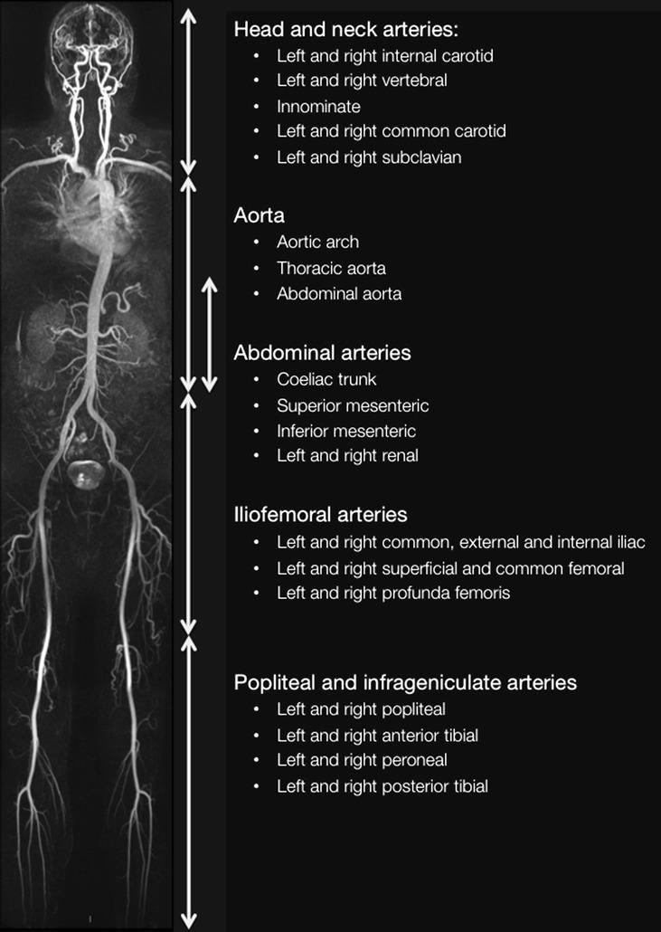
A whole-body diagnostic image presented as a Maximum Intensity Projection (MIP) of a Magnetic Resonance Angiogram (MRA). The visual provides a comprehensive coronal overview of the human arterial system from the cranium to the lower extremities, accompanied by descriptive labels of vascular territories. The content is divided into five anatomical stations: (1) Head and neck arteries, including carotids, vertebrals, and subclavians; (2) Aorta, encompassing the arch, thoracic, and abdominal segments; (3) Abdominal arteries, detailing the celiac trunk, superior/inferior mesenteric, and renal arteries; (4) Iliofemoral arteries, showing the common/external/internal iliac and femoral systems; and (5) Popliteal and infrageniculate arteries, highlighting the trifurcation into anterior/posterior tibial and peroneal vessels. The image serves as an educational reference for whole-body vascular anatomy and systematic assessment of arterial patency, atherosclerosis, or stenotic lesions. The visualization highlights the continuous, tapering morphology of the arterial tree and its major branch points, essential for radiology trainees and vascular specialists.
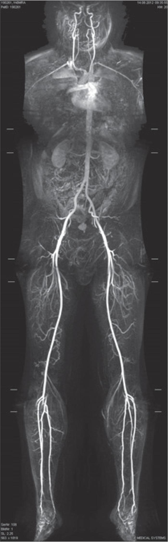
This diagnostic image is a coronal maximum intensity projection (MIP) reconstruction from a contrast-enhanced whole-body Magnetic Resonance Angiography (MRA). It demonstrates the entire human arterial system from the head to the feet in a 52-year-old asymptomatic male. Key visible structures include the bilateral carotid and vertebral arteries in the neck, the aortic arch with its primary branches (brachiocephalic, left common carotid, and left subclavian arteries), and the descending thoracic and abdominal aorta. In the abdomen, the renal arteries are visible supplying the kidneys. The aorta bifurcates at the pelvis into the common iliac arteries, which further divide into the internal and external iliac arteries. The arterial tree continues into the lower extremities via the femoral arteries, popliteal arteries at the knees, and the trifurcation into the anterior tibial, posterior tibial, and peroneal arteries in the calves. The image serves as a comprehensive educational overview of systemic arterial anatomy, showcasing normal vessel continuity, caliber, and branching patterns in a screening context.
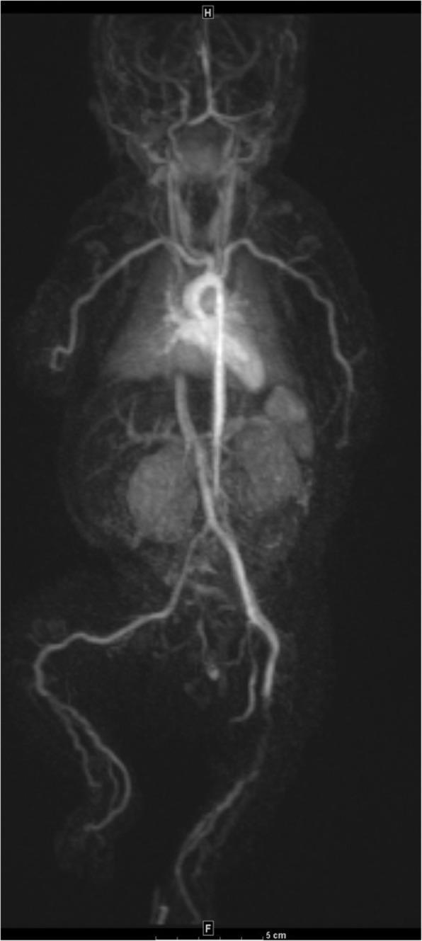
This diagnostic image is a whole-body magnetic resonance angiography (MRA) of a one-month-old neonate, illustrating systemic arterial abnormalities. The study reveals diffuse, bilateral tortuosity and gadolinium enhancement of several medium-sized arteries. In the upper extremities, the distal axillary and brachial arteries exhibit marked irregularity and undulating courses. Similarly, in the lower extremities, the distal femoral, popliteal, posterior tibialis, and anterior tibialis arteries demonstrate significant tortuosity and uniform wall enhancement. The gadolinium uptake along these vessel walls indicates active inflammation or increased vascular permeability, highly suggestive of a neonatal vasculitis or systemic vasculopathy. Notably, the major central vessels, including the aorta, pulmonary arteries, renal arteries, and celiac trunk, appear normal in caliber and course without abnormal enhancement. The contrast between the affected peripheral medium-sized vessels and the spared large vessels is a key diagnostic feature for pediatric rheumatological or inflammatory vessel disorders.
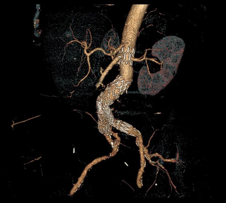
This diagnostic image is a 3D computed tomography (CT) angiography reconstruction of the abdominal aorta and its branches, specifically demonstrating a late complication of endovascular aneurysm repair (EVAR). The image reveals a complete structural failure of the EVAR stent-graft system. Key findings include the total disconnection and distal migration of the main body stent-graft from the bare suprarenal metal stent, which remains anchored near the renal arteries. The migrated main body of the endograft is positioned lower in the abdominal aorta and displays a sharp, focal kinking in the left iliac extension, likely compromising luminal flow. Anatomically, the renal arteries are visible as landmarks, with the right renal artery appearing partially covered by the displaced graft material. The bifurcation into the common iliac arteries is clearly visualized, showing the longitudinal extent of the migrated device. This image serves as a significant clinical example of device migration and component separation, highlighting the necessity of long-term radiological surveillance for post-EVAR patients.
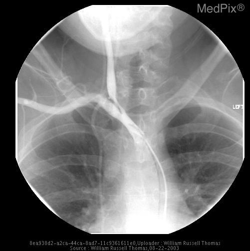
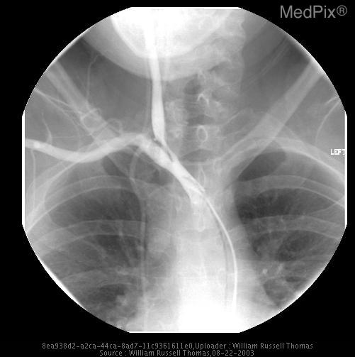
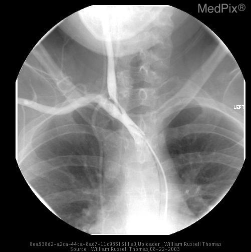
aortic arch branches brachiocephalic common carotid subclavian
1. The big picture
- Systemic circuit: The left ventricle feeds the aorta. Arteries branch to the tissues, and veins return blood through the SVC and IVC to the right atrium.
- Pulmonary circuit: The right ventricle feeds the pulmonary trunk, then the right and left pulmonary arteries to the lungs. Four pulmonary veins return blood to the left atrium.
- Rule of thumb: Deep veins accompany arteries and share their names. Superficial veins run alone in the subcutaneous tissue. Perforating veins connect the two.
2. Aorta and its segments
| Segment | Branches |
|---|---|
| Ascending | Right and left coronary arteries |
| Arch | Brachiocephalic trunk, left common carotid, left subclavian |
| Descending thoracic | Posterior intercostals, bronchial, esophageal, mediastinal, subcostal and superior phrenic arteries |
| Abdominal (T12 to L4) | Paired: inferior phrenic, lumbar, middle suprarenal, renal, gonadal. Unpaired: celiac (T12), SMA (L1), IMA (L3), median sacral |
| Terminal (L4) | Right and left common iliac arteries |
- The arch becomes the descending aorta at about the T4-T5 level. The brachiocephalic trunk, left common carotid and left subclavian arise from it (Fischer's Mastery of Surgery, "Arch of Aorta").
- Variants are common. The left common carotid can arise from the brachiocephalic trunk (the "bovine arch"). The vertebral artery can arise directly from the arch (Imaging Anatomy Text and Atlas Vol 3, "Aortic Arch"; Gray's Anatomy for Students).
3. Head and neck
- Common carotid: The right arises from the brachiocephalic trunk and the left from the arch. Each bifurcates at about C4 (upper border of the thyroid cartilage).
- External carotid branches: superior thyroid, ascending pharyngeal, lingual, facial, occipital, posterior auricular, then terminally the maxillary and superficial temporal.
- Internal carotid: It has no neck branches. It enters through the carotid canal, passes through the cavernous sinus, and gives off the ophthalmic, posterior communicating, anterior choroidal, anterior cerebral and middle cerebral arteries.
- Vertebral: It arises from the 1st part of the subclavian and ascends through the C6 to C1 transverse foramina. It enters via the foramen magnum and the two vertebrals join to form the basilar artery, which ends as the posterior cerebral arteries.
- Circle of Willis: It joins the anterior and posterior circulations.
- Dural venous sinuses: They drain to the sigmoid sinus, then the internal jugular vein. The internal jugular exits via the jugular foramen and runs in the carotid sheath.
- Joining the subclavian: The internal jugular joins the subclavian to form the brachiocephalic vein.
- Other drainage: The external jugular, anterior jugular and vertebral veins also drain the region.
- Danger area: The facial vein connects via the angular and ophthalmic veins to the cavernous sinus. This is a route for spread of infection.
4. Upper limb
- Subclavian: The branches are the vertebral, internal thoracic, thyrocervical trunk and costocervical trunk. It ends at the lateral border of the 1st rib.
- Axillary: It runs from the 1st rib to the lower border of teres major. The branches are the superior thoracic, thoracoacromial, lateral thoracic, subscapular and the anterior and posterior circumflex humeral arteries.
- Brachial: The main branch is the profunda brachii. It runs in the medial arm to the cubital fossa, where it divides.
- Radial and ulnar: The ulnar gives off the common interosseous artery. The radial pulse is felt at the wrist.
- Palmar arches: The superficial arch is mainly ulnar. The deep arch is mainly radial.
- Deep veins: The venae comitantes accompany the arteries. They form the brachial vein, then the axillary vein, then the subclavian vein.
- Cephalic vein: It runs from the anatomical snuffbox up the lateral arm. It lies in the deltopectoral groove and pierces the clavipectoral fascia to drain into the axillary vein.
- Basilic vein: It runs up the medial arm, pierces the deep fascia in mid-arm, and joins the brachial veins.
- Median cubital vein: It links the cephalic and basilic veins in the cubital fossa and is used for venepuncture.
5. Thorax
- Internal thoracic artery: It arises from the subclavian and gives off the anterior intercostal arteries. It ends as the superior epigastric and musculophrenic arteries.
- Posterior intercostals: They arise from the descending aorta. The chest wall is supplied by both the posterior and anterior sets (Thieme Atlas, General Anatomy and Musculoskeletal System).
- Azygos system: The azygos, hemiazygos and accessory hemiazygos veins drain the posterior chest wall. The azygos arches over the right main bronchus into the SVC.
- SVC: It is formed by the two brachiocephalic veins and enters the right atrium.
- Pulmonary vessels: The pulmonary arteries carry deoxygenated blood and the pulmonary veins carry oxygenated blood. The bronchial arteries (from the aorta) supply the lung tissue itself.
- Heart:
- The left coronary artery divides into the LAD and circumflex. The right coronary artery gives off the marginal and posterior interventricular (PDA) branches.
- The cardiac veins (great, middle and small) drain into the coronary sinus, which opens into the right atrium.
6. Abdomen
- Celiac trunk: It gives the left gastric, splenic and common hepatic arteries. The common hepatic gives the proper hepatic, gastroduodenal and right gastric arteries.
- SMA: It gives the inferior pancreaticoduodenal, jejunal and ileal, ileocolic, right colic and middle colic arteries.
- IMA: It gives the left colic, sigmoid and superior rectal arteries.
- Renal arteries: They arise at L1-L2 below the SMA.
- IVC: It forms at L5 from the two common iliac veins and passes through the diaphragm at T8 to reach the right atrium.
- IVC tributaries: lumbar, right gonadal, renal, right suprarenal, inferior phrenic and hepatic veins. The left gonadal and left suprarenal veins drain into the left renal vein instead.
- Hepatic portal system: The SMV and splenic vein join behind the neck of the pancreas to form the portal vein. The IMV drains into the splenic vein.
- Blood passes through the liver sinusoids, then the hepatic veins, then the IVC.
- Portosystemic anastomoses: These open up in portal hypertension.
- Esophageal: left gastric to azygos (varices).
- Rectal: superior rectal to middle and inferior rectal (hemorrhoids).
- Umbilical: paraumbilical to epigastric veins (caput medusae).
- Retroperitoneal.
7. Pelvis and perineum
- Common iliac: It divides into the external and internal iliac arteries.
- Internal iliac, anterior division: umbilical (superior vesical), obturator, inferior vesical or uterine, middle rectal, internal pudendal, inferior gluteal.
- Internal iliac, posterior division: iliolumbar, lateral sacral, superior gluteal.
- Ovarian and testicular arteries: They arise from the aorta, not the iliacs.
- Uterine artery: It crosses over the ureter ("water under the bridge").
- Pelvic veins: They form plexuses (rectal, vesical, uterine) that drain to the internal iliac vein, then the common iliac vein, then the IVC.
8. Lower limb
- External iliac: It becomes the femoral artery at the inguinal ligament.
- Femoral: In the femoral triangle the order from lateral to medial is nerve, artery, vein. The main branch is the profunda femoris, which gives the circumflex femoral and perforating arteries.
- Popliteal: The femoral becomes the popliteal at the adductor hiatus. It lies in the popliteal fossa and gives off the genicular branches.
- Anterior tibial: It passes through the interosseous membrane and becomes the dorsalis pedis.
- Tibioperoneal trunk: It gives the posterior tibial artery, which passes behind the medial malleolus and splits into the medial and lateral plantar arteries. It also gives the peroneal artery.
- Deep veins: They run with the arteries (tibial, popliteal, femoral, then external iliac).
- Great saphenous vein: It starts in front of the medial malleolus and runs up the medial leg and thigh. It drains into the femoral vein at the saphenofemoral junction, through the saphenous opening about 3 to 4 cm below and lateral to the pubic tubercle.
- Small saphenous vein: It runs behind the lateral malleolus and up the back of the calf to join the popliteal vein.
- Perforators and valves: Perforating veins connect the superficial and deep systems. One-way valves direct flow toward the heart. Valve failure causes varicose veins.
9. Venous return summary
Recent evidence note
- Vertebral artery variants (PMID 40542940)
- Hepatic arterial variants (PMID 41192686)
- Brachial artery variants (PMID 41305981)