femoral, umbilical and paraumbilical hernias
Reading File
Finding Sources
Finding Sources
Finding Sources
Reading File
Reading File
Finding Sources
Reading File
Finding Sources
Reading File
Reading File
Reading File
Reading File
Reading File
Loading Image
I now have comprehensive content from the textbooks. Let me compile the full answer.
Femoral, Umbilical, and Paraumbilical Hernias
FEMORAL HERNIA
Anatomy & Pathophysiology
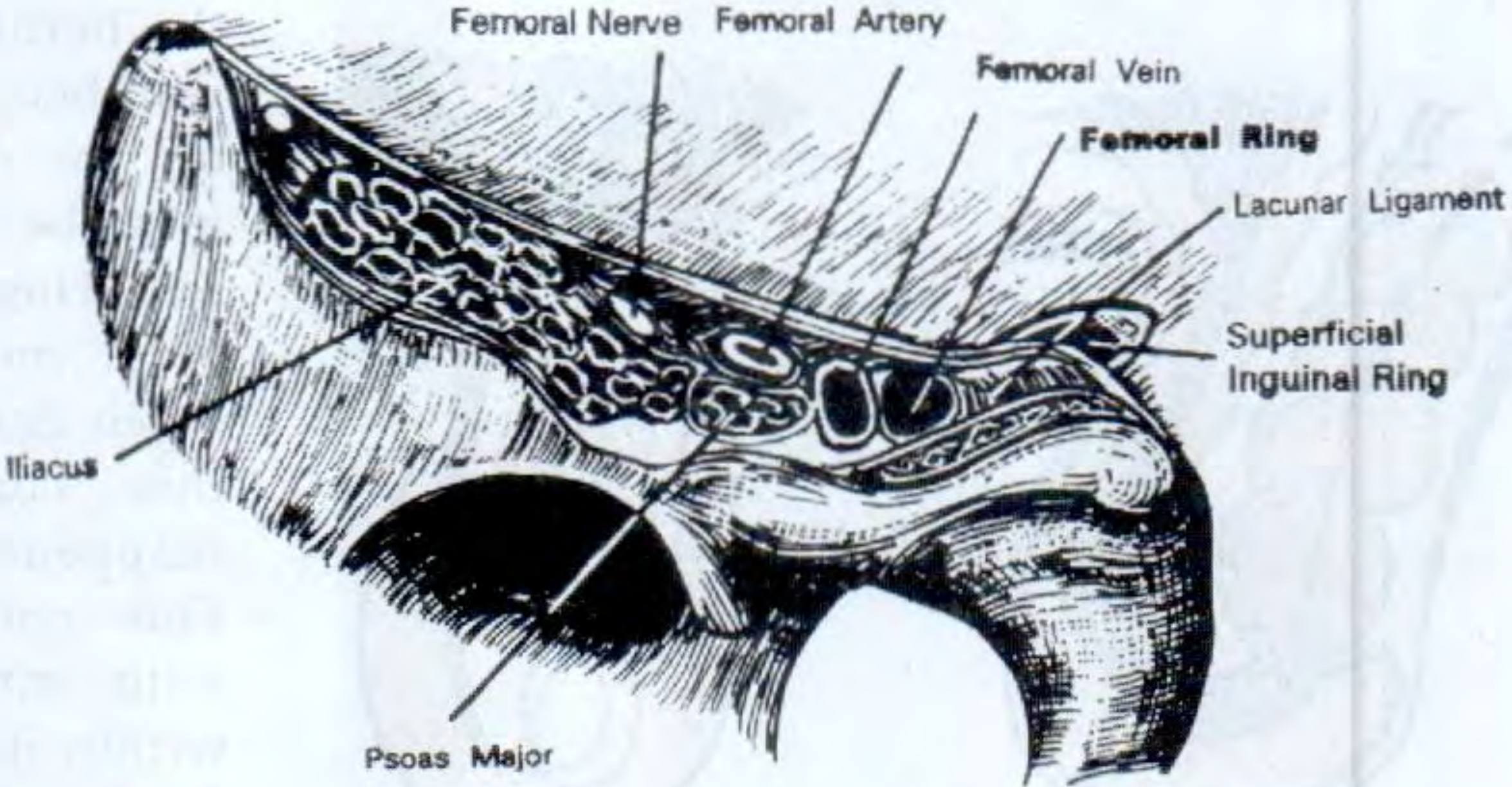
A femoral hernia is a protrusion of extraperitoneal tissue, peritoneum and sometimes abdominal contents through the femoral canal. The boundaries of the femoral canal are:
| Border | Structure |
|---|---|
| Lateral | Femoral vein |
| Anterior | Inguinal ligament |
| Posterior | Iliopectineal (Astley Cooper's) ligament / pelvic bone |
| Medial | Lacunar (Gimbernat's) ligament — sharp, unyielding edge |
The hernia passes through the femoral canal and emerges superficially through the saphenous opening (~4 cm below and lateral to the pubic tubercle). Once past this opening into loose areolar tissue, it expands, creating the classic retort shape — with the bulbous extremity curving upward, sometimes above the inguinal ligament.
Epidemiology
- Rare before age 20; peak incidence >50 years
- More common in women (2:1), but even in women the commonest groin hernia is still inguinal
- Right side affected twice as often as left
- 20% bilateral
- 50% present as an emergency with strangulation — the rigid femoral canal makes this the highest-risk hernia for strangulation
Diagnosis
- Swelling appears below and lateral to the pubic tubercle (vs. inguinal hernia which is medial to the pubic tubercle and above the inguinal ligament)
- Often rapidly irreducible — loses cough impulse early
- May be only 1–2 cm; easily mistaken for a lymph node
- As it enlarges, curves superiorly — can mimic a direct inguinal hernia
- Invagination test: inguinal canal will be empty
- Diagnostic error is common, leading to delayed treatment
Differential Diagnosis
- Saphena varix — disappears completely on lying down; fluid thrill on percussion (Schwartz's test); venous hum on auscultation
- Enlarged lymph nodes — including node of Cloquet (within femoral canal), which mimics an irreducible femoral hernia exactly
- Psoas abscess — cold abscess tracking from Pott's disease; reducible with cough impulse; lies lateral to femoral vessels (femoral hernia is medial)
- Inguinal hernia (direct)
Surgical Repair
Surgery is mandatory — there is no alternative — and should be treated with urgency given high strangulation risk.
| Approach | Description | Indication |
|---|---|---|
| Low (Lockwood) | Transverse incision over hernia; suitable for local anaesthesia; sutures between inguinal and pectineal ligaments | Elective, no bowel resection risk |
| Inguinal (Lotheissen) | Inguinal canal incision; transversalis fascia opened; access to extraperitoneal space | Elective |
| High (McEvedy/Nyhus) | Transverse incision above inguinal canal, preperitoneal space; best access for bowel inspection and resection | Emergency with strangulation risk |
| Laparoscopic (TEP/TAPP) | Standard mesh in extraperitoneal plane | Elective; preferred in women (reduces misdiagnosis of inguinal vs. femoral) |
Caution with Lockwood approach: if bowel resection is needed, the anastomosis cannot be returned through the narrow femoral canal — use McEvedy instead.
UMBILICAL HERNIAS
Classification — Four Varieties
Any hernia closely related to the umbilicus falls under this heading:
| Type | Description |
|---|---|
| Exomphalos | Abdominal contents protrude into the umbilical cord covered by a transparent diaphanous membrane — congenital defect |
| Congenital umbilical hernia | Through centre of umbilical scar; common in Black infants; wide neck; rarely strangulates; ~90% resolve spontaneously in first 5 years |
| Acquired umbilical hernia | Adults; protrudes through the umbilical scar; usually due to raised intra-abdominal pressure (pregnancy, ascites, bowel distension, ovarian cyst, fibroid) |
| Para-umbilical hernia | Most common acquired variety — through a defect adjacent to the umbilicus, usually just above it |
UMBILICAL HERNIA IN INFANTS
- Occurs in up to 10% of infants; more common in prematures
- Incidence in Black infants is up to 8× higher than in white infants
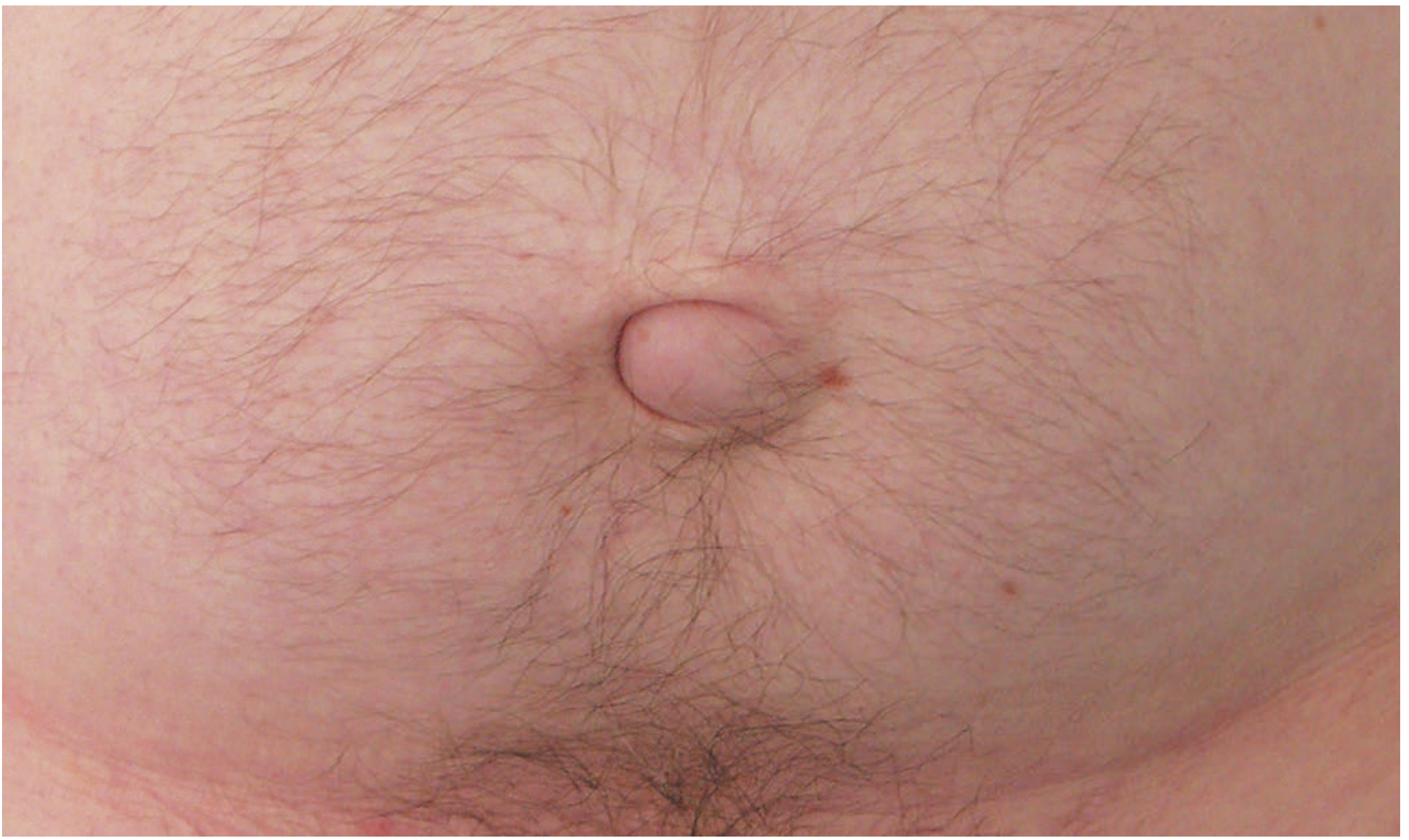
- Classic conical shape on crying
- Obstruction/strangulation extremely uncommon under 3 years
- Treatment: Conservative below age 2 — 95% resolve spontaneously; surgical repair if persists beyond age 2
Surgery (children): Small curved incision below umbilicus → define neck of sac → reduce contents → close sac → close linea alba defect with interrupted slowly absorbable sutures.
UMBILICAL HERNIA IN ADULTS
Predisposing Factors
- Obesity, pregnancy, liver cirrhosis with ascites (stretch and thin the linea alba)
- Under current guidelines, hernias in the immediate vicinity of the umbilicus (including paraumbilical) are all termed "umbilical"
Clinical Features
- Overweight men or postpartum women
- Bulge typically slightly to one side of umbilical depression → crescent-shaped umbilicus
- Small hernias contain extraperitoneal fat or omentum; larger ones may contain small or large bowel
- Prone to becoming irreducible, obstructed, and strangulated (narrow neck relative to sac size)
Treatment
- Surgery advised when hernia contains bowel (high strangulation risk)
- Small asymptomatic hernias may be observed
- Open repair options:
- Defects <1 cm: simple suture repair (fascia not under tension)
- Defects up to 2 cm: Mayo repair (transverse incision; "waistcoat over trousers" overlapping fascial closure with non-absorbable sutures)
- Defects >2 cm: mesh repair strongly recommended (high recurrence with suture alone); current evidence supports mesh even for small defects
- Laparoscopic repair: ports placed laterally, well away from defect; mesh fixed to cover defect
Special Circumstances
- Pregnancy-related: advise weight loss, abdominal exercises, and delay surgery until postpartum; may resolve spontaneously within months
- Liver cirrhosis (Child's B/C): extremely high surgical mortality/morbidity; careful patient selection; use fine continuous sutures to minimise post-operative ascites leakage
PARAUMBILICAL HERNIA
Definition & Anatomy
The most common acquired umbilical hernia. Occurs through a defect adjacent to (usually just above) the umbilicus, between the two recti in the linea alba. The lower half of the fundus of the sac is typically covered by the umbilicus.
Key Diagnostic Features
- Develops in middle and old age
- Obese women most commonly affected
- Main symptoms: pain and swelling — if small, pain/discomfort may dominate
- Surface smooth, edge distinct (unless patient is very obese)
- Contents: omentum (firm lump) or bowel (soft, resonant to percussion)
- Many are irreducible (contents adhere to sac or narrow neck); when reducible, the firm fibrous edge of the linea alba defect can be palpated
- Intermittent abdominal pain due to rigid linea alba defect that does not enlarge proportionately — strangulation is not common despite the narrow neck
Treatment
- Same principles as adult umbilical hernia
- Mayo repair (overlap technique) or mesh repair depending on defect size
- Mesh repair preferred for larger defects given high recurrence rates
Summary Comparison
| Feature | Femoral | Umbilical (adult) | Paraumbilical |
|---|---|---|---|
| Sex predominance | Women (2:1) | Women > men | Obese women |
| Location | Below & lateral to pubic tubercle | Through umbilical scar | Adjacent to umbilicus (above) |
| Strangulation risk | Very high (rigid canal) | High (narrow neck) | Less common |
| Emergency presentation | 50% | Less common | Less common |
| Common content | Bowel, omentum | Omentum → bowel | Omentum → bowel |
| Surgery urgency | Urgent (mandatory) | When bowel involved | Elective |
| Preferred repair | Lockwood (elective), McEvedy (emergency), Laparoscopic in women | Mayo (<2cm) / Mesh (>2cm) | Mayo / Mesh |
Sources: Bailey & Love's Short Practice of Surgery 28th Ed., S Das Manual of Clinical Surgery 13th Ed.
This is a shared conversation. Sign in to Orris to start your own chat.