
Management of laceration on forehead and knee
laceration forehead wound closure suture technique
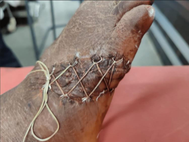
This clinical photograph shows a superior view of a traumatic laceration on the dorsolateral aspect of a human foot, illustrating a specialized wound closure technique. The wound is managed using a 'shoelace' suture technique, where a thin, light-colored suture material (Ethibond) is threaded in a criss-cross, zig-zag pattern through surgical skin staples placed along the wound margins. This technique allows for gradual approximation of skin edges to manage tension and skin loss. The underlying wound bed appears dark and granulated, consistent with a healing stage involving platelet-rich plasma (PRP) infiltration. The surrounding skin is dry, hyperkeratotic, and slightly desquamated, with no evident signs of acute purulent infection. This image demonstrates an orthopedic surgical approach for managing complex soft tissue defects associated with high-energy trauma and open fractures, specifically highlighting tension-reduced primary closure methods in areas with limited skin mobility.
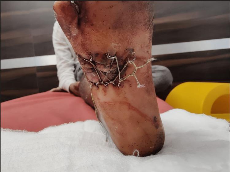
This clinical photograph shows the plantar aspect of a human foot featuring a complex wound closure. The image demonstrates the 'shoelace suture technique' applied to a large laceration. A heavy, braided suture material is woven in a criss-cross pattern across the wound bed, anchored at the margins by surgical skin staples. This tension-distributing method is used to gradually approximate wound edges in cases of significant skin loss or soft tissue trauma, such as a Gustilo-Anderson type IIIB fracture. The surrounding tissue shows signs of post-traumatic changes, including localized hyperpigmentation, bruising, and inflammatory erythema. Frayed ends of the suture material are visible at the anchoring points. The plantar skin appears thickened and exhibits some superficial abrasions. This visual serves as an educational example of advanced wound management in orthopedic trauma, specifically illustrating mechanical skin stretching combined with biological adjuncts like platelet-rich plasma (PRP) for secondary closure.
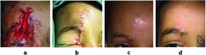
This clinical progression series comprises four photographs documenting the repair and healing of a traumatic facial laceration. Panel (a) shows the initial injury: a vertical, deep 6-cm laceration on the forehead involving the skin, subcutaneous fat, and muscle, characterized by tissue gaping and fresh blood. Panel (b) depicts the wound immediately postoperative after primary closure using autologous hair filament sutures; the edges are well-approximated with visible interrupted stitches and minimal peri-wound erythema. Panel (c) shows the follow-up at 5 weeks, demonstrating a maturing, slightly erythematous linear scar without suture track marks or infection. Panel (d) illustrates the 6-month long-term outcome, displaying a soft, flat, hypopigmented scar that blends with the surrounding skin texture. The series highlights surgical techniques aimed at minimizing permanent scarring and suture-induced ‘railroad’ marks in pediatric plastic surgery and emergency medicine contexts.
Management of Lacerations: Forehead and Knee
General Principles of Wound Management
| Phase | Steps |
|---|---|
| Preparation | Analgesia/anaesthesia, tetanus prophylaxis, antibiotic prophylaxis (if indicated), wound irrigation |
| Wound | Exploration, debridement, haemostasis, repair structures |
| Closure | Skin closure without tension, suture choice, optimal dressings |
| Follow-up | Suture removal, physiotherapy, scar management |
Step-by-Step Management
1. Initial Assessment
- Take history: mechanism, time of injury, immunization status, allergies, comorbidities (diabetes, immunosuppression)
- Perform neurovascular examination of the involved area
- For knee lacerations: specifically assess for joint cavity communication (see below)
- For forehead lacerations: assess for intracranial injury, skull fracture, galeal involvement
- X-ray / CT if bony injury or foreign body is suspected
2. Anaesthesia
- Forehead: Topical agents (lidocaine-epinephrine-tetracaine or EMLA) reduce pain of injection. Local infiltration or supraorbital/supratrochlear nerve block. Epinephrine-containing agents are acceptable on the forehead and face (rich vascularity helps control bleeding).
- Knee: Bupivacaine 0.5% without epinephrine, regional or local infiltration. For extremity wounds, a bloodless field with a tourniquet or sphygmomanometer is useful.
3. Wound Preparation
- Do NOT shave surrounding hair - cut if visualization needed (shaving increases infection risk)
- Prepare surrounding skin with chlorhexidine-alcohol solution
- Irrigate copiously with normal saline (high-pressure irrigation) or 1% povidone-iodine solution to reduce contamination
- Debride all devitalized tissue and foreign material; excise non-viable tissue until healthy bleeding margins are seen
4. Tetanus Prophylaxis
| Immunization Status | DTaP (0.5 mL) | TIG (250 IU) |
|---|---|---|
| Fully immunized, last booster < 10 years | No | No |
| Fully immunized, last booster > 10 years | Yes | No |
| Incomplete series (< 3 injections) | Yes | Yes |
Forehead Laceration - Specific Management
Classification
- Superficial: Frontalis muscle NOT involved
- Deep: Frontalis muscle IS involved - requires layered closure
Closure Technique (Tintinalli's Emergency Medicine, p. 328)
- Key stitches first - align skin tension lines and the hairline before general closure
- Superficial lacerations: Close skin with 6-0 nonabsorbable interrupted sutures or tissue adhesive
- Deep lacerations (muscle involved):
- Close the muscle fascia: buried 5-0 absorbable suture
- Approximate the dermis: buried subcuticular stitch
- Close the epidermal layer: 6-0 nonabsorbable simple interrupted sutures, or skin closure strips, or tissue adhesive
- Tape/adhesive strips (Steri-Strips): The forehead is one of the areas MOST suited to tape closure - it is low tension, relatively flat, and highly vascular. Use when the wound is superficial, straight, and under little tension.
Suturing Guidelines for Forehead
| Layer | Suture Type | Size | Removal |
|---|---|---|---|
| Muscle fascia | Absorbable | 5-0 | Not removed |
| Dermis | Absorbable | 5-0 | Not removed |
| Skin | Nonabsorbable monofilament | 6-0 | 5-7 days |
- Use adhesive strips or tissue adhesive if the patient is at risk for keloids or hypertrophic scars
- Cosmetic note: Wounds parallel to skin tension lines (horizontal forehead lines) give best cosmetic outcomes. These run perpendicular to the frontalis muscle.
Knee Laceration - Specific Management
Key Concern: Joint Communication
- This becomes an orthopaedic emergency - requires joint washout, usually in theatre
- Perform the saline load test (inject 60-100 mL saline into the joint through a separate needle; if saline leaks from the wound, joint penetration is confirmed)
- Refer to orthopaedics if joint is involved
Closure Considerations
- The knee is a high-tension, high-movement area. Tape closure is NOT ideal over joint areas.
- Use 3-0 or 4-0 nonabsorbable monofilament interrupted sutures for skin closure
- Deep/fascial layers: 3-0 or 4-0 absorbable suture
- Wound over the patella or patellar tendon: assess for tendon integrity before closure
- Consider splinting post-closure to reduce tension and movement at the wound site
- Suture removal: 10-14 days (joint-area wounds are removed later than facial wounds due to tension)
Wound Closure Summary for Extremity Wounds
| Feature | Management |
|---|---|
| Superficial, no tension | Tape (Steri-Strips) or interrupted sutures |
| Deep, high tension | Layered closure - deep absorbable + skin nonabsorbable |
| Joint involvement | Do NOT close primarily - orthopaedic referral, joint washout |
| Contaminated wound | Leave open, delayed primary closure at 4-5 days |
Aftercare and Follow-up
- Dressings: Non-adherent dressing for 24-48 hours; keep wound dry
- Pressure dressing: Consider over deep lacerations for first 24 hours to prevent haematoma
- Antibiotics: Generally not required for clean lacerations. Consider for contaminated wounds, bites, immunocompromised patients, or wounds >6 hours old
- Suture removal:
- Face/forehead: 5-7 days (to minimize scarring)
- Knee/extremities over joints: 10-14 days
- Scar management: Sunscreen, silicone gel/sheets after healing; monitor for keloids
- Physiotherapy: For knee lacerations, gentle range of motion once wound is stable
- Red flags to return: Increasing pain, redness, swelling, discharge, fever (signs of infection)
Wound Closure Options - Summary
| Method | Best For | Notes |
|---|---|---|
| Simple interrupted sutures | Most lacerations | Versatile; allows individual tension control |
| Tissue adhesive (cyanoacrylate) | Superficial, low-tension, facial wounds | Good cosmesis; no suture removal needed |
| Steri-Strips / tape | Forehead, chin, thorax; non-joint areas | Not suitable over joints or wet/hairy areas |
| Staples | Scalp | Fast, good haemostasis |
| Hair apposition | Scalp | No sutures needed; requires hair strands |
| Delayed primary closure | Contaminated, infected, bite wounds | Wound left open 4-5 days, then closed |