
Tell me everything about acne
acne vulgaris treatment management
acne vulgaris types comedones papules pustules
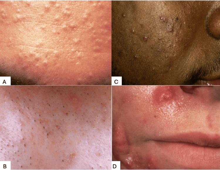
This composite of four clinical photographs illustrates different clinical morphologies of acne vulgaris, highlighting the spectrum from non-inflammatory to inflammatory lesions. Panel A displays closed comedones (whiteheads), appearing as small, skin-colored, dome-shaped papules without an apparent follicular opening, densely clustered on the forehead. Panel B shows open comedones (blackheads), characterized by dilated follicular orifices containing dark, oxidized keratinous plugs. Panel C demonstrates mixed inflammatory acne, presenting as a combination of erythematous papules and pustules on pigmented skin, indicating moderate follicular inflammation. Panel D captures severe nodulocystic acne, featuring large, deep-seated, painful-appearing inflammatory nodules and cysts with significant surrounding erythema and edema, which carry a higher risk of permanent scarring. These images serve as a dermatological reference for classifying acne severity and type, which is critical for determining appropriate therapeutic interventions ranging from topical retinoids for comedonal types to systemic therapies for nodulocystic presentations.
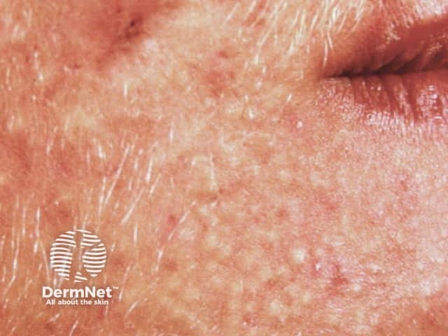
Clinical photograph of adult facial skin showing acne vulgaris. Modality: color digital photography; close-up frontal view of left cheek and perioral region. The skin shows numerous small inflammatory papules and closed comedones within erythematous background. Surface shows rough texture, post-inflammatory hyperpigmentation; no visible pustules or nodules beyond papules; sebaceous prominence; mild focal scarring. The distribution is predominantly in the central to lower cheek and perioral zones; symmetric involvement is suspected though lateralization not certain in this single frame. The visual features correspond to acne vulgaris, predominantly non-inflammatory comedones and mild inflammatory papules; differential includes rosacea (erythema with flushing and telangiectasia), folliculitis, perioral dermatitis. Clinically significant due to risk of scarring if untreated; management typically includes topical retinoids (tretinoin, adapalene), benzoyl peroxide, salicylic acid, topical/oral antibiotics, hormonal therapy in women; isotretinoin in severe cases. This image provides educational reference for adult-onset facial acne assessment, severity grading (milDer-type with comedones and papules), and therapy planning. Notable clinical context: identify pilosebaceous unit involvement, comedone formation, and post-inflammatory hyperpigmentation. Educational value includes recognizing lesion morphology, distinguishing acne from similar mimics, and selecting appropriate imaging-assisted documentation for follow-up, patient education, and clinical trials evaluating novel anti-acne therapies. This supports standardized severity assessment and treatment planning.
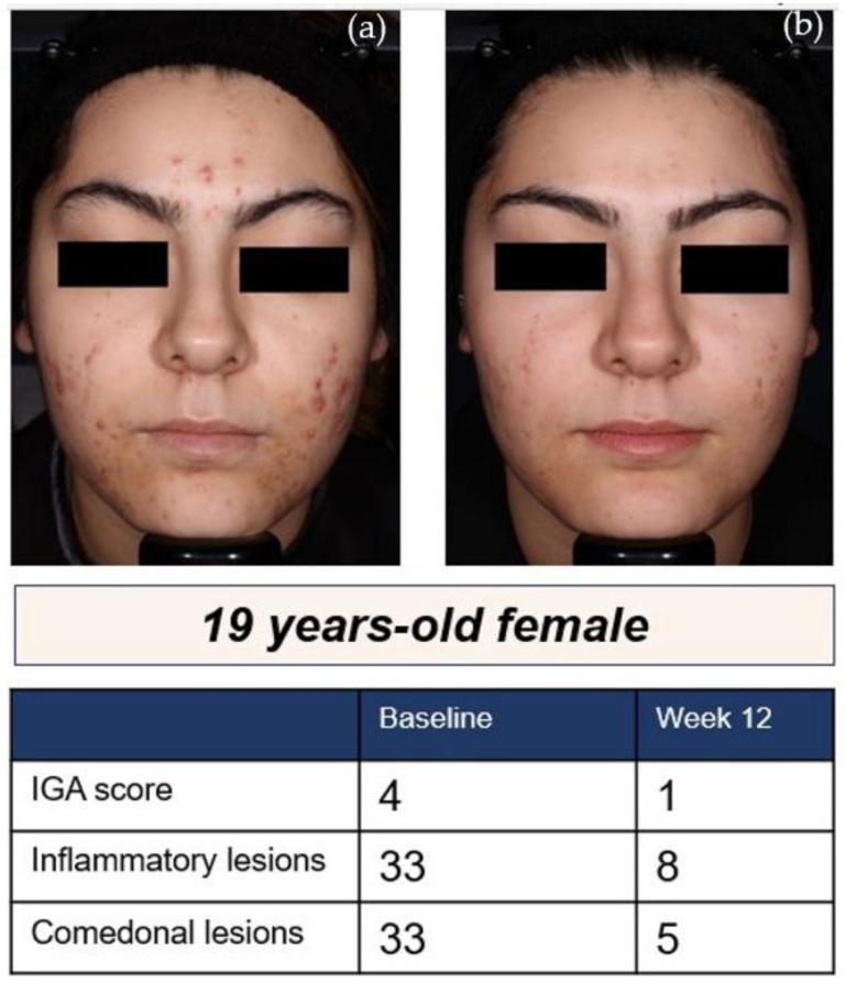
This clinical photograph set demonstrates the therapeutic response of moderate-to-severe acne vulgaris in a 19-year-old female patient. The composite image includes a baseline photograph (a) and a follow-up photograph (b) after 12 weeks of treatment with the narrow-spectrum tetracycline antibiotic, sarecycline. At baseline, the patient presents with numerous inflammatory papules, pustules, and comedones distributed across the forehead, cheeks, and chin, corresponding to an Investigator's Global Assessment (IGA) score of 4. The 12-week post-treatment image shows a significant reduction in lesion count and severity, reflecting an IGA score of 1 (almost clear). An accompanying data table quantifies this improvement, showing a reduction in inflammatory lesions from 33 to 8 and comedonal lesions from 33 to 5. The images highlight clinical features such as erythema, inflammatory morphology, and post-inflammatory changes, serving as a visual case study for dermatological pharmacological efficacy and acne management.
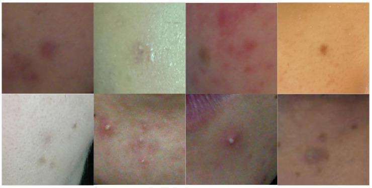
This composite clinical photograph displays multiple cropped views of facial skin demonstrating the morphological diversity of acne vulgaris. The images showcase a range of lesions across different skin phototypes, from fair to dark tones. Visible pathology includes erythematous papules (inflamed red bumps), pustules characterized by central white purulent material (whiteheads), and closed comedones. Some regions exhibit post-inflammatory hyperpigmentation, appearing as brownish or purplish macules following lesion resolution. The distribution varies from discrete, isolated lesions to clusters of inflamed, irregularly shaped papules. Textural irregularities are evident, ranging from smooth elevations to rough, crusting surfaces and prominent follicular openings. This collection serves as a dermatological reference for identifying acne manifestations, highlighting variations in lesion color (red, pink, white, brown) and morphology (papular, pustular, comedonal) essential for diagnostic training and the development of automated medical imaging segmentation algorithms.
acne vulgaris treatment guidelines 2024 2025 update
Acne Vulgaris - A Complete Guide
1. Definition and Epidemiology
-
Affects approximately 85-95% of all teenagers to some degree
-
Greatest frequency between ages 15-18 years in both sexes
-
More common in males, but more persistent in females
-
For 12% of women and 3% of men, it persists into the mid-40s
-
Can begin as adult-onset acne in post-adolescent women (ages 20-35) who had no teenage acne
-
Primarily affects the face, neck, chest, back, and trunk - areas rich in sebaceous glands
-
Swanson's Family Medicine Review, p. 2438
2. Pathogenesis - The Four Pillars
Pillar 1: Increased Sebum Production
Pillar 2: Abnormal Follicular Keratinization (Comedogenesis)
Pillar 3: Cutibacterium acnes (C. acnes) Proliferation
- Hyaluronidase, lipases, proteases - enzymes that damage follicular walls
- Free fatty acids from sebum breakdown
- Chemotactic factors that attract neutrophils
- Interacts with toll-like receptors, triggering inflammation
Pillar 4: Inflammation
- Swanson's Family Medicine Review, p. 2459-2466; Goodman & Gilman's Pharmacological Basis of Therapeutics, p. 2091
3. Lesion Types and Classification
Non-Inflammatory Lesions
| Lesion | Description |
|---|---|
| Open comedone (blackhead) | Dilated follicular opening with dark, oxidized keratin plug. Dark color is from melanin + oxidation, NOT dirt |
| Closed comedone (whitehead) | Blocked follicle with no surface opening; appears as a small, skin-colored dome-shaped papule |
Inflammatory Lesions
| Lesion | Description |
|---|---|
| Papule | Small (<5 mm), solid, raised, red bump; no pus |
| Pustule | Raised lesion with visible pus (white/yellow center) |
| Nodule | Large (>5 mm), solid, deep-seated, painful; high risk of scarring |
| Cyst | Deep, pus-filled, fluctuant; most likely to leave permanent scars |
Severity Grading
- Mild: Predominantly comedones; few papules/pustules; no nodules
- Moderate: Mix of comedones and inflammatory lesions; possible scarring
- Severe: Extensive papules/pustules + nodules/cysts; significant scarring risk
4. Special Forms of Acne
| Type | Features |
|---|---|
| Acne conglobata | Severe, interconnected nodules and cysts; mostly in males; significant scarring |
| Acne fulminans | Sudden onset severe inflammatory acne with systemic features (fever, arthralgia); rare |
| Neonatal acne | Appears within first weeks of life; maternal hormones; usually resolves spontaneously |
| Hormonal/adult female acne | Cyclic flares around menses; distribution along jawline/chin; linked to androgen excess or PCOS |
| Acne excoriee | Psychologically driven picking/scratching of acne lesions |
| Drug-induced acne (acneiform eruption) | From steroids, lithium, iodides, phenytoin, EGFR inhibitors; typically lacks comedones |
5. Triggers and Aggravating Factors
- Hormonal fluctuations: Menstrual cycles, PCOS, adrenal tumors
- Medications: Corticosteroids, anabolic steroids, lithium, phenytoin, some antiepileptics
- Cosmetics: Comedogenic products (pomade acne, acne cosmetica)
- Mechanical trauma: Pressure, friction, rubbing (acne mechanica - from helmets, chinstraps)
- Occupational exposures: Oils, tars, chlorinated compounds (chloracne)
- Diet: High glycemic index foods and dairy have some evidence for aggravation (evidence moderate)
- Stress: Induces androgen-like hormones and neuropeptides
6. Treatment - A Systematic Approach
Treatment Philosophy (2024 AAD Guidelines)
Topical Treatments
Retinoids (First-Line for all Acne)
- Tretinoin (prototype; apply in evening; UV-inactivated)
- Adapalene (comparable efficacy, better tolerability, available OTC)
- Tazarotene (most potent; most irritating)
- Trifarotene (FDA-approved; targets RAR-gamma; good for truncal acne)
Benzoyl Peroxide (BPO)
- Bactericidal: generates reactive oxygen species that kill C. acnes
- Does NOT cause antibiotic resistance - key advantage
- Available as 2.5%, 5%, 10% wash or gel
- Side effects: dryness, bleaching of fabric/hair
- Cornerstone of combination therapy
Topical Antibiotics
-
Clindamycin and erythromycin (topical)
-
Should never be used as monotherapy due to resistance risk
-
Must always be combined with benzoyl peroxide
-
Newer: topical minocycline (foam/gel) and topical dapsone
-
Azelaic acid: antibacterial + comedolytic + anti-inflammatory; safe in pregnancy; also treats post-inflammatory hyperpigmentation
-
Dermatology 2-Volume Set 5e, p. 543-545; Goodman & Gilman's, p. 2093
Oral Antibiotics (Moderate to Severe Acne)
Tetracyclines (Most Commonly Used)
- Doxycycline: better GI absorption than tetracycline; can take with food; photosensitizing
- Minocycline: less photosensitizing; risks include vestibular toxicity, drug-induced lupus, skin hyperpigmentation
- Sarecycline: narrow-spectrum tetracycline; FDA-approved specifically for acne; less disruption of gut/vaginal microbiome
- Tetracycline: must take on empty stomach; being replaced by doxycycline/minocycline
Macrolides
- Azithromycin, erythromycin (second-line when tetracyclines contraindicated)
Duration
-
Limit to 3-6 months maximum; taper once controlled
-
Transition to topical maintenance therapy to avoid prolonged antibiotic use
-
Goodman & Gilman's, p. 2094-2102
Hormonal Therapy (Females)
Combined Oral Contraceptives (COCs)
- FDA-approved for acne: Ortho Tri-Cyclen, Estrostep, Yaz (ethinyl estradiol + drospirenone), Alesse, Yasmin
- Reduce androgen-driven sebum production
- Take 3-6 months to show significant effect
Spironolactone
- Androgen receptor blocker; reduces sebum production
- Typical dose: 50-200 mg/day
- First-line hormonal option for adult female acne
- Requires monitoring for hyperkalemia
Other Antiandrogens
- Cyproterone acetate (available outside the US): potent antiandrogen
- Clascoterone (Winlevi): topical androgen receptor inhibitor; FDA-approved 2020; works locally without systemic hormonal effects; can be used in males too
Oral Isotretinoin (13-cis-Retinoic Acid) - Gold Standard for Severe Acne
- Massively suppresses sebum production (unique among retinoids)
- Normalizes follicular keratinization
- Reduces C. acnes colonization indirectly
- Has anti-inflammatory effects
- Severe nodulocystic/conglobate acne
- Moderate acne with scarring
- Acne resistant to conventional therapy (topical + oral antibiotics)
- Acne fulminans (with low-dose systemic corticosteroid)
- Psychologically devastating acne at any severity
- Starting dose: 0.5 mg/kg/day; titrated to 1 mg/kg/day as tolerated
- Low-dose regimens (0.3-0.5 mg/kg/day) can be effective with longer treatment
- Cumulative dose target: 120-150 mg/kg to minimize relapse
- Treatment duration: typically 4-6 months
- Lag period of 1-3 months before visible improvement
- ~1/3 of patients require a second course
- Teratogenicity (Category X) - causes severe birth defects; requires two forms of contraception
- Cheilitis (lip dryness) - nearly universal
- Xerosis (dry skin, eyes, nose)
- Initial acne flare (first 1-4 weeks)
- Elevated triglycerides and liver enzymes (monitoring required)
- Photosensitivity
- Myalgias
- Isotretinoin and neuropsychiatric effects: The 2024 AAD guidelines state it is unlikely to be associated with neuropsychiatric disorders or inflammatory bowel disease - reassuring update
- Dermatology 2-Volume Set 5e, p. 681-700; Swanson's Family Medicine Review, p. 2507-2511
Treatment Algorithm by Severity
| Severity | First-Line | Second-Line |
|---|---|---|
| Comedonal | Topical retinoid | Azelaic acid |
| Mild papulopustular | BPO + topical retinoid OR BPO + topical antibiotic | Azelaic acid, salicylic acid |
| Moderate (no scarring) | BPO + topical retinoid + topical antibiotic | Oral antibiotic + BPO + topical retinoid |
| Moderate (with scarring) - males | Oral antibiotic + BPO or topical retinoid | Oral isotretinoin |
| Moderate (with scarring) - females | Oral antiandrogen contraceptive + BPO + topical antibiotic | Oral isotretinoin |
| Severe papulopustular / Nodulocystic | Oral isotretinoin | - |
| Acne fulminans | Oral isotretinoin + low-dose oral corticosteroid | - |
Light and Laser-Based Therapies (Second-Line)
- Blue light (415 nm): Activates porphyrins in C. acnes, bactericidal
- Blue/red light combination: Anti-inflammatory (red) + bactericidal (blue)
- Photodynamic therapy (PDT): Aminolevulinic acid + light; more effective but more downtime
- 1450-nm diode laser: Targets sebaceous glands
- Energy-based devices + isotretinoin: A 2024 systematic review supports combining these for severe acne and acne scars
7. Acne Scars
Types of Acne Scars
| Type | Description |
|---|---|
| Ice-pick scars | Deep, narrow, pitted; most common |
| Boxcar scars | Broad, rectangular depressions with sharp edges |
| Rolling scars | Broad, shallow; undulating appearance |
| Hypertrophic/keloid scars | Raised; more common in skin of color |
| Post-inflammatory hyperpigmentation (PIH) | Not true scars; dark macules; especially prominent in darker skin tones |
Scar Treatments
- Fractional laser resurfacing
- Chemical peels
- Microneedling
- Subcision
- Dermal fillers
- Intralesional corticosteroids (for keloids/hypertrophic scars)
8. Psychosocial Impact
-
Depression, anxiety, and reduced self-esteem are common
-
Interpersonal and work-related difficulties
-
Isotretinoin treatment has been shown to significantly improve sociability and self-esteem
-
The psychological burden should always factor into treatment decisions - even mild acne can warrant aggressive treatment if psychological distress is significant
-
Dermatology 2-Volume Set 5e, p. 694
9. Special Populations
Pregnancy
- Safe: Azelaic acid, glycolic acid, topical erythromycin, topical clindamycin
- Use with caution: Benzoyl peroxide (limited data but generally considered safe)
- Avoid: All retinoids (topical and oral), tetracyclines, doxycycline, spironolactone
Adolescents
- Tetracyclines contraindicated under age 8
- Emphasize gentle skincare routine; avoid scrubbing
Skin of Color
- Higher risk of post-inflammatory hyperpigmentation (PIH)
- Azelaic acid is particularly useful (both anti-acne and depigmenting)
- Avoid overly aggressive treatments that worsen PIH
- Keloid scar formation risk is higher
10. Recent Evidence
- A 2025 systematic review (PMID: 39269130) highlights the role of the cutaneous microbiome beyond just C. acnes - diversity disruption may contribute to acne, and probiotic/prebiotic approaches are under investigation.
- A 2024 meta-analysis (PMID: 39179105) confirms increasing prevalence of antibiotic-resistant C. acnes, reinforcing the 2024 AAD guideline to limit antibiotic monotherapy.
- The 2024 AAD guidelines (Reynolds et al., J Am Acad Dermatol 2024;90(5):1006) represent the most comprehensive update in years, emphasizing: combination therapy over monotherapy, limiting systemic antibiotic duration, and reassurance that isotretinoin is not linked to IBD or depression.
Quick Summary Table
| Aspect | Key Points |
|---|---|
| Cause | Sebum overproduction + comedogenesis + C. acnes + inflammation |
| Primary lesion | Microcomedone |
| Most common age | 15-18 years; can persist into adulthood |
| First-line topical | Retinoid (adapalene/tretinoin) + benzoyl peroxide |
| Best for resistance | Always add BPO to any antibiotic regimen |
| Systemic antibiotic | Doxycycline or minocycline (3-6 months max) |
| Hormonal (female) | Spironolactone, COCs, clascoterone |
| Most effective overall | Oral isotretinoin (only drug hitting all 4 pillars) |
| Teratogenic | Isotretinoin - requires iPLEDGE/REMS |
| Safe in pregnancy | Azelaic acid, topical clindamycin |