
Clinical Features, Investigations, and Treatment of Pulmonary TB.
"pulmonary tuberculosis"[MeSH Terms] AND "diagnosis"[MeSH Terms]
pulmonary tuberculosis chest X-ray cavitation
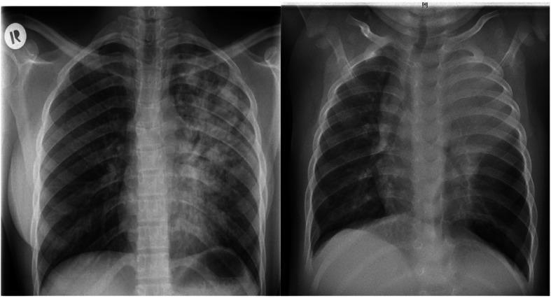
This side-by-side comparison displays two posteroanterior (PA) chest X-rays illustrating the diverse radiographic presentations of pulmonary tuberculosis (TB) in adult versus pediatric patients. The adult image (left) demonstrates extensive, heterogeneous, patchy infiltrates and areas of consolidation predominantly involving the left lung field, typical of post-primary or reactivation TB. Visible radiolucencies suggest cavitation within the upper lobe. In contrast, the pediatric image (right) shows a different pattern characterized by diffuse, relatively homogeneous opacification throughout both lung fields. Notable features in the pediatric X-ray include significant widening of the superior mediastinum, which is highly suggestive of bilateral mediastinal lymphadenopathy, a hallmark of primary TB in children. Both cases exhibit reduced lung clarity, but the pediatric case shows more pronounced bronchial involvement and hilar prominence without the distinct cavitary lesions seen in the adult. These images serve as a comparative educational resource for identifying age-specific diagnostic markers of Mycobacterium tuberculosis infection.
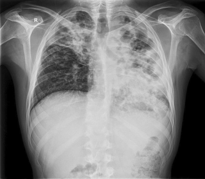
This posteroanterior (PA) chest X-ray demonstrates extensive bilateral pulmonary pathology, highly characteristic of advanced post-primary tuberculosis with superinfection. The left lung field shows a significantly distorted architecture with large, irregular, and oval-shaped lucencies in the upper and middle zones, consistent with pulmonary cavitation. The left lower zone exhibits dense alveolar opacification with visible air bronchograms and spotty shadowing, indicating consolidation. The right lung displays inhomogeneous opacities in the upper and middle lobes, accompanied by volume loss evidenced by the retraction of the right hilum and horizontal fissure. The mediastinal structures appear shifted, and there is visible distortion of the normal hilar anatomy. This diagnostic image illustrates severe parenchymal destruction, cavitary disease, and bilateral infiltrates, which are key clinical markers for evaluating disease severity and treatment response in infectious respiratory medicine.
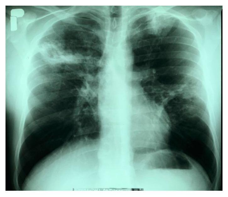
This diagnostic image is a posteroanterior (PA) chest X-ray demonstrating bilateral pulmonary pathology consistent with active tuberculosis. In the right lung, there is a large, heterogeneous infiltrative lesion in the upper field containing a distinct cavitary lesion measuring approximately 35 x 30 mm with irregular borders. The left lung shows an infiltrative opacity at the apex and multiple nodular, fibrous lesions distributed throughout the middle lung field, suggesting a chronic or granulomatous process. The cardiac silhouette appears within normal limits, and the left costophrenic angle is sharp and clear. This radiograph serves as a classic educational example of secondary pulmonary tuberculosis, highlighting hallmark features such as apical predilection, parenchymal infiltration, and cavitation. These findings are clinically significant for the assessment of disease severity and the risk of hemoptysis in patients with Mycobacterium tuberculosis complex infections.
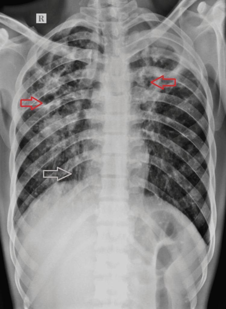
Posteroanterior (PA) chest X-ray demonstrating extensive bilateral pulmonary pathology, highly characteristic of chronic or post-primary pulmonary tuberculosis. The imaging shows widespread, asymmetrical air-space opacities scattered throughout both lung fields, with a greater density and involvement on the right side. Multiple well-defined cavitary lesions are visible, predominantly located in the bilateral upper zones and the right mid-zone (highlighted by red arrows), appearing as radiolucent areas surrounded by thick, irregular walls. In the right lower and upper zones, there is evidence of tractional bronchiectasis (highlighted by white arrows), indicated by tubular lucencies and architectural distortion. The trachea shows a mild shift toward the right, suggesting volume loss or fibrotic changes in the right lung parenchyma. These findings illustrate the chronic sequelae of mycobacterial infection, including lung destruction, cavitation, and secondary airway remodeling.
tuberculosis granuloma histopathology Ghon complex
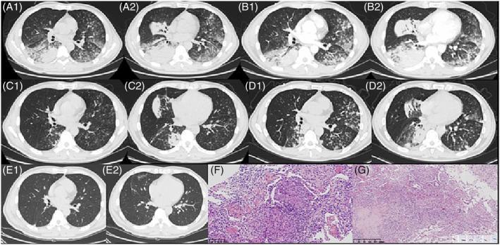
This composite educational graphic illustrates the longitudinal progression and histopathology of a complex pulmonary condition, later identified as tuberculosis presenting as acute fibrinous and organizing pneumonia (AFOP). Panels A1 through E2 are axial non-contrast chest CT scans showing a temporal series of pulmonary changes. Initial scans (A1, A2) reveal bilateral consolidations, ground-glass opacities (GGO) along bronchioles, and centrilobular nodules. Subsequent scans (B1, B2) show worsening density and distribution, followed by temporary resolution (C1, C2) and subsequent relapse (D1, D2) in multiple lobes. Final scans (E1, E2) demonstrate significant absorption of lesions. Panel F shows histopathology (H&E stain) indicating AFOP, characterized by fibrin deposition in alveolar spaces and granulation tissue proliferation. Panel G displays a necrotizing granuloma; the inset confirms acid-fast bacilli via Ziehl-Neelsen staining. This material demonstrates the radiographic evolution of infectious pneumonia and the diagnostic role of lung biopsy in distinguishing organizing pneumonia patterns from underlying mycobacterial infection.
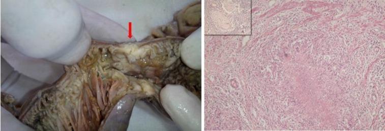
A composite image demonstrating clinical and histopathological findings of abdominal tuberculosis. The left panel is a clinical photograph of a resected small bowel segment during a laparotomy. A prominent red arrow indicates a focal stricture with significant luminal narrowing and mucosal thickening, consistent with granulomatous ileitis. The right panel displays a histopathological section (H&E stain, 100x magnification) showing an epithelioid cell granuloma. Key features include central caseous necrosis and a Langhans giant cell located adjacent to the muscularis mucosae. An inset in the upper left corner of the histopathology panel shows a similar granulomatous reaction within a dissected mesenteric lymph node. These findings collectively illustrate the classic morphological and microscopic features of intestinal tuberculosis, highlighting the progression from macroscopic obstruction to microscopic necrotizing granulomatous inflammation.
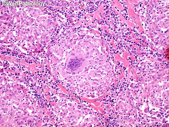
Histopathology via light microscopy of an H&E-stained lymph node biopsy demonstrates a granulomatous inflammatory response characterized by an epithelioid cell nodular formation with a surrounding lymphocytic infiltrate. Within the central core, a multinucleated Langhans-type giant cell is present, displaying nuclei arranged in a horseshoe pattern at the periphery. The granuloma is composed predominantly of well-differentiated epithelioid histiocytes with scant cytoplasm and tight cellular packing, producing a palisaded appearance that integrates with a mildly eosinophilic to translucent extracellular matrix. The surrounding paracortex contains a dense lymphoid crowding with scattered plasma cells and occasional eosinophils, consistent with chronic granulomatous inflammation. In this image, the granuloma lacks conspicuous caseous necrosis, a feature that supports noncaseating granulomatous processes; however, logical caveats apply since granulomas in sarcoidosis can mimic infectious etiologies. The presence of Langhans-type giant cells within nonnecrotizing granulomas is classically associated with sarcoidosis but can be seen in tuberculosis, atypical mycobacterial infections, fungal infections, berylliosis, and other granulomatous diseases. Additional diagnostic workup would include acid-fast bacilli staining and fungal stains, microbial cultures, and clinical correlation. Recognizing this histologic pattern guides differential diagnosis toward sarcoid-type granulomatous lymphadenitis and informs subsequent clinical management and exclusion of granulomatous infections. Correlation with imaging and laboratory data essential.
Pulmonary Tuberculosis (PTB)
Spectrum of Infection
1. Clinical Features
Symptoms
| Category | Features |
|---|---|
| Respiratory | Persistent cough (productive or dry) - the most common symptom; present in 75-90% of culture-confirmed cases. Hemoptysis (in advanced disease), dyspnea, chest pain |
| Constitutional | Fever, night sweats, weight loss, fatigue, anorexia, malaise |
| Advanced | Massive hemoptysis (from Rasmussen aneurysm - dilated vessel in cavity wall), breathlessness, hoarseness (laryngeal involvement) |
Signs on Examination
- Generally non-specific in early disease
- Post-tussive rales (crackles) in upper lung zones
- Amphoric (hollow) breath sounds over a cavity
- Reduced breath sounds / dullness in pleural effusion
- Lymphadenopathy - more common with HIV co-infection or primary TB
- Clubbing (chronic disease with fibrosis)
- Wasting and pallor in advanced disease
Laboratory Findings
- Leukocytosis or leukopenia (latter with HIV)
- Normochromic normocytic anaemia
- Raised ESR
- Hyponatraemia (SIADH - in extensive disease)
- Elevated alkaline phosphatase (hepatic involvement)
Radiological Features
| Pattern | Comment |
|---|---|
| Upper lobe infiltrates | Apical and posterior segments - classic reactivation pattern |
| Cavitation | Thick-walled cavities with air-fluid levels; hallmark of post-primary TB |
| Nodules / patchy consolidation | Acinar/lobular pattern |
| Miliary pattern | 1-2 mm uniform nodules throughout both lungs |
| Pleural effusion | Often exudative; lymphocyte-predominant |
| Hilar/mediastinal lymphadenopathy | More prominent in primary TB in children |
| Fibrosis/calcification | Healed TB; Ranke complex (Ghon focus + calcified hilar node) |
2. Investigations
A. Microbiological (Definitive Diagnosis)
- Collect at least 2 sputum specimens (same-day collection acceptable)
- Sensitivity: ~60-70% in smear-positive disease; lower in early/HIV-related disease
- Rapid (results within hours), cheap, widely available
- Does NOT distinguish M. tuberculosis from non-tuberculous mycobacteria (NTM)
- Lowenstein-Jensen (solid) medium: results in 3-8 weeks
- BACTEC MGIT (liquid): results in 1-3 weeks; more sensitive
- Essential for drug susceptibility testing (DST)
- 10-25% of bacteriologically confirmed TB cases have smear-negative disease
- WHO first-line recommended test (preferred over smear in many settings)
- Simultaneously detects M. tuberculosis DNA AND rifampin resistance mutations in 90 minutes
- Sensitivity: 88% in smear-positive, ~68% in smear-negative cases
- Rifampin resistance detected = strongly predictive of MDR-TB
- For smear-positive patients: positive NAAT confirms TB; negative NAAT suggests NTM
- For smear-negative patients with intermediate-high probability: positive NAAT = presumptive TB
- A negative NAAT does NOT exclude active TB (insufficient sensitivity to replace culture)
- All confirmed TB isolates should have DST performed
- Phenotypic DST: liquid culture systems (MGIT) - considered gold standard
- Genotypic DST: detects resistance mutations:
- rpoB mutations in >96% of rifampin-resistant strains
- katG / inhA mutations in 65-75% of isoniazid-resistant strains
- gyrA / gyrB mutations in 42-85% of fluoroquinolone-resistant strains
- Next-generation sequencing (NGS): Deeplex Myc-TB, DeepChek-TB - provides resistance profiles for 13 anti-TB drugs within 48 hours
- Sputum induction with 3-5% hypertonic saline (preferred - well tolerated)
- Bronchoscopy with BAL (bronchoalveolar lavage)
- Gastric lavage (especially in children)
B. Immunological Tests
- Intradermal injection of 5 TU PPD; read at 48-72 hours
- Positive: ≥5 mm (HIV+, close contact, immunosuppressed), ≥10 mm (high-risk groups), ≥15 mm (low-risk)
- Indicates immune sensitization (past or present infection); cannot distinguish LTBI from active disease
- False negatives: HIV, malnutrition, miliary TB, steroid use
- False positives: prior BCG, NTM infection
- QuantiFERON-TB Gold Plus (blood test) or T-SPOT.TB
- More specific than TST (not affected by BCG)
- Preferred in BCG-vaccinated individuals or NTM-endemic areas
- Limitation: cannot distinguish LTBI from active TB
C. Histopathology
- Biopsy of lymph nodes, pleura, or lung (via TBLB/VATS)
- Shows caseating (necrotizing) granulomas with Langhan's giant cells and central caseous necrosis
- AFB staining (ZN) of tissue sections; culture of tissue specimen
D. Pleural Fluid Analysis (if effusion present)
- Exudate (Light's criteria)
- Lymphocyte-predominant (>70% lymphocytes)
- Raised ADA (adenosine deaminase) >40 U/L - highly suggestive
- Low glucose, raised protein
- Culture positive in only 20-40%; biopsy increases yield significantly
3. Treatment
General Principles
- All confirmed TB must have DST performed before or during treatment
- Directly Observed Therapy (DOT) is strongly recommended to ensure adherence
- All TB patients must be notified to public health authorities
- Monitor monthly for clinical response (fever, cough, weight, sputum)
- Sputum culture should be negative by month 2 in 75-80%, and by month 3 in ~95% of patients
- Persistent culture positive at 4 months = treatment failure
Standard Regimen for Drug-Susceptible Pulmonary TB
| Phase | Drugs | Duration | Minimum Doses |
|---|---|---|---|
| Intensive (Initial) | Isoniazid (H) + Rifampin (R) + Pyrazinamide (Z) + Ethambutol (E) | 8 weeks (2 months) | 5-7 days/week |
| Continuation | Isoniazid (H) + Rifampin (R) | 18 weeks (4.5 months) | 5-7 days/week |
| Total duration | 6 months (26 weeks) |
Mnemonic: 2HRZE / 4HR
- Cavitary disease on CXR with sputum culture still positive at 2 months
- Situations where pyrazinamide cannot be used in the intensive phase
Standard Adult Doses (Oral)
| Drug | Daily Dose | Max Dose |
|---|---|---|
| Isoniazid (H) | 5 mg/kg | 300 mg/day |
| Rifampin (R) | 10 mg/kg | 600 mg/day |
| Pyrazinamide (Z) | 20-25 mg/kg | 2000 mg/day |
| Ethambutol (E) | 15-20 mg/kg | 1600 mg/day |
Key Adverse Effects to Monitor
| Drug | Major Adverse Effects |
|---|---|
| Isoniazid (H) | Hepatotoxicity, peripheral neuropathy, drug-induced lupus |
| Rifampin (R) | Hepatotoxicity, orange discolouration of secretions, drug interactions (CYP450 inducer), flu-like syndrome |
| Pyrazinamide (Z) | Hepatotoxicity, hyperuricaemia/gout, arthralgia |
| Ethambutol (E) | Optic neuritis (dose-dependent, reversible if detected early) - check visual acuity before and during treatment |
Drug-Resistant TB
- Group A drugs (use all if possible): Levofloxacin/Moxifloxacin, Bedaquiline, Linezolid
- Group B drugs (add next): Clofazimine, Cycloserine/Terizidone
- Group C drugs (add if needed): Delamanid, Amikacin, Ethambutol, Pyrazinamide, etc.
- Duration: typically 18-24 months (or 9-12 months for shorter regimens if criteria met)
- BPaL regimen (Bedaquiline + Pretomanid + Linezolid) - approved for XDR-TB / treatment-intolerant MDR-TB
Special Situations
| Situation | Modification |
|---|---|
| HIV co-infection | Start ART within 2-8 weeks of starting TB treatment (immediately if CD4 <50); rifabutin substituted for rifampin if on certain ARVs |
| Pregnancy | Rifampin, isoniazid, ethambutol safe; pyrazinamide: use if benefit outweighs risk; streptomycin CONTRAINDICATED (ototoxicity) |
| Hepatic disease | Avoid pyrazinamide (most hepatotoxic); consider HR-E regimen with extended duration |
| Renal impairment | Adjust ethambutol and pyrazinamide doses (renally cleared); rifampin and isoniazid largely safe |
Infectivity and Isolation
- At least 2 weeks of effective therapy
- Clinical improvement (reduced cough, no fever, weight gain)
- Three sputum specimens ≥8 hours apart showing no AFB on microscopy
Refer Archit Boloor
What would you like me to do with Archit Boloor's material?
Archit Boloor medicine notes book tuberculosis
- Archit Boloor may refer to a popular Indian medical educator/YouTuber known for rapid revision notes for MBBS/PG exams - but his material is not indexed in this library.
- The name may be slightly different - for example, could you be thinking of Arvind Boloor, or another author?
- His notes/lectures may exist only as YouTube videos or privately distributed PDFs not available here.
- Cover Pulmonary TB from any of the major textbooks available in the library (Goldman-Cecil, Murray & Nadel, Harrison's-equivalent, Robbins Pathology, Park's Preventive Medicine, etc.) - as I just did above.
- If you have Archit Boloor's PDF/notes, you can upload the file and I can summarize or extract information from it directly.
- If you can share a YouTube link to his lecture, I can help you work with that content.