
Explain visual pathway not lesion but pathway
The Visual Pathway
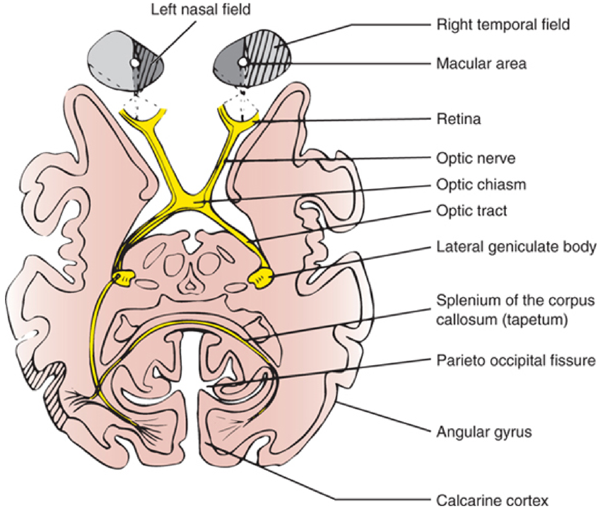
Step 1 - The Retina (First-Order Neurons)
- Photoreceptors (rods and cones) - detect light
- Bipolar cells - relay signals from photoreceptors
- Retinal ganglion cells (RGCs) - the output neurons whose axons form the optic nerve
- Midget (P) cells - small receptive fields, sensitive to fine detail and color; project to parvocellular layers of the LGN
- Parasol (M) cells - larger receptive fields, sensitive to motion and low contrast; project to magnocellular layers of the LGN
Step 2 - The Optic Nerve (CN II)
| Portion | Length | Notes |
|---|---|---|
| Intraocular (optic nerve head) | ~1 mm | Axons become myelinated here |
| Intraorbital | ~25 mm | S-shaped, allows eye movement |
| Intracanalicular | ~9 mm | Travels through the optic canal |
| Intracranial | ~4-16 mm | Between optic canal and chiasm |
- Localization in Clinical Neurology, 8e
Step 3 - The Optic Chiasm
- Fibers from the nasal (medial) retina of each eye cross to the opposite side
- Fibers from the temporal (lateral) retina of each eye stay ipsilateral
- The nasal retina of the left eye sees the right visual field
- The temporal retina of the right eye also sees the right visual field
- After the chiasm, ALL information about the right visual field travels in the left optic tract (and vice versa)
Step 4 - The Optic Tract
- Pretectal area and superior colliculus - for the pupillary light reflex and visual orienting
- Hypothalamus (suprachiasmatic nucleus) - for circadian rhythm regulation
Step 5 - The Lateral Geniculate Nucleus (LGN) of the Thalamus
| Layers | Cell Type | Input |
|---|---|---|
| 1 and 2 | Magnocellular | Motion, depth, low contrast |
| 3, 4, 5, 6 | Parvocellular | Color, fine detail |
| Between each | Koniocellular | Color (blue-yellow channel) |
- Contralateral eye projects to layers 1, 4, and 6
- Ipsilateral eye projects to layers 2, 3, and 5
Step 6 - The Optic Radiation (Geniculocalcarine Tract)
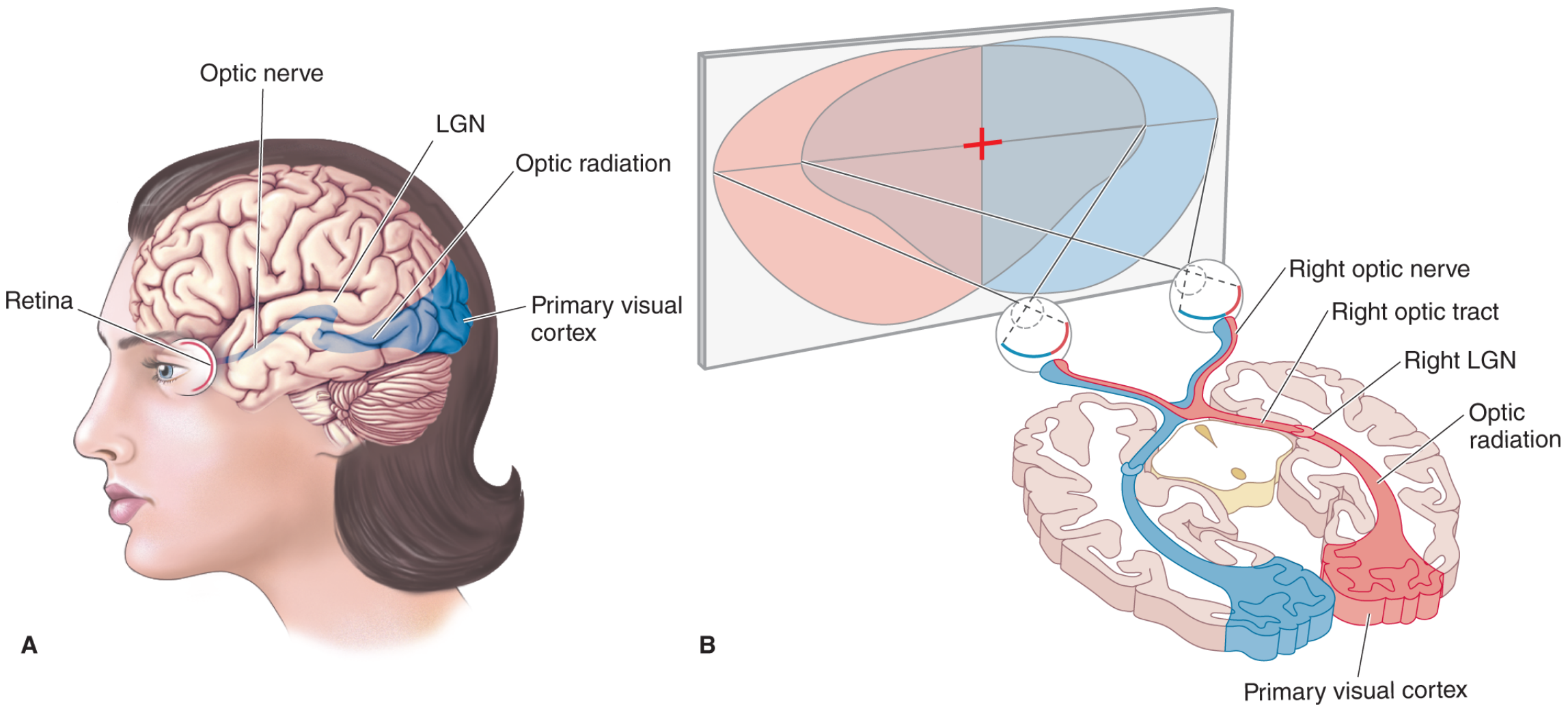
Upper division (parietal / dorsal fibers)
- Carries information from the superior retina (representing the inferior visual field)
- Travels directly posteriorly through the parietal lobe
- Terminates on the superior bank (cuneus) of the calcarine fissure
Lower division - Meyer's Loop (temporal fibers)
- Carries information from the inferior retina (representing the superior visual field)
- Loops anteriorly into the temporal lobe first, then swings back posteriorly (this loop is called Meyer's loop or Flechsig's loop)
- Terminates on the inferior bank (lingual gyrus) of the calcarine fissure
Step 7 - The Primary Visual Cortex (V1 / Area 17 / Striate Cortex)
- Striate cortex - named after the Stria of Gennari, a thick myelinated band in layer 4 visible to the naked eye
- Brodmann area 17
- Each hemisphere's V1 receives input from the contralateral visual field
- Information is projected inverted: superior retina → superior (dorsal) calcarine cortex; inferior retina → inferior (ventral) calcarine cortex
- The macula (central vision) has disproportionately large representation at the occipital pole (cortical magnification)
- LGN parvocellular neurons project to cortical layer IVCβ; magnocellular to layer IVCα; koniocellular to "blobs" in layers 2 and 3
- Inputs from both eyes remain in separate ocular dominance columns within V1
Step 8 - Higher Visual Cortex (V2, V3, V4, V5)
| Stream | Direction | Function |
|---|---|---|
| Dorsal "where?" stream | V1 → Parietal lobe | Motion, spatial location, visually guided action |
| Ventral "what?" stream | V1 → Temporal lobe | Form recognition, color, object identity, faces |
- Neuroanatomy through Clinical Cases, 3rd ed.
Summary Flowchart
Photoreceptors (rods/cones)
        ↓
Bipolar cells (retina)
        ↓
Retinal Ganglion Cells → axons form OPTIC NERVE (CN II)
        ↓
OPTIC CHIASM
  - Nasal fibers cross → contralateral optic tract
  - Temporal fibers stay → ipsilateral optic tract
        ↓
OPTIC TRACT (wraps around midbrain)
  - Side branch → pretectum/superior colliculus (pupillary reflex)
        ↓
LATERAL GENICULATE NUCLEUS (thalamus)
  - 6 layers; eye inputs remain segregated
        ↓
OPTIC RADIATION
  - Upper division (parietal): inferior visual field
  - Lower division (Meyer's loop, temporal): superior visual field
        ↓
PRIMARY VISUAL CORTEX (V1 / striate cortex)
  - Calcarine fissure, occipital lobe
        ↓
ASSOCIATION CORTEX (V2–V5)
  - Dorsal stream: "where?"
  - Ventral stream: "what?"
Flow chart of visual pathway from authentic source only
A detailed medical flowchart of the visual pathway, styled like a textbook diagram with clean boxes, arrows, and annotation labels. The flowchart flows top to bottom with the following sequential steps, each in a labeled box: 1. RETINA - Photoreceptors (Rods & Cones) → Bipolar Cells → Retinal Ganglion Cells (RGCs). Two parallel cell types labeled: P-cells (Midget) and M-cells (Parasol). Axons converge at optic disc (blind spot). 2. OPTIC NERVE (CN II) - ~1.2 million ganglion cell axons. CNS tract (myelinated by oligodendrocytes). Four portions: Intraocular (1mm) → Intraorbital (25mm) → Intracanalicular (9mm) → Intracranial (4–16mm). 3. OPTIC CHIASM - Partial decussation. Nasal retinal fibers CROSS to contralateral optic tract. Temporal retinal fibers STAY ipsilateral. Result: each optic tract carries contralateral hemifield from BOTH eyes. 4. OPTIC TRACT - Wraps posterolaterally around midbrain. Side branch arrows: (a) Pretectum + Superior Colliculus → Pupillary Light Reflex & visual orienting. (b) Suprachiasmatic Nucleus → Circadian rhythm. 5. LATERAL GENICULATE NUCLEUS (LGN) of Thalamus - 6 layers. Layers 1–2: Magnocellular (M pathway – motion, depth). Layers 3–6: Parvocellular (P pathway – color, detail). Between each: Koniocellular (K pathway – blue/yellow color). Eye segregation: Contralateral eye → layers 1, 4, 6. Ipsilateral eye → layers 2, 3, 5. 6. OPTIC RADIATION (Geniculocalcarine Tract) - Upper division (Parietal): carries inferior visual field → superior calcarine bank (Cuneus). Lower division – MEYER'S LOOP (Temporal): loops anteriorly into temporal lobe, carries superior visual field → inferior calcarine bank (Lingual Gyrus). 7. PRIMARY VISUAL CORTEX V1 (Area 17 / Striate Cortex) - Calcarine fissure, occipital lobe. Stria of Gennari visible in layer 4. Macular vision (central) → occipital pole (large representation). Parvocellular → Layer IVCβ. Magnocellular → Layer IVCα. Koniocellular → Layers 2/3 blobs. Ocular dominance columns. 8. VISUAL ASSOCIATION CORTEX (V2, V3, V4, V5) - Two streams labeled with arrows: Dorsal "WHERE?" stream → Parietal lobe (motion, spatial). Ventral "WHAT?" stream → Temporal lobe (form, color, object recognition). The chart is on a white background, with alternating light blue and white boxes, bold black text, clean directional arrows, and side-branch annotations in a slightly smaller font. Title at top: "VISUAL PATHWAY" in large bold font. Source citation at bottom: "Based on: Gray's Anatomy for Students | Kandel: Principles of Neural Science, 6th ed. | Neuroanatomy through Clinical Cases, 3rd ed."
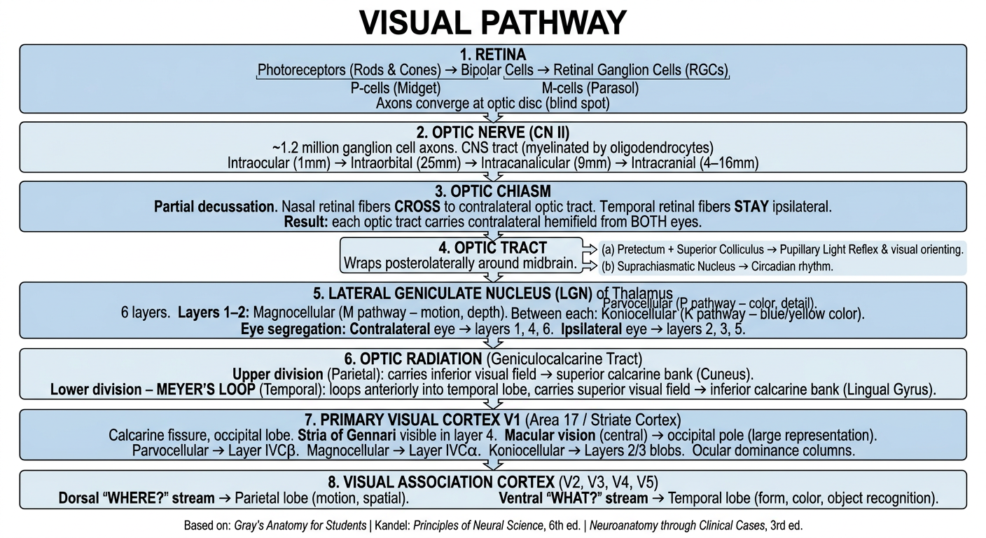
Visual Pathway - Sequential Flowchart
┌─────────────────────────────────────────────────────────────┐
│                        RETINA                               │
│  Photoreceptors (Rods & Cones)                              │
│         ↓                                                   │
│  Bipolar Cells                                              │
│         ↓                                                   │
│  Retinal Ganglion Cells (RGCs)                              │
│  ├─ Midget (P) cells → fine detail, color                   │
│  └─ Parasol (M) cells → motion, contrast                   │
│  Axons converge at Optic Disc (= blind spot, no receptors)  │
└───────────────────────────┬─────────────────────────────────┘
                            ↓
┌─────────────────────────────────────────────────────────────┐
│                  OPTIC NERVE (CN II)                        │
│  ~1.2 million RGC axons; CNS tract (oligodendrocyte myelin) │
│  4 portions:                                                │
│  Intraocular (1mm) → Intraorbital (25mm)                    │
│  → Intracanalicular (9mm) → Intracranial (4–16mm)           │
└───────────────────────────┬─────────────────────────────────┘
                            ↓
┌─────────────────────────────────────────────────────────────┐
│                    OPTIC CHIASM                             │
│  Located anterior to pituitary infundibular stalk           │
│  PARTIAL DECUSSATION:                                       │
│  Nasal retina fibers → CROSS to contralateral optic tract   │
│  Temporal retina fibers → STAY ipsilateral                  │
│  Result: each optic tract = contralateral hemifield         │
│          from BOTH eyes                                     │
└───────────────────────────┬─────────────────────────────────┘
                            ↓
┌─────────────────────────────────────────────────────────────┐
│                     OPTIC TRACT                             │
│  Courses posterolaterally around the midbrain               │
│  Side branches:                                             │
│  ├─ Pretectum + Superior Colliculus → Pupillary light reflex│
│  │                                  + visual orienting      │
│  └─ Suprachiasmatic nucleus → Circadian rhythm              │
│  Majority continue to LGN                                   │
└───────────────────────────┬─────────────────────────────────┘
                            ↓
┌─────────────────────────────────────────────────────────────┐
│         LATERAL GENICULATE NUCLEUS (LGN) - Thalamus         │
│  6 layers (ventral → dorsal):                               │
│  Layers 1–2: Magnocellular (M) → motion, depth, contrast    │
│  Layers 3–6: Parvocellular (P) → color, fine detail         │
│  Between each: Koniocellular (K) → blue/yellow color        │
│                                                             │
│  Eye segregation:                                           │
│  Contralateral eye → Layers 1, 4, 6                         │
│  Ipsilateral eye   → Layers 2, 3, 5                         │
│  (Both eyes remain SEGREGATED within LGN)                   │
└───────────────────────────┬─────────────────────────────────┘
                            ↓
┌─────────────────────────────────────────────────────────────┐
│         OPTIC RADIATION (Geniculocalcarine Tract)           │
│  Upper division (Parietal / dorsal fibers):                 │
│  → Carries INFERIOR visual field                            │
│  → Terminates on SUPERIOR calcarine bank (Cuneus)           │
│                                                             │
│  Lower division – MEYER'S LOOP (Temporal fibers):           │
│  → Loops anteriorly into temporal lobe first               │
│  → Carries SUPERIOR visual field                            │
│  → Terminates on INFERIOR calcarine bank (Lingual Gyrus)    │
└───────────────────────────┬─────────────────────────────────┘
                            ↓
┌─────────────────────────────────────────────────────────────┐
│         PRIMARY VISUAL CORTEX V1 (Area 17)                  │
│         Striate Cortex / Calcarine Fissure, Occipital Lobe  │
│  Stria of Gennari = thick myelinated band in Layer 4        │
│  Macula (central vision) → large area at occipital pole     │
│  P-cells (LGN) → Layer IVCβ                                 │
│  M-cells (LGN) → Layer IVCα                                 │
│  K-cells (LGN) → Layers 2/3 "blobs"                         │
│  Ocular dominance columns (eyes still segregated here)      │
└───────────────────────────┬─────────────────────────────────┘
                            ↓
┌─────────────────────────────────────────────────────────────┐
│        VISUAL ASSOCIATION CORTEX (V2, V3, V4, V5)           │
│                                                             │
│  Dorsal "WHERE?" stream → Parietal lobe                     │
│  (motion, spatial relations, visually guided action)        │
│                                                             │
│  Ventral "WHAT?" stream → Temporal lobe                     │
│  (form, color, object/face recognition)                     │
└─────────────────────────────────────────────────────────────┘
- Gray's Anatomy for Students - optic nerve portions, chiasm decussation, optic radiation
- Kandel: Principles of Neural Science, 6th ed. - LGN layers, eye segregation, cortical projections
- Neuroanatomy through Clinical Cases, 3rd ed. - dorsal/ventral streams, LGN layer inputs
- Grainger & Allison's Diagnostic Radiology - Meyer's loop, striate cortex, calcarine fissure
- Localization in Clinical Neurology, 8e - optic nerve portions, retinal cell types
What is frist ad second order neuron in retina
First & Second Order Neurons in the Retina
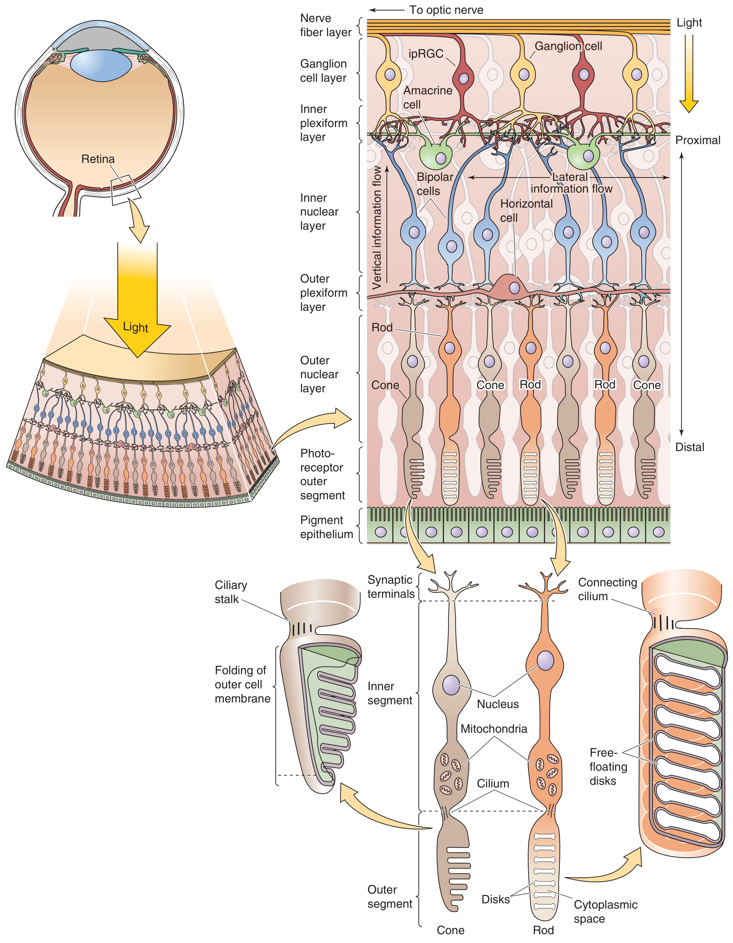
The Three-Neuron Chain in the Retina
| Neuron | Cell | Layer |
|---|---|---|
| 0th / Receptor | Photoreceptors (Rods & Cones) | Outer nuclear layer |
| 1st Order | Bipolar Cells | Inner nuclear layer |
| 2nd Order | Retinal Ganglion Cells (RGCs) | Ganglion cell layer |
"These synaptic terminals [of rods and cones] contact bipolar cells, the first-order neurons in the visual pathway. Bipolar cells then synapse with ganglion cells, the second-order neurons in the visual pathway."
- Gray's Anatomy for Students (verbatim)
In Detail
Photoreceptors (Rods & Cones) - the Receptor/0th neuron
- Light hits the retina and must pass through ganglion and bipolar cell layers first before reaching the photoreceptors (the retina is inverted)
- Rods - for low-light/scotopic vision; 120 million
- Cones - for color/photopic vision; concentrated at the fovea; 6 million
- They do NOT fire action potentials; they communicate by graded synaptic potentials
- They synapse in the outer plexiform layer
First-Order Neuron: Bipolar Cell
- Receives input from photoreceptors in the outer plexiform layer
- Two types relevant here:
- ON bipolar cells - depolarize when light hits (glutamate decreases → cell activates)
- OFF bipolar cells - hyperpolarize when light hits
- Also communicate by graded potentials (no action potentials)
- Synapse with ganglion cells in the inner plexiform layer
Second-Order Neuron: Retinal Ganglion Cell (RGC)
- The output neuron of the retina
- The first cell in the visual pathway to fire action potentials
- Their axons form the optic nerve (CN II)
- Two main types:
- Midget (P) cells - color, fine detail
- Parasol (M) cells - motion, contrast
Modulating Interneurons (not numbered in the order)
| Cell | Location | Function |
|---|---|---|
| Horizontal cells | Outer plexiform layer | Laterally connect photoreceptors to each other and to bipolar cells; create surround inhibition |
| Amacrine cells | Inner plexiform layer | Laterally connect bipolar cells to ganglion cells; modulate their activity |
"Two other cell types present in the retina are horizontal and amacrine cells. These transversely oriented interneurons moderate the excitation level of bipolar and ganglion cells."
- Gray's Anatomy for Students
Simple Summary
Light
  ↓
Photoreceptors (Rods/Cones)  ← Receptor / 0th neuron
  ↓  [outer plexiform layer]
Bipolar Cells               ← 1st Order Neuron
  ↓  [inner plexiform layer]
Retinal Ganglion Cells      ← 2nd Order Neuron
  ↓
Optic Nerve (CN II) → rest of visual pathway