
Diptheria
Reading File
Now let me search the medical library for diphtheria content in parallel.
Finding Sources
Searching PubMed
diphtheria
Good. Now let me read the key textbook sections in parallel - Rosen's Emergency Medicine, Tintinalli's, and Robbins Pathology.
Reading File
Reading File
Reading File
Excellent content. Let me now read the Rosen's section more fully and also fetch the pseudomembrane image from Robbins.
Reading File
Loading Image
I now have comprehensive content. Let me compile the full answer.
Diphtheria
Overview
Diphtheria is an acute toxin-mediated infection caused by Corynebacterium diphtheriae - an unencapsulated, nonmotile, gram-positive bacillus (named from korynee = "club," for its club-shaped ends). The disease name comes from the Greek diphtheria = "leather," describing the characteristic throat membrane. Hippocrates described it in the 5th century BCE; Pierre Bretonneau named it in 1821. Von Behring and Kitasato developed the first antitoxin in 1890.
- Rosen's Emergency Medicine, p. 2582
- Robbins & Cotran Pathologic Basis of Disease, p. 344
Epidemiology
- Humans are the only reservoir. Spread is via respiratory droplets, direct contact with secretions, skin exudate, or rarely fomites/food.
- Crowded conditions favor transmission; carriers (symptomatic, convalescent, or asymptomatic) all spread the disease.
- US cases fell from 206,000 in 1921 to just 2 between 2004 and 2017 - a direct result of vaccination.
- More than 50% of adults over 40 in industrialized countries now lack protective antibodies, because widespread childhood vaccination reduces natural booster exposure.
- Recent outbreaks have occurred in Yemen, Venezuela, and cases linked to travelers in Spain (disrupted vaccination programs).
- WHO reported >5,000 global cases in 2022, with large outbreaks in areas with displaced populations.
Microbiology & Toxin Mechanism
C. diphtheriae is lysogenized by a bacteriophage that encodes the diphtheria A-B exotoxin. The mechanism:
- The B (binding) fragment attaches to host cell receptors and enables entry.
- The A (active) fragment catalyzes ADP-ribosylation of Elongation Factor-2 (EF-2) - irreversibly inactivating it.
- This blocks mRNA translation (protein synthesis) in all host cells.
- One toxin molecule can ADP-ribosylate >1 million EF-2 molecules, killing the cell.
Toxigenic strains cause both local (membrane) and systemic disease. Non-toxigenic strains cause local infection only.
Pathology & Morphology
The hallmark is the pseudomembrane (also called the diphtheritic membrane):
- Composed of leukocytes, erythrocytes, fibrin, epithelial cells, and bacteria
- Forms from coagulation of fibrinopurulent exudate over necrotic, ulcerated mucosa
- Tough, dirty gray to black, leathery in texture
- Beneath it: intense neutrophilic infiltrate, vascular congestion, interstitial edema
- When the membrane sloughs off its vascularized bed: bleeding and asphyxiation may result
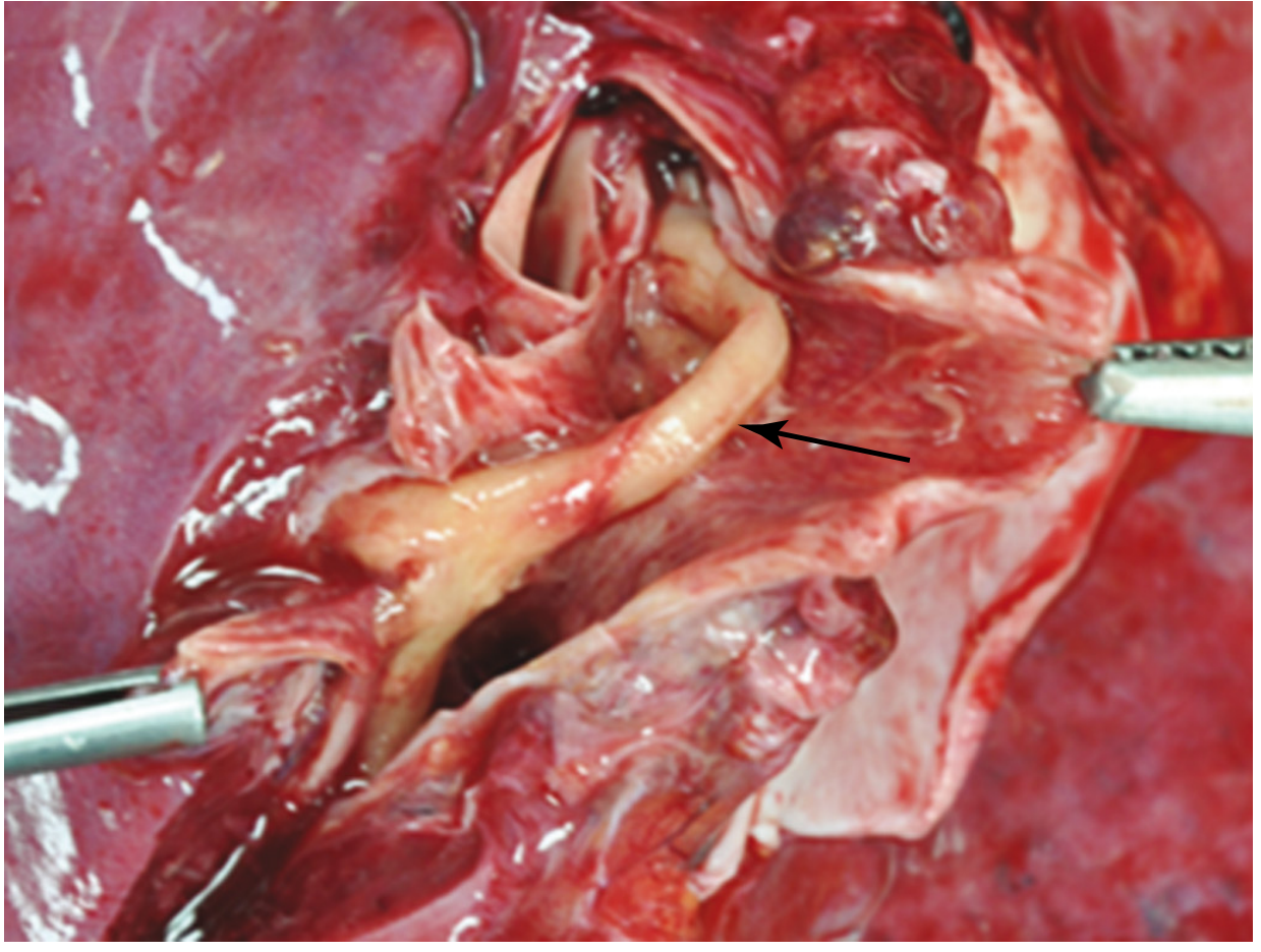
Pseudomembrane of diphtheria (arrow) lying within a transverse bronchus - Robbins & Cotran, p. 345
Systemic toxin effects (from bloodstream entry):
- Myocardium: fatty change, isolated myofiber necrosis
- Nerves: demyelination with degeneration of myelin sheaths and axis cylinders (polyneuritis)
- Liver, kidneys, adrenals: fatty change and focal necrosis (less common)
Clinical Forms
1. Faucial (Pharyngeal/Tonsillar) Diphtheria - Most Common
- Incubation: 2-4 days (range 1-8 days)
- Symptoms: sore throat, low-grade fever, malaise, dysphagia, weakness, headache, voice changes
- A membrane on the tonsils/pharynx is seen in >50% of patients
- Extent of membrane parallels clinical toxicity:
- Limited to tonsils = mild disease
- Covering entire pharynx = severe, abrupt onset
- "Bull neck": cervical lymphadenopathy + tissue infiltration so extensive it distorts the neck contour - sign of malignant diphtheria
- Malignant form: high fever, severe weakness, vomiting, diarrhea, restlessness, delirium
2. Laryngeal Diphtheria
- Classic "barking" cough, stridor, hoarseness, difficulty breathing
- Marked neck edema ("bull neck")
- High risk of airway obstruction
3. Nasal Diphtheria
- Serous or serosanguineous nasal discharge
- Usually mild; membrane on nasal septum
- Acts as source of spread
4. Cutaneous Diphtheria
- Chronic, non-healing ulcers with a dirty gray membrane
- Primary or secondary infection of pre-existing wounds
- Does not cause systemic toxin damage
- Least toxicity of all forms
Complications
Cardiac (appears 1-2 weeks post-onset, earlier in severe cases)
- ECG changes (myocarditis pattern): up to 2/3 of patients
- Clinical myocarditis: 10-25% of cases
- Can cause fatal cardiac failure
Neurological
- ~5% of respiratory infection cases develop polyneuritis
- 75% of patients with severe disease have some neuropathy
- Palatal muscles affected first (nasal voice, regurgitation)
- Then: cranial nerves, peripheral nerves, spinal cord (dorsal root + ventral horn ganglia)
- Cortical cells are spared
- Proximal muscle groups affected first
- Severe cases: paralysis within first few days
- Duration: typically <10 days, up to 3 months; complete recovery is the rule
Respiratory
- Membrane extension causing obstruction
- Diaphragmatic paralysis (from neuritis) leading to death
Diagnosis
- Clinical: characteristic pseudomembrane + systemic toxicity
- Confirmation: culture of nasopharyngeal swab - isolation of C. diphtheriae
- Culture requires special media (Loeffler's or tellurite medium)
- Toxigenicity confirmed by Elek test or PCR for the tox gene
Treatment
The three pillars:
| Treatment | Details |
|---|---|
| Diphtheria Antitoxin (DAT) | Given as soon as clinical diagnosis is made - neutralizes circulating toxin (not cell-bound toxin). Equine-derived; pre-test for hypersensitivity. Dose based on site and duration of infection |
| Antibiotics | Erythromycin or penicillin G - eliminate bacteria, terminate toxin production, reduce carrier state. Does NOT replace antitoxin |
| Respiratory support | Airway management, mechanical ventilation if needed; management of myocarditis/arrhythmias |
- Isolation is mandatory. Patient remains infectious until 2 consecutive negative cultures.
Prevention & Vaccination
- Toxoid vaccine (formalin-fixed toxin) stimulates neutralizing antibodies
- Part of DTP/DTaP (with tetanus, pertussis) series
- Standard childhood schedule: 3 primary doses + boosters
- Booster as Td or Tdap in adults every 10 years
- Immunization dramatically reduced global incidence (see global trend data from WHO 1980-2016)
Key Memory Points
| Feature | Detail |
|---|---|
| Organism | Gram-positive bacillus, C. diphtheriae |
| Toxin mechanism | ADP-ribosylation of EF-2 → protein synthesis block |
| Membrane composition | Fibrin, leukocytes, RBCs, epithelium, bacteria |
| First muscle affected by neuritis | Soft palate |
| "Bull neck" | Massive cervical lymphadenopathy + edema |
| Cardiac complication timing | 1-2 weeks post-illness onset |
| Treatment | Antitoxin + penicillin/erythromycin + airway support |
| Vaccine | Diphtheria toxoid (part of DTaP/Tdap) |
Sources: Rosen's Emergency Medicine, p. 2582-2584; Tintinalli's Emergency Medicine, p. 839; Robbins & Cotran Pathologic Basis of Disease, p. 344-345
What are the diagnostic criteria and timeline for treating suspected diphtheria in the ED?
Diphtheria is a clinical diagnosis - do not wait for culture confirmation before treating. Antitoxin must be given as soon as the diagnosis is suspected, as it only neutralizes free (unbound) toxin. The key diagnostic features are the pseudomembrane plus systemic toxicity in a patient with incomplete vaccination history.
Searching sources...
Finding Sources
Searching PubMed
diphtheria diagnosis treatment antitoxin emergency
Now let me pull the full ED management sections from Rosen's and Tintinalli's - the most EM-focused sources.
Reading File
Finding Sources
Excellent - very rich content captured. Let me read the rest of the management section.
Reading File
I have everything needed. Let me compile the full, detailed ED-focused answer now.
Diagnostic Criteria & ED Management Timeline for Diphtheria
The Core Principle
Diphtheria is a clinical diagnosis. Treatment must not wait for culture confirmation. Diphtheria antitoxin only neutralizes free, circulating toxin - once toxin is bound to cells, it cannot be reversed. Every hour of delay increases irreversible organ damage.
Diagnostic Classification (CDC Framework)
Rosen's reproduces the CDC diagnostic checklist which divides cases into three tiers:
Suspect Case (minimum threshold to begin treatment)
All of the following:
- Pharyngitis, nasopharyngitis, tonsillitis, laryngitis, or tracheitis (any combination)
- Absent or low-grade fever (high fever is actually atypical)
- Grayish adherent pseudomembrane present
- Membrane bleeds when manipulated or dislodged - this is a key distinguishing feature from other exudates
Probable Case
Suspect case criteria PLUS one or more of:
| Feature | Clinical Significance |
|---|---|
| Stridor | Laryngeal/tracheobronchial extension |
| Bull-neck (cervical edema) | Malignant diphtheria - high mortality |
| Toxic circulatory collapse | Myocarditis or septic shock |
| Acute renal insufficiency | Systemic toxin effect |
| Submucosal/subcutaneous petechiae | Toxin-mediated vascular damage |
| Myocarditis | Toxin effect on myocardium |
| Death | - |
| Travel to endemic area within <2 weeks | Yemen, Venezuela, South Asia |
| Contact with confirmed case/carrier within <2 weeks | - |
| Contact with visitor from endemic area within <2 weeks | - |
| Contact with dairy/farm animals or domestic pets | Rare zoonotic source |
| Incomplete vaccination (no DTaP/DT/Tdap/Td within past 10 years) | Major risk factor |
Laboratory Confirmed Case
- Positive culture of C. diphtheriae or C. ulcerans
- PLUS positive Elek test (toxin detection) OR
- PCR positive for tox gene (subunit A and B)
Important: A positive group A beta-hemolytic strep culture does not exclude diphtheria - up to 30% of diphtheria patients co-test positive for strep, which can mislead the clinician.
Differential Diagnosis to Work Through in the ED
These must be considered but should not delay treatment if diphtheria features are present:
- Streptococcal pharyngitis (exudate is white/yellow, not gray-black; does not bleed)
- Viral pharyngitis (EBV, adenovirus, HSV)
- Tonsillitis
- Gonococcal pharyngitis
- Acute necrotizing ulcerative gingivitis (ANUG) - involves gingivae, spared in diphtheria
- Acute epiglottitis - more rapid onset; laryngoscopy shows erythematous epiglottis without membrane
- Infectious mononucleosis
- Candida (thrush) - white patches, less adherent, immunocompromised patient
The diphtheritic membrane is specifically darker, grayer, more fibrous, and more firmly attached than membranes in other conditions.
Diagnostic Workup in the ED
Specimen Collection (do before starting antibiotics)
- Throat AND nasopharyngeal swabs for respiratory diphtheria
- Membrane material itself if present - send a piece
- Skin lesion swab/biopsy for cutaneous form
- Notify the lab explicitly - routine throat culture media will miss C. diphtheriae; tellurite selective medium is required
Labs
| Test | Finding | Utility |
|---|---|---|
| CBC | Leukocytosis, mild thrombocytopenia | Common but non-specific |
| Urinalysis | Proteinuria | Common but non-specific |
| ECG | ST-T wave changes, AV block (any degree), dysrhythmias; may be normal even with myocarditis | Monitor for cardiac toxicity |
| Echocardiogram | Dilated or hypertrophic cardiomyopathy | Assess degree of myocardial involvement |
| Troponin | Elevated; correlates with severity of myocarditis | Prognostic |
ED Treatment Timeline
Immediate (0-15 minutes) - Simultaneously
1. Airway
- Assess immediately on arrival
- Early intubation for any laryngeal involvement - do not wait for obstruction to develop
- Membrane can detach and cause acute asphyxiation
2. Respiratory Isolation
- Place in respiratory isolation immediately upon suspicion
- Patient is infectious until 2 consecutive negative cultures
3. IV Access + Resuscitation
- Patients are often dehydrated (fever + dysphagia + neurologic impairment)
- Assess for fluid responsiveness - the myocardial toxin effect can cause heart failure; over-aggressive fluids can worsen it
Early (within 1 hour) - Do Not Delay
4. Notify Public Health
- Mandatory reportable disease - contact state/local health department immediately
- They facilitate access to DAT (held at CDC in the US)
5. Administer Diphtheria Antitoxin (DAT) - The Most Time-Sensitive Intervention
Equine-derived antitoxin is the only specific therapy. It neutralizes only unbound toxin.
Before administration:
- Test for equine serum hypersensitivity (conjunctival or intradermal test)
- Have epinephrine at bedside - anaphylaxis risk
Dosing by clinical severity:
| Presentation | DAT Dose | Route |
|---|---|---|
| Nasal diphtheria only | 20,000-40,000 units | IM |
| Tonsillar/pharyngeal (mild, <48 h) | 40,000-60,000 units | IM or slow IV |
| Pharyngeal/laryngeal (moderate, <48 h) | 80,000-100,000 units | IV |
| Severe/bull-neck or disease >3 days | 80,000-120,000 units | IV |
| Cutaneous diphtheria | 20,000-40,000 units | IM or IV |
The longer the delay from symptom onset to DAT administration, the higher the dose required and the worse the outcome.
6. Antibiotics (start in the ED)
Antibiotics eliminate the organism, halt toxin production, and reduce carrier transmission. They do NOT substitute for antitoxin.
- Erythromycin 500 mg IV/PO q6h x 14 days (preferred)
- Penicillin G 100,000-150,000 units/kg/day IV in divided doses x 14 days (alternative)
- Duration: 14 days total; confirm eradication with 2 negative cultures after completion
Ongoing (first 24-48 hours) - In-Hospital
7. Cardiac Monitoring
- Continuous ECG monitoring for all patients with pharyngeal diphtheria
- Serial troponin and echo
- Cardiology consult for myocarditis - pacemaker may be needed for high-degree AV block
8. Neurology Monitoring
- Assess palatal function (nasal voice, regurgitation = soft palate neuropathy = first sign)
- Monitor for respiratory muscle weakness; early ICU/ventilator planning if cranial neuropathy progresses
9. Post-exposure Prophylaxis for Contacts
- All close contacts: culture + prophylactic antibiotics (erythromycin x 7-10 days OR single dose benzathine penicillin G)
- Unimmunized contacts: give diphtheria toxoid
- Contacts with incomplete vaccination: booster dose
Mortality Reference Points
| Clinical Form | Mortality |
|---|---|
| Overall | <3% |
| With myocarditis | ~7% |
| Malignant (bull-neck) form | ~26% |
These figures underscore why the bull-neck and early cardiac signs must trigger maximal early intervention.
Summary: The ED Decision Tree
Sore throat + low-grade fever + adherent gray membrane
            ↓
Does membrane bleed when touched?
            ↓ YES
SUSPECT DIPHTHERIA
            ↓
→ Isolate immediately
→ Airway assessment (intubate if laryngeal involvement)
→ Swab cultures (before antibiotics)
→ ECG + troponin
→ Call public health for DAT access
→ Test for equine hypersensitivity → Administer DAT
→ Start erythromycin or penicillin G
→ Admit to ICU/monitored bed
DO NOT WAIT FOR CULTURE CONFIRMATION
Sources: Rosen's Emergency Medicine, pp. 2583-2585; Tintinalli's Emergency Medicine, p. 839; CDC Diphtheria Diagnostic Checklist
diagnostic criteria for chronic rhinosinusitis
Your credits are exhausted. You can purchase a top-up or subscribe to a plan to continue using Orris. If you're already subscribed, your daily credits will reset tomorrow.
adenoids enlargement
Your credits are exhausted. You can purchase a top-up or subscribe to a plan to continue using Orris. If you're already subscribed, your daily credits will reset tomorrow.
This is a shared conversation. Sign in to Orris to start your own chat.