
Lacrimal puncta explain bds 1st yr anatomy
Reading File
The lacrimal puncta are the tiny openings at the medial end of each eyelid that drain tears from the eye surface into the lacrimal drainage system. Let me pull the full anatomy from the textbooks.
Searching sources...
Finding Sources
Finding Sources
Good results. Now let me read the detailed anatomy sections from Gray's Anatomy for Students and Kanski's Clinical Ophthalmology, plus fetch the diagram image.
Reading File
Reading File
Finding Sources
Excellent content retrieved. Now let me fetch the key diagram images to include in the answer.
Loading Image
Reading File
Reading File
I now have comprehensive content from multiple authoritative textbooks. Let me also fetch the Kanski diagram for a complete visual.
Loading Image
Loading Image
I have everything needed for a thorough BDS 1st year level answer. Here it is:
Lacrimal Puncta - BDS 1st Year Anatomy
Overview of the Lacrimal Apparatus
The lacrimal apparatus consists of structures that produce and drain tears. The drainage pathway runs:
Lacrimal gland → Tear film on eye surface → Lacrimal lake → Lacrimal puncta → Lacrimal canaliculi → Lacrimal sac → Nasolacrimal duct → Inferior nasal meatus
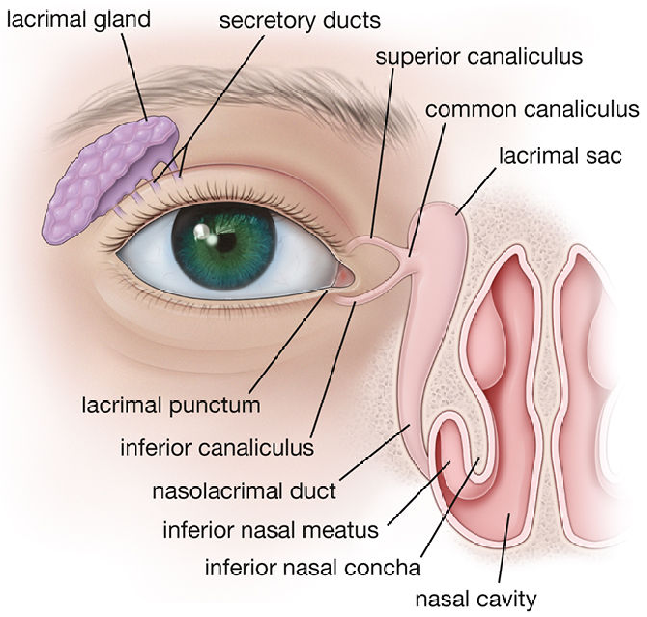
Lacrimal Puncta - Definition
The lacrimal puncta (singular: punctum) are the small openings of the lacrimal canaliculi through which tears enter from the lacrimal lake. They are the beginning of the lacrimal drainage pathway.
Position / Location (Important for exams)
- There are two puncta - one on each eyelid (upper and lower lid)
- Located at the medial angle (medial canthus) of each eye
- Situated at the posterior edge of the lid margin, at the junction of:
- The lash-bearing lateral 5/6 of the lid margin (pars ciliaris)
- The medial non-ciliated 1/6 (pars lacrimalis)
- Normally, the puncta face slightly posteriorly (toward the eyeball), which is why you need to evert the medial lid to inspect them properly
- The punctum and the vertical portion of the canaliculus are part of the tarsal plate
Structure
Each punctum leads into a lacrimal canaliculus, which has two parts:
| Part | Description |
|---|---|
| Ampulla (vertical part) | ~2 mm long, runs vertically downward from the punctum before turning |
| Horizontal part | ~8 mm long, runs medially toward the lacrimal sac |
- The superior and inferior canaliculi usually unite (in >90% of people) to form a common canaliculus
- The common canaliculus opens into the lateral wall of the lacrimal sac
- A small mucosal flap called the Valve of Rosenmüller (internal punctum) covers this junction and prevents reflux of tears back into the canaliculi
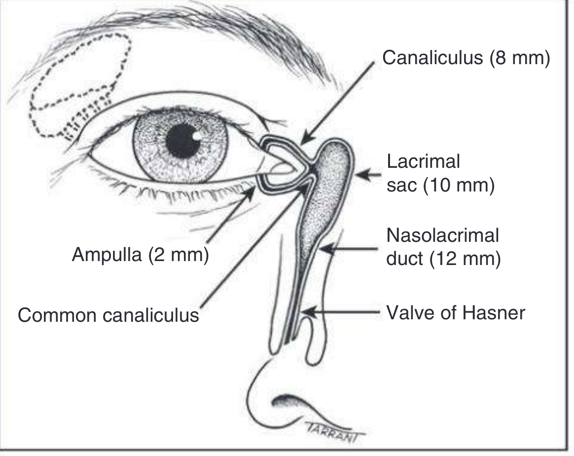
How Tears Flow Through the Puncta (Physiology)
- Tears are secreted by the lacrimal gland (upper lateral orbit) via ~12 ducts into the superior fornix of the conjunctiva
- Blinking spreads tears across the cornea from lateral to medial
- Tears accumulate in the lacrimal lake (medial angle of the eye)
- From the lacrimal lake, tears enter the puncta by capillary action and suction
- With each blink, the pretarsal orbicularis oculi muscle:
- Compresses the ampullae
- Shortens and compresses the horizontal canaliculi
- Closes and moves the puncta medially - preventing reflux
- Simultaneously, the lacrimal part of orbicularis oculi creates positive pressure that forces tears down the nasolacrimal duct
- When the eyes open, the canaliculi and sac expand, creating a negative pressure that draws fresh tears from the puncta into the sac
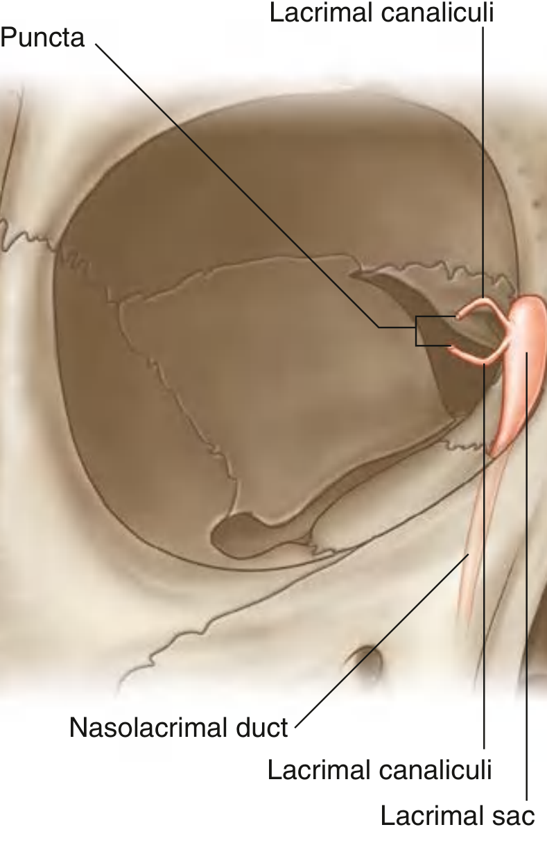
Complete Drainage Pathway with Measurements
| Structure | Length |
|---|---|
| Vertical canaliculus (Ampulla) | ~2 mm |
| Horizontal canaliculus | ~8 mm |
| Lacrimal sac | ~10-12 mm |
| Nasolacrimal duct | ~12-18 mm |
The nasolacrimal duct opens into the inferior nasal meatus (below the inferior nasal concha), and its opening is partially covered by the Valve of Hasner (a mucosal fold).
Related Muscles
The lacrimal part of orbicularis oculi (Horner's muscle) surrounds the horizontal canaliculus. Its contraction during blinking is the key pump mechanism that drives tears through the puncta and down into the nasal cavity.
Clinical Relevance (Important for BDS viva)
| Condition | Key Point |
|---|---|
| Punctal stenosis | Narrowing of the punctum causing watery eye (epiphora) |
| Punctal malposition (ectropion) | Punctum turns outward, cannot contact the lacrimal lake |
| Dacryocystitis | Inflammation of the lacrimal sac, often from obstruction of the nasolacrimal duct |
| Congenital nasolacrimal duct obstruction | Valve of Hasner fails to open at birth; most common cause of watery eye in neonates |
| Epiphora | Overflow of tears onto the cheek due to obstruction or malposition anywhere in the drainage system |
Quick Exam Recall Points
- Two puncta per eye - one upper, one lower
- Located at the medial canthus, on the posterior edge of the lid margin
- Each leads into a canaliculus with a 2 mm vertical part (ampulla) + 8 mm horizontal part
- Upper and lower canaliculi join as a common canaliculus in >90% cases
- Valve of Rosenmüller - prevents reflux at entry to lacrimal sac
- Valve of Hasner - at opening of nasolacrimal duct into inferior meatus
- Drain ultimately into the inferior nasal meatus
- Blinking is the active pump - driven by orbicularis oculi (Horner's muscle)
Sources: Gray's Anatomy for Students; Kanski's Clinical Ophthalmology 10th Ed.; Histology: A Text and Atlas (Pawlina)
This is a shared conversation. Sign in to Orris to start your own chat.