
Morphology of emphysema breakdown from Robbins
Reading File
Emphysema in Robbins covers the four classic types and their morphological features in detail. Let me pull the full content now.
Searching sources...
Finding Sources
Finding Sources
The user asked specifically for Robbins. Let me search for emphysema morphology in Robbins specifically.
Finding Sources
Finding Sources
Both Robbins books have emphysema sections. The larger "Pathologic Basis of Disease" (PBD) will have the most detailed morphology. Let me read both simultaneously.
Reading File
Reading File
I have rich content from both Robbins PBD and Robbins Basic Pathology. Let me also read the MORPHOLOGY box from PBD and fetch the key diagram images.
Reading File
Reading File
Loading Image
I now have the MORPHOLOGY box from Basic Pathology being cut off. Let me grab the rest of it.
Reading File
Loading Image
I also need to fetch the microscopic emphysema histology image and the PBD macroscopic images. Let me get those.
Loading Image
Reading File
I now have all the information needed for a comprehensive answer. Let me also fetch the histology image from Basic Pathology.
Loading Image
Loading Image
I now have all the content needed for a thorough, well-illustrated answer directly from Robbins. Here is the full morphology breakdown:
Morphology of Emphysema - Robbins
Definition
Emphysema is defined as irreversible enlargement of airspaces distal to the terminal bronchiole, accompanied by destruction of alveolar walls, but without significant fibrosis. Classification is based on the anatomic part of the acinus involved.
The acinus is the functional unit distal to the terminal bronchiole, consisting of respiratory bronchioles, alveolar ducts, and alveolar sacs. A cluster of 3-5 acini forms a lobule.
The Four Types
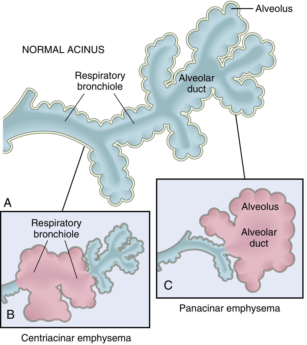
1. Centriacinar (Centrilobular) Emphysema
- Most common type - >95% of clinically significant cases
- Part affected: Central/proximal parts of the acinus - the respiratory bronchioles - are destroyed; distal alveoli are spared
- Result: Both emphysematous AND normal airspaces coexist within the same acinus and lobule
- Distribution: Upper lobes predominate, particularly the apical segments
- Association: Heavy cigarette smokers; commonly accompanied by chronic bronchitis
- In severe cases, the distal acinus is also involved, making distinction from panacinar difficult
2. Panacinar (Panlobular) Emphysema
- Part affected: Acini are uniformly enlarged from the respiratory bronchiole all the way to the terminal blind alveoli - the entire acinus is involved
- Distribution: Lower lung zones and anterior margins; most severe at the lung bases
- Association: Alpha-1 antitrypsin (A1AT) deficiency - present in >80% of A1AT-deficient patients, occurring earlier and more severely if they also smoke. Exacerbated by smoking.
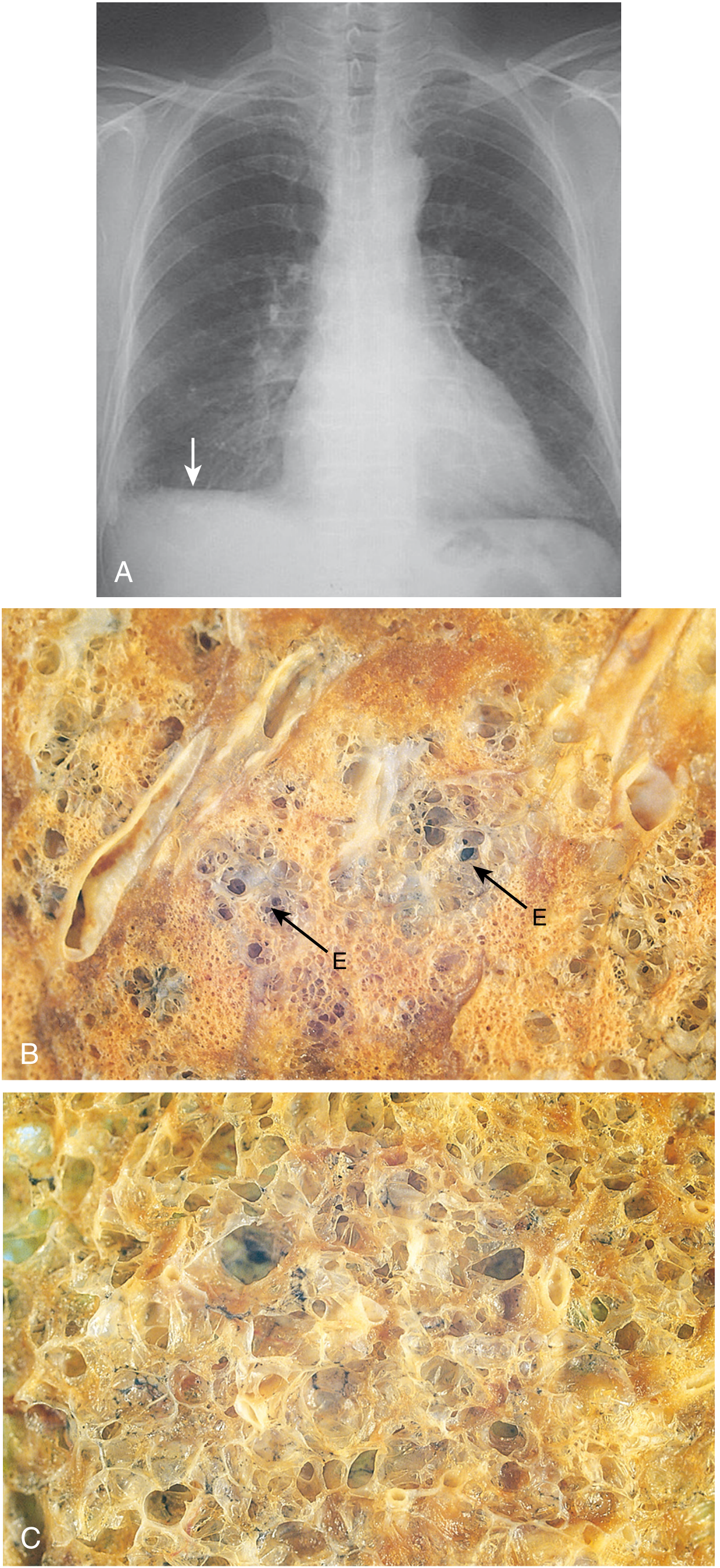
- Grossly: Produces pale, voluminous lungs that may obscure the heart when the chest is opened at autopsy
3. Distal Acinar (Paraseptal) Emphysema
- Part affected: Proximal portion of the acinus is normal; distal part is predominantly involved
- Location: Adjacent to the pleura, along lobular connective tissue septa, at the margins of lobules - often adjacent to areas of fibrosis, scarring, or atelectasis
- Usually more severe in the upper half of the lungs
- Characteristic finding: Multiple enlarged airspaces ranging from <0.5 mm to >2.0 cm in diameter, sometimes forming cyst-like structures - with further enlargement these become bullae
- Clinical significance: Underlies many cases of spontaneous pneumothorax in young adults (rupture of subpleural bullae)
4. Irregular (Paracicatricial) Emphysema
- Part affected: The acinus is irregularly involved - hence the name
- Association: Almost invariably associated with scarring (hence also called paracicatricial)
- Usually occurs in small foci
- Clinically insignificant in most instances
Summary Comparison Table
| Feature | Centriacinar | Panacinar | Distal Acinar | Irregular |
|---|---|---|---|---|
| Part of acinus | Respiratory bronchioles (central) | Entire acinus | Distal part | Irregular |
| Lobar distribution | Upper lobe (apex) | Lower lobes/bases | Upper half, subpleural | Near scars |
| Main cause | Cigarette smoking | A1AT deficiency | Unknown | Scarring |
| Clinically significant? | Yes (most common COPD type) | Yes (A1AT deficiency COPD) | Spontaneous pneumothorax | No |
| Frequency in COPD | >95% of cases | Less common | - | - |
MORPHOLOGY Box (Robbins Basic Pathology)
Gross:
- Panacinar emphysema produces pale, voluminous lungs that often obscure the heart at autopsy
- Centriacinar emphysema: lungs are a deeper pink and less voluminous than in panacinar; upper two-thirds more severely affected
Histology:
- Destruction of alveolar walls without fibrosis - leads to enlarged airspaces
- Reduced number of alveolar capillaries (due to alveolar loss)
- Terminal and respiratory bronchioles may be deformed because septa that normally tether them in the parenchyma are destroyed
- Bronchiolar inflammation and submucosal fibrosis often present in advanced disease
Other Forms (Not COPD-Related)
Robbins also lists these distinct conditions under "other forms of emphysema":
- Compensatory hyperinflation - alveolar dilation in response to loss of lung substance elsewhere (e.g., post-lobectomy); not true emphysema
- Obstructive overinflation - air trapping from subtotal airway obstruction by tumor or foreign body; can be life-threatening
- Bullous emphysema - large subpleural blebs/bullae (>1 cm diameter); can occur in any form; rupture causes pneumothorax
- Interstitial emphysema - air enters connective tissue stroma of the lung, mediastinum, or subcutaneous tissue; caused by alveolar tears from transient increases in intra-alveolar pressure (coughing) or chest wounds
Sources: Robbins, Cotran & Kumar Pathologic Basis of Disease (9780443264528), pp. 634-638; Robbins & Kumar Basic Pathology (9780323790185), pp. 447-449
Cataract for clinical postings
Reading File
This is a broad clinical topic. Searching for cataract in ophthalmology textbooks across headings and key clinical subtopics simultaneously.
Finding Sources
Finding Sources
Kanski's Clinical Ophthalmology is the primary source. Let me pull the main cataract section and the Wills Eye Manual simultaneously.
Reading File
Reading File
Good - I have the ToC. Now let me read the main clinical content sections in parallel - age-related cataract, management, and Wills Eye Manual.
Reading File
Finding Sources
Reading File
Reading File
Reading File
Reading File
Excellent. Now let me read the management/surgery section and the Wills treatment section.
Reading File
Reading File
Now let me fetch the key clinical images - the slit lamp photos of cataract types, and the maturity stages.
Loading Image
Reading File
Now let me fetch the key slit lamp photos and the maturity stages image.
Loading Image
Reading File
I now have comprehensive content for a clinical postings-level answer. Let me compile the full response.# Cataract - Clinical Postings Guide
Definition
Cataract is any opacity of the normally clear crystalline lens. It is the commonest cause of reversible visual disability in the world. There is no medical treatment to prevent its development; phacoemulsification with IOL implantation is the definitive treatment and is the most commonly performed surgical procedure globally.
Lens Anatomy (Relevant to Understanding Cataracts)
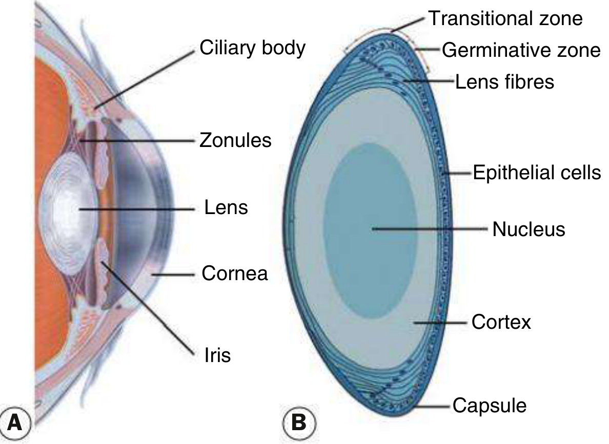
- Capsule: Acellular membrane enclosing all lens material
- Epithelial cells: Cuboid, under the anterior capsule; divide continuously in the germinative zone to form new lens fibres
- Nucleus: Central, surrounded by cortex; oldest fibres
- Cortex: Peripheral surrounding the nucleus
- Zonules: Hold the lens in position, attached to the ciliary body
Classification
By Etiology
| Type | Features |
|---|---|
| Age-related (senile) | Most common overall |
| Congenital | Present at birth; must treat early to prevent amblyopia |
| Secondary (complicated) | Due to other ocular disease |
| Traumatic | Penetrating injury, concussion, electric shock, radiation |
| Metabolic/Systemic | Diabetes, hypocalcemia, myotonic dystrophy, galactosaemia, Wilson disease |
| Drug-induced | Steroids (any route), miotics, phenothiazines |
Age-Related Cataract - By Location
1. Posterior Subcapsular (PSC) Cataract
- Opacity lies just in front of the posterior capsule
- Granular/plaque-like on oblique slit lamp; black and vacuolated on retroillumination (vacuoles = Wedl/bladder cells = swollen migratory lens epithelial cells)
- Profound effect on vision due to location at the nodal point of the eye
- Symptoms: Glare (especially night driving), worse in bright light and near vision (miosis brings opacity into optical axis)
- Also occurs with: steroid use, diabetes, trauma, radiation, uveitis - typically in patients <50 years, more rapid onset
2. Nuclear Sclerotic Cataract
- Exaggeration of normal ageing change in the nucleus
- Yellowish hue (urochrome pigment deposition); when very advanced, brown (brunescent) or rarely black
- Best assessed with oblique slit lamp beam; retroillumination shows subtle nucleus-cortex distinction
- Associated with myopia (increase in refractive index of nucleus) - causes "second sight of the aged" - previously presbyopic elderly patient can read without glasses again
- Hypermetropic shift occurs in the healthy ageing eye
3. Cortical Cataract
- Involves anterior, posterior, or equatorial cortex
- Begins as clefts and vacuoles from cortical hydration
- Classic pattern: cuneiform (wedge-shaped) or radial spoke-like opacities - often starting in the inferonasal quadrant
- Glare is a common symptom; often asymptomatic until central
4. Christmas Tree Cataract
- Uncommon; polychromatic needle-like formations in deep cortex and nucleus
- Associated with myotonic dystrophy
Cataract Maturity Stages
| Stage | Features |
|---|---|
| Immature | Lens partially opaque; iris shadow present on oblique illumination |
| Mature | Lens completely opaque; no iris shadow; fundus view lost; absent red reflex |
| Hypermature | Shrunken, wrinkled anterior capsule (water leaks out) |
| Morgagnian | Hypermature + cortex liquefied; nucleus sinks inferiorly within the capsule |
Important: A mature cataract with leaking cortex can cause phacolytic glaucoma (lens proteins block trabecular meshwork).
Secondary (Complicated) Cataract
| Cause | Features |
|---|---|
| Chronic anterior uveitis | Most common cause; earliest sign = polychromatic posterior pole lustre; progresses to PSC + anterior opacities; worsened by posterior synechiae |
| Acute angle closure glaucoma | Glaukomflecken - small grey-white anterior subcapsular opacities in pupillary area; represent focal lens epithelial infarcts; pathognomonic of previous acute angle closure |
| High myopia | PSC opacity + early nuclear sclerosis |
| Retinitis pigmentosa, Leber CA, gyrate atrophy, Stickler syndrome | PSC opacities |
Cataract in Systemic Disease
| Disease | Type of Cataract |
|---|---|
| Diabetes mellitus | Young: classic snowflake cortical opacities (rare, matures in days); Older: accelerated age-related cataract, rapid nuclear sclerosis. 1 in 5 cataract surgery patients are diabetic |
| Myotonic dystrophy | Fine iridescent cortical opacities in 3rd decade → star-shaped/wedge subcapsular opacities by 5th decade; ~90% affected |
| Hypocalcaemia | Small white iridescent cortical opacities; seen with tetany |
| Wilson disease | Sunflower cataract (anterior capsular copper deposits) |
| Atopic dermatitis | Anterior subcapsular ("shield") cataract |
| Galactosaemia | Oil-droplet cataract in infancy |
Symptoms
- Slowly progressive blurred vision, usually over months-years (one or both eyes)
- Glare - especially bright sunlight or oncoming headlights at night
- Altered colour perception (yellowing/browning with nuclear cataract)
- Monocular diplopia (irregular refraction through opacified lens)
- Second sight - nuclear sclerosis causing myopic shift; patient can read without presbyopic glasses
- Cataract does NOT cause a RAPD (relative afferent pupillary defect) - if RAPD present, look for optic nerve or retinal disease
Clinical Signs
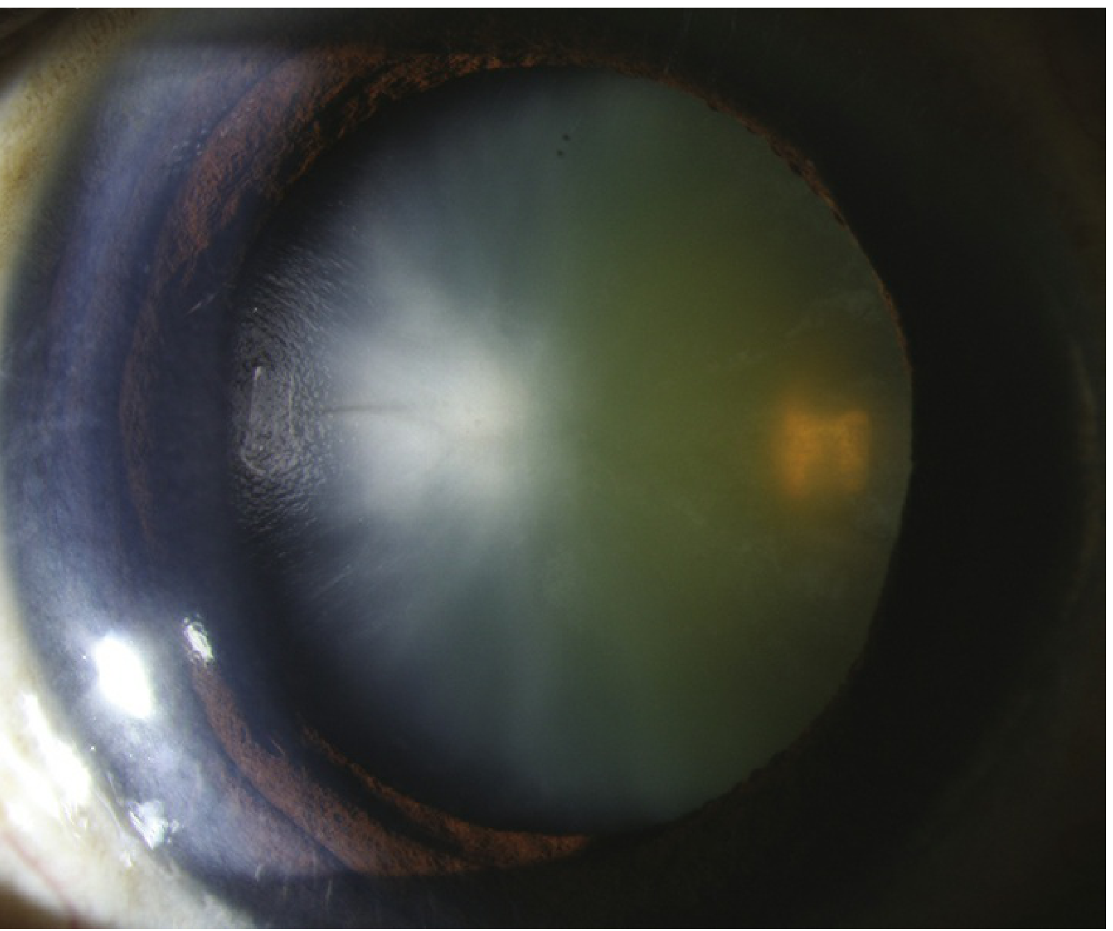
- Opacification or discoloration of the crystalline lens (critical sign)
- Diminished/absent red reflex on ophthalmoscopy or retinoscopy
- Blurred fundal view
- Myopic shift (nuclear sclerosis)
- Reduced visual acuity
Examination at Slit Lamp
- Direct illumination (oblique beam): Best for nuclear sclerosis and cortical opacities
- Retroillumination: Best for PSC (dark shadow against red reflex) and cortical opacities
- Dilated pupil examination is required for full assessment
Workup
- History: Drug history (steroids, tamsulosin - risk of intraoperative floppy iris syndrome (IFIS)), systemic diseases, trauma, previous ocular disease
- Visual acuity - distance and near, before and after refraction
- Slit lamp examination - dilated, both direct and retroillumination
- Pupil exam (no RAPD in pure cataract)
- Glare testing - useful when BCVA is 20/30 or better to demonstrate functional impact
- Fundus examination - to rule out concurrent macular/retinal disease
- B-scan ultrasound - if fundus not visible (mature cataract) to rule out posterior segment pathology (e.g., retinal detachment, vitreous haemorrhage)
- Potential Acuity Meter (PAM) / Laser interferometry - estimate visual potential when posterior segment disease coexists
- Preoperative biometry - to calculate IOL power (keratometry + axial length measurement)
Treatment
Non-surgical (temporising)
- Correct refractive error (spectacles/contact lenses)
- Trial of mydriasis (cyclopentolate 1% BD-TDS) - useful mainly for PSC cataracts (dilated pupil moves opacity out of axis); only temporary benefit
Surgical - Indications
- To improve visual function - symptomatic visual disability that affects quality of life
- As surgical therapy - lens-related glaucoma (phacolytic, phacomorphic), lens-induced uveitis
- To facilitate management - improve fundal view for monitoring/treating diabetic retinopathy or glaucoma
Surgical Techniques
Phacoemulsification (standard in high-income countries)
- Ultrasound probe fragments and aspirates the lens nucleus through a small self-sealing incision (2.2-2.8 mm)
- Followed by cortex aspiration and foldable IOL implantation into the capsular bag
- No sutures needed; fast visual recovery; low astigmatism induction
Manual Small-Incision Cataract Surgery (MSICS)
- Variant of ECCE for high-volume settings (e.g., India)
- Self-sealing sclero-corneal tunnel; nucleus expressed manually; comparable outcomes to phaco but faster and cheaper
Extracapsular Cataract Extraction (ECCE)
- Large limbal incision (8-10 mm), nucleus expressed after capsulotomy, cortex aspirated
- Requires sutures; now used mainly when phaco not feasible (dense brunescent cataract, no phaco machine)
Intracapsular Cataract Extraction (ICCE)
- Cryoprobe removes entire lens including capsule
- Largely abandoned; no capsular bag for posterior chamber IOL support
Anaesthesia Options
| Type | Details |
|---|---|
| Topical | Proxymetacaine 0.5%, tetracaine 1%; augmented with intracameral lidocaine 0.2-1%; no akinesia but adequate for cooperative patients |
| Sub-Tenon block | Blunt cannula through conjunctival incision; good anaesthesia, minimal complications; variable akinesia |
| Peribulbar block | 1-inch needle; effective anaesthesia + akinesia; rare globe perforation risk (avoid in long eyes/high myopes) |
| General anaesthesia | Children, young adults, very anxious patients, tremor, epilepsy, dementia |
Complications of Cataract Surgery
Intraoperative
- Posterior capsular rupture + vitreous loss (~1% experienced surgeons, higher for trainees)
- Zonular dehiscence
- Dropped nucleus (~0.2%)
Postoperative - Common/Mild
- IOP spike
- Iridocyclitis
- Periocular ecchymosis
- Posterior capsular opacification (PCO) - commonest late complication
Postoperative - Serious
- Endophthalmitis (0.1%) - most feared; presents with pain, reduced vision, hypopyon
- Cystoid macular oedema (CMO)
- Retinal detachment (<1%)
- Corneal decompensation
- IOL dislocation
- Unexpected refractive outcome
- Suprachoroidal haemorrhage (0.04%) - rare but devastating
Posterior Capsular Opacification (PCO) - "After-Cataract"
- Most common late complication of cataract surgery
- Residual lens epithelial cells proliferate on the posterior capsule
- Presents with gradual deterioration of vision weeks-months-years post-op
- Treatment: Nd:YAG laser posterior capsulotomy - creates a central clear opening; safe, outpatient procedure
Key Clinical Points for Postings
- Cataract alone does NOT cause RAPD - always investigate if RAPD present
- Glaukomflecken = PSC opacities in the pupillary zone = pathognomonic of previous acute angle closure
- Second sight of the aged = nuclear sclerosis inducing myopia; presbyopic patient temporarily reads without glasses
- Morgagnian cataract = nucleus sinks in liquefied cortex - the classic hypermature cataract
- PSC most visually disabling per unit size - at nodal point; worst with miosis (near work, bright light)
- Tamsulosin (alpha-1 blocker) use must be flagged pre-op - causes intraoperative floppy iris syndrome (IFIS)
- Diabetes - check and monitor for diabetic macular oedema before and after surgery (oedema can worsen post-op)
- B-scan US mandatory before operating on a white mature cataract - rule out retinal detachment
- Biometry (IOL power calculation) requires keratometry + axial length measurement
Sources: Kanski's Clinical Ophthalmology, 10th Ed. (9780443110993), Ch. 10; Wills Eye Manual, 8th Ed. (9781975160753), Ch. 13.1
This is a shared conversation. Sign in to Orris to start your own chat.