Here is a thorough overview of postural (dependent) edema, drawn from multiple authoritative textbooks.
Postural (Dependent) Edema
Definition
Edema is the presence of excess fluid within the interstitial space of the body. Postural or dependent edema is edema that accumulates specifically in the lowest gravity-dependent regions of the body - the distal lower extremities (ankles, feet, pretibial region) in ambulatory patients, and the sacral area in bedridden patients. Finger pressure over edematous subcutaneous tissue displaces interstitial fluid and leaves a temporary depression, giving it the name pitting edema. - Robbins, Cotran & Kumar Pathologic Basis of Disease
Pathophysiology - Starling Forces
Fluid movement between the intravascular and interstitial spaces is governed by the balance of four Starling forces:
| Force | Direction of Effect |
|---|
| Capillary hydrostatic pressure (Pc) | Pushes fluid OUT of capillary |
| Interstitial hydrostatic pressure (Pif) | Pushes fluid INTO capillary |
| Capillary oncotic pressure (Oc) | Pulls fluid INTO capillary |
| Interstitial oncotic pressure (Oif) | Pulls fluid OUT of capillary |
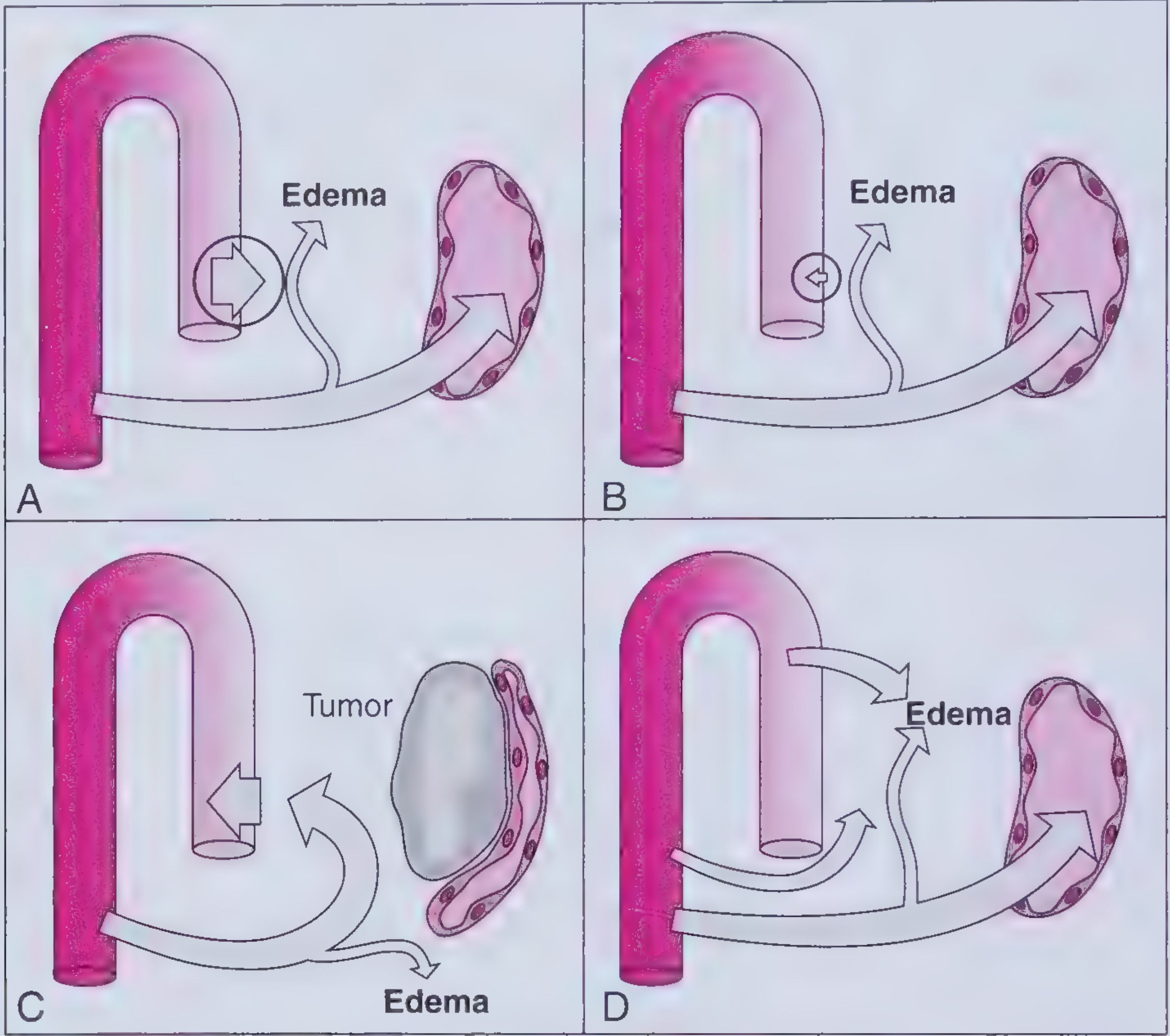
Edema forms when net outward filtration exceeds the maximal drainage capacity of the lymphatic system. The gravitational column of blood in dependent limbs raises local capillary hydrostatic pressure, which is why standing or sitting for long periods worsens ankle swelling. - Frameworks for Internal Medicine
Figure: Four mechanisms of edema formation - Frameworks for Internal Medicine
Four Core Mechanisms
1. Increased Capillary Hydrostatic Pressure
- Raises outward filtration pressure
- Results in localized or bilateral, dependent, pitting edema
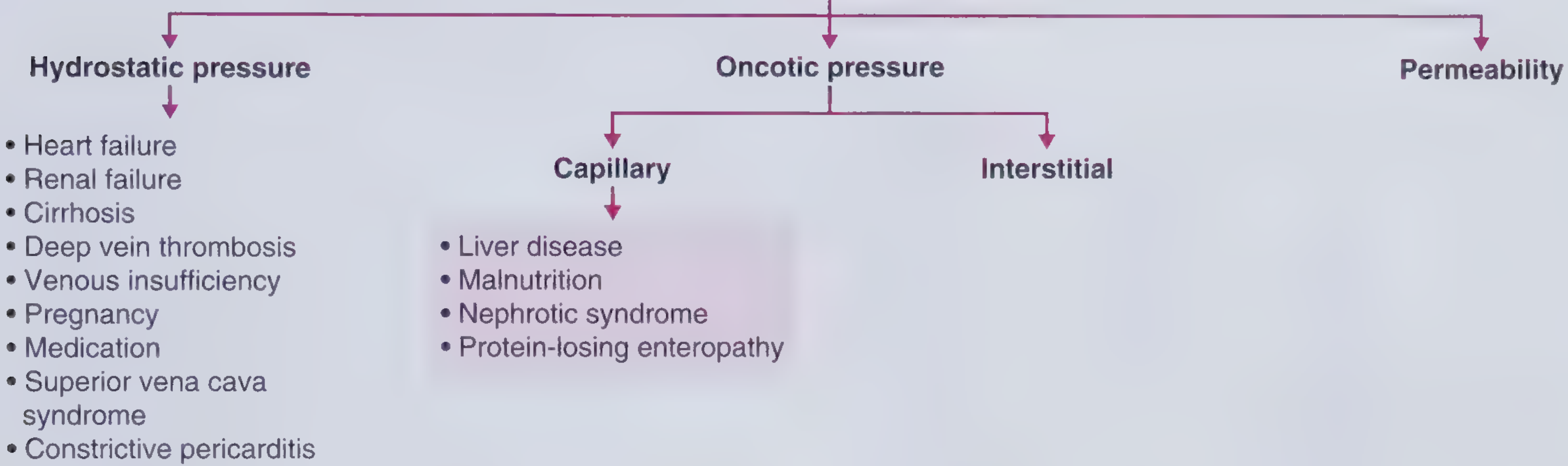
- Key causes: right heart failure, renal failure, cirrhosis (portal hypertension), DVT, chronic venous insufficiency, pregnancy, superior vena cava syndrome, medications (calcium channel blockers, NSAIDs, steroids), constrictive pericarditis
- Note: systemic arterial hypertension does NOT cause peripheral edema because precapillary sphincter autoregulation prevents arterial pressures from transmitting into the capillary bed
2. Decreased Capillary Oncotic Pressure
- Plasma albumin < 2 g/dL is typically required for edema to manifest
- Results in generalized, dependent, pitting edema, often with ascites and pleural effusions
- Key causes: liver disease (reduced synthesis), malnutrition, nephrotic syndrome (protein loss in urine), protein-losing enteropathy
3. Increased Interstitial Oncotic Pressure (Lymphatic Obstruction)
- Lymphatic blockage allows proteins to accumulate in the interstitium, raising oncotic pressure there
- Causes: malignancy (e.g., lymphoma), infection (filariasis), surgical lymph node dissection, radiation
- Produces non-pitting edema (lymphedema) - protein-rich fluid does not pit well
4. Increased Capillary Permeability
- Fluid AND proteins leak into the interstitium, diminishing the oncotic gradient
- Can produce pitting or non-pitting edema
- Causes: local inflammation, trauma, cellulitis, burns, preeclampsia, ACE inhibitor-induced angioedema, systemic capillary leak syndrome
Figure: Classification of peripheral edema causes by mechanism - Frameworks for Internal Medicine
Distribution
| Body Position | Location of Dependent Edema |
|---|
| Ambulatory / upright | Ankles, feet, pretibial region |
| Bedridden / supine | Sacral area |
| Severe (generalized) = anasarca | Throughout peripheral tissues, lungs, peritoneum |
Edema from renal disease often appears first around the eyelids (loose periorbital connective tissue), rather than in the legs. - Robbins, Cotran & Kumar
Grading of Pitting Edema
| Grade | Depth of Pit | Recovery Time |
|---|
| 1+ | ~2 mm | Immediate |
| 2+ | ~4 mm | < 15 seconds |
| 3+ | ~6 mm | 15-60 seconds |
| 4+ | ~8 mm | > 60 seconds |
A more precise method measures the depth of indentation in millimeters over a bony prominence (e.g., tibial surface). - Frameworks for Internal Medicine
Clinical Consequences
- Subcutaneous edema: signals underlying cardiac, renal, or hepatic disease; impairs wound healing and infection clearance
- Pulmonary edema: impedes gas exchange (hypoxemia), promotes bacterial infection - most often from left ventricular failure
- Brain edema: life-threatening - can cause transtentorial herniation or brainstem compression
- Peritoneal effusion (ascites): risk of spontaneous bacterial peritonitis
- Robbins, Cotran & Kumar Pathologic Basis of Disease
Key Clinical Pearls
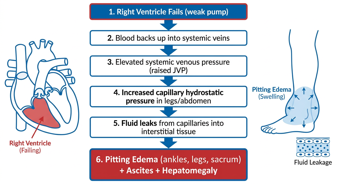
- Bilateral pitting ankle edema + elevated JVP = think right heart failure or constrictive pericarditis
- Bilateral edema + ascites + spider angiomas + caput medusae = think cirrhosis
- Edema + proteinuria + hypoalbuminemia = think nephrotic syndrome
- Unilateral leg edema + pain + erythema = rule out DVT immediately
- Non-pitting edema = think lymphedema (lymphatic obstruction)
- Angioedema (ACE inhibitor): non-pitting, non-dependent, involves lips/tongue/face - a medical emergency
Management Principles
Treatment targets the underlying cause. General measures include:
-
Sodium restriction (dietary)
-
Leg elevation above the level of the heart
-
Compression stockings (graduated compression)
-
Diuretics: loop diuretics (furosemide) for most causes; spironolactone + furosemide combination preferred for cirrhosis (starting ratio 100:40 mg to preserve potassium balance)
-
Treat the primary disease: heart failure management, albumin replacement, lymphedema physiotherapy (manual drainage), antibiotics for cellulitis, etc.
-
Frameworks for Internal Medicine, p. 314
Sources: Frameworks for Internal Medicine (Wolters Kluwer); Robbins, Cotran & Kumar Pathologic Basis of Disease (Elsevier); Robbins & Kumar Basic Pathology.