
Meliodosis
I'll start by reading the medical library skill, then search for melioidosis content in parallel.
Reading File
Now let me search the library and PubMed in parallel.
Finding Sources
Finding Sources
Searching PubMed
melioidosis Burkholderia pseudomallei
Good. Now let me read the most comprehensive sections in parallel - Fitzpatrick's dermatology (which has the most content), Murray & Nadel's respiratory, and the Clinical Pathology compendium.
Reading File
Reading File
Reading File
Reading File
Let me now get the cutaneous lesions, laboratory findings, and treatment sections from Fitzpatrick's, plus search for images.
I now have comprehensive information to write a detailed, well-sourced answer on melioidosis.
Melioidosis (Whitmore Disease)
Definition and Causative Organism
Melioidosis is an infectious disease caused by Burkholderia pseudomallei (formerly Pseudomonas pseudomallei), a motile, pleomorphic, Gram-negative, obligately aerobic, non-spore-forming bacillus. The name derives from the Greek melis ("donkey distemper") and eidos ("resembles") - reflecting its resemblance to glanders (B. mallei). It is phylogenetically close to B. mallei, and both diseases have similar clinical presentations.
- Dermatology 2-Volume Set 5e, p. 1541
Microbiology Characteristics
| Feature | Detail |
|---|---|
| Morphology | Gram-negative bacillus, motile |
| Staining | Bipolar staining ("safety pin" appearance) |
| Culture | Wrinkled colonies on MacConkey and blood agar |
| Biochemistry | Oxidase positive |
| Growth | Grows on routine media; gentamicin agar prevents overgrowth |
| Niche | Environmental saprophyte in soil, water, rice paddies |
- Quick Compendium of Clinical Pathology 5e
Epidemiology
-
Endemic regions: Southeast Asia (especially northeastern Thailand, Singapore, Malaysia) and northern Australia (Northern Territory). Cases increasingly reported in Africa, Middle East, Central and South America.
-
B. pseudomallei has been identified in the environment along the Gulf Coast of the USA; contaminated imported products (e.g., aromatherapy spray from India) have caused outbreaks in the US.
-
Hyperendemic areas: may be the most common cause of severe community-acquired pneumonia (CAP) in northeastern Thailand and northern Australia.
-
Seasonality: Over 75% of cases occur during the rainy monsoon season, likely due to aerosolization of bacteria from water and soil.
-
Occupational: Rice farmers in Thailand; male-to-female ratio 4:1 (occupational exposure).
-
Serologic surveys in rural Thailand show 5-20% of inhabitants have antibodies, suggesting frequent subclinical infection.
-
Post-tsunami (December 2004): hundreds of thousands exposed to flood waters; cases of severe post-immersion pneumonic melioidosis were reported across the region.
-
Fitzpatrick's Dermatology Vol. 1&2, p. 2874
Routes of Infection
- Percutaneous inoculation - direct contact of abraded/lacerated skin with contaminated soil or water (most common)
- Inhalation - of contaminated droplets, soil, or dust
- Ingestion
- Sexual intercourse (rare)
Risk Factors for Severe Disease
- Diabetes mellitus (most significant)
- Alcohol use disorder / alcoholic liver disease
- Chronic renal disease
- Chronic lung disease / COPD
- Heart disease, smoking
- Cystic fibrosis (in those who live/travel in endemic areas)
- Near-drowning / tsunami exposure
- Note: HIV infection does NOT significantly increase risk of acquiring disease or severity
Pathogenesis
B. pseudomallei uses a type III secretion system to inhibit autophagy in host cells (epithelia, macrophages). After macrophage ingestion, it subverts phagolysosomal maturation and can cause cell lysis via caspase-1-dependent pyroptosis, releasing bacteria to infect neighboring cells. Reactive oxygen/nitrogen species and Th1 responses are important for control.
- Murray & Nadel's Textbook of Respiratory Medicine
Clinical Presentations
The incubation period is 1-21 days (mean ~9 days; can be as brief as 2-3 days).
Melioidosis is described as a "great imitator" with a wide spectrum from asymptomatic to fulminant sepsis.
1. Acute Melioidosis
- Pneumonic form (most common - ~50% of all cases): abrupt onset fever, chills, cough, dyspnea, chest pain, hemoptysis; can progress to septic shock in 33%.
- Septicemic form: bacteremia - often no identifiable skin source (though many patients have minor foot abrasions); leads to jaundice, hepatosplenomegaly, myocarditis, severe gastroenteritis, miliary pulmonary disease.
2. Chronic/Subacute Melioidosis
- Defined as symptoms present for >2 months.
- Upper lobe cavitary disease resembling tuberculosis (clinically and radiologically).
- Presenting features: fever, weight loss, productive cough, hemoptysis.
- CXR: diffuse miliary nodules that may expand and cavitate; ~50% upper lobe changes, one-third multilobe involvement.
- Draining sinuses, subcutaneous abscesses (especially scalp).
3. Localized/Cutaneous Melioidosis
- ~15-20% of cases have cutaneous manifestations.
- Primary lesion (especially children): ulcer at inoculation site, possibly with purulent exudate.
- Disseminated: multiple superficial pustules, ecthyma gangrenosum, necrotizing fasciitis.
- Chronic: draining sinuses, subcutaneous abscesses (especially scalp).
- Acute suppurative parotitis (usually unilateral) - classic presentation in children from endemic areas.
- Other skin manifestations: cellulitis, granulomatous lesions, purpura, pustules, Sweet syndrome, urticaria.
4. Extrapulmonary Disease
- Genitourinary infection
- Bone/joint: septic arthritis, osteomyelitis
- CNS disease
- Visceral abscesses (liver, spleen, prostate)
- Suppurative lymphadenopathy
Latent Melioidosis
- B. pseudomallei can establish latency and reactivate decades later when the host becomes debilitated. Presentations as unexplained persistent fever years after leaving endemic areas have been reported.
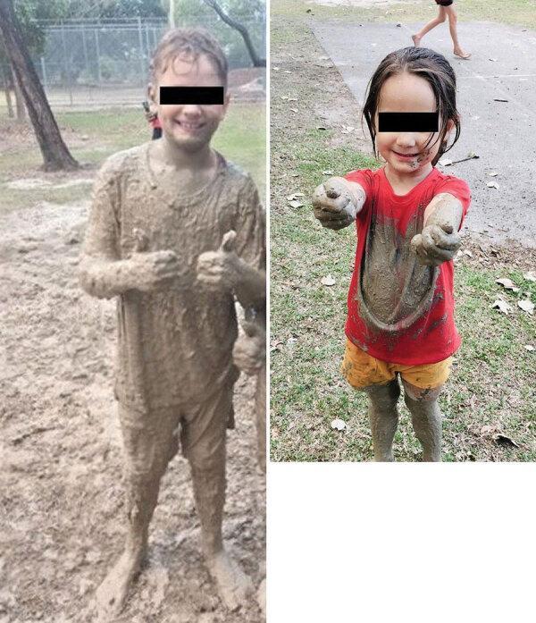
Skin Exposure Risk
Diagnosis
| Method | Notes |
|---|---|
| Bacterial culture (gold standard) | Sensitivity ~60%; grows on routine media; alert lab to use gentamicin-supplemented agar. Culture from blood, sputum, or abscess aspirate. |
| PCR | More sensitive and species-specific than culture; increasingly used |
| Serology | Complement-fixing and agglutinating antibodies appear 4-6 weeks post-infection; inadequate alone in endemic regions. Indirect hemagglutination assay (IHA) is most commonly used in Southeast Asia. |
| Histopathology | Sharply circumscribed abscesses in organs/subcutaneous tissues, often with surrounding granulomatous response. Bipolar-staining Gram-negative bacilli on smear support diagnosis. |
| Automated systems (2024 meta-analysis [PMID 38820898]) | Diagnostic accuracy of automation vs. non-automation techniques systematically reviewed |
Differential Diagnosis
| Presentation | Mimics |
|---|---|
| Acute melioidosis | Typhoid fever, staphylococcal pneumonia, disseminated fungal infections (Penicillium marneffei), glanders, septicemia |
| Chronic pulmonary | Pulmonary tuberculosis, nocardiosis, deep fungal infection, bacterial lung abscess |
| Cutaneous/draining sinuses | Actinomycosis, mycobacterial infection |
Treatment
Treatment is in two phases:
Phase 1: Intensive (Induction) Phase
- IV ceftazidime 50 mg/kg up to 2 g three times daily, OR
- IV meropenem 25 mg/kg up to 1 g three times daily (preferred for: ICU-requiring disease, persistent bacteremia, CNS disease)
- Duration: minimum 10-14 days (at least 2 weeks)
Phase 2: Eradication (Maintenance) Phase
- Oral TMP-SMX (co-trimoxazole) - preferred, OR
- Oral amoxicillin-clavulanate
- Duration: ≥3 months (longer for osteomyelitis or multiple suppurative foci)
Key Resistance Profile
- B. pseudomallei is intrinsically resistant to quinolones, macrolides, and aminoglycosides.
- Susceptibility testing must be performed for each isolate.
Prognosis
| Form | Mortality |
|---|---|
| Septicemic + shock + dissemination to skin/viscera | ~50% |
| Primary pulmonary melioidosis (Northern Australia) | ~20% |
| Overall (with intensive supportive care) | ~20% |
| Localized cutaneous without bacteremia | <9% |
| Melioidosis-associated sepsis (without intensive care) | 50-90% |
- Relapse rate: ~10%
- B. pseudomallei can remain latent for decades - recrudescence may occur with any subsequent immune compromise.
Bioterrorism Significance
B. pseudomallei (along with B. mallei) is classified as a potential bioterrorism agent. Efforts are underway to develop vaccines. - Dermatology 2-Volume Set 5e, p. 1542
Summary Table
| Feature | Detail |
|---|---|
| Causative organism | Burkholderia pseudomallei (Gram-neg rod, bipolar staining) |
| Synonym | Whitmore disease |
| Reservoir | Wet soil, water (not animals) |
| Transmission | Skin contact > inhalation > ingestion |
| Endemic zones | SE Asia, N. Australia; Gulf Coast USA (emerging) |
| Peak season | Rainy/monsoon season |
| Key risk factor | Diabetes mellitus |
| Classic presentation in children | Acute suppurative parotitis |
| "TB mimic" | Chronic upper-lobe cavitary disease |
| Key resistance | Quinolones, macrolides, aminoglycosides |
| Treatment backbone | Ceftazidime or carbapenem → TMP-SMX |
| Latency | Yes - can reactivate after decades |
| Bioterrorism potential | Yes (Category B agent) |
Sources: Fitzpatrick's Dermatology Vol. 1&2; Dermatology 2-Volume Set 5e; Murray & Nadel's Textbook of Respiratory Medicine; Andrews' Diseases of the Skin; Quick Compendium of Clinical Pathology 5e
This is a shared conversation. Sign in to Orris to start your own chat.