
Harmone act via intracellular pathway
Reading File
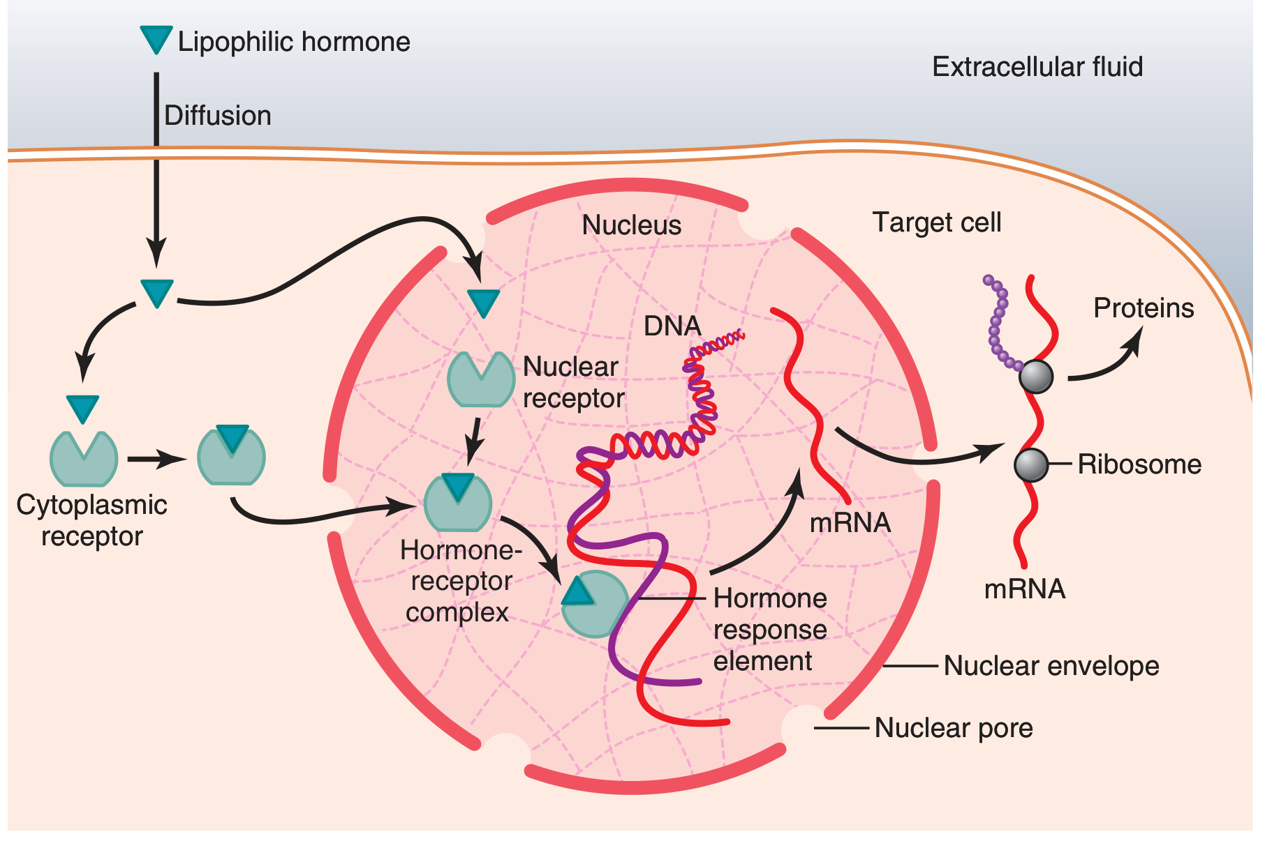
Hormones acting via intracellular receptors are mainly lipophilic (steroid hormones, thyroid hormones, and vitamin D). They cross the plasma membrane and bind to cytoplasmic or nuclear receptors, which then act as transcription factors to regulate gene expression.
Searching sources...
Finding Sources
Finding Sources
Reading File
Reading File
Loading Image
Loading Image
Reading File
Reading File
Loading Image
Here is a complete explanation of hormones acting via the intracellular pathway, sourced from Guyton & Hall and Costanzo Physiology.
Hormones Acting via Intracellular Receptors
Which Hormones Use This Pathway?
Hormones that are lipid-soluble (lipophilic) readily cross the phospholipid cell membrane and bind to receptors inside the cell rather than on its surface. These include:
| Hormone Class | Examples |
|---|---|
| Glucocorticoids | Cortisol |
| Mineralocorticoids | Aldosterone |
| Sex steroids | Testosterone, Estrogen, Progesterone |
| Thyroid hormones | T3, T4 |
| Vitamin D (active form) | 1,25-dihydroxycholecalciferol |
| Retinoids | Retinoic acid |
The Intracellular Receptor (Structure)
These receptors are monomeric phosphoproteins belonging to a gene superfamily of nuclear receptors. Each receptor has six domains (A-F):
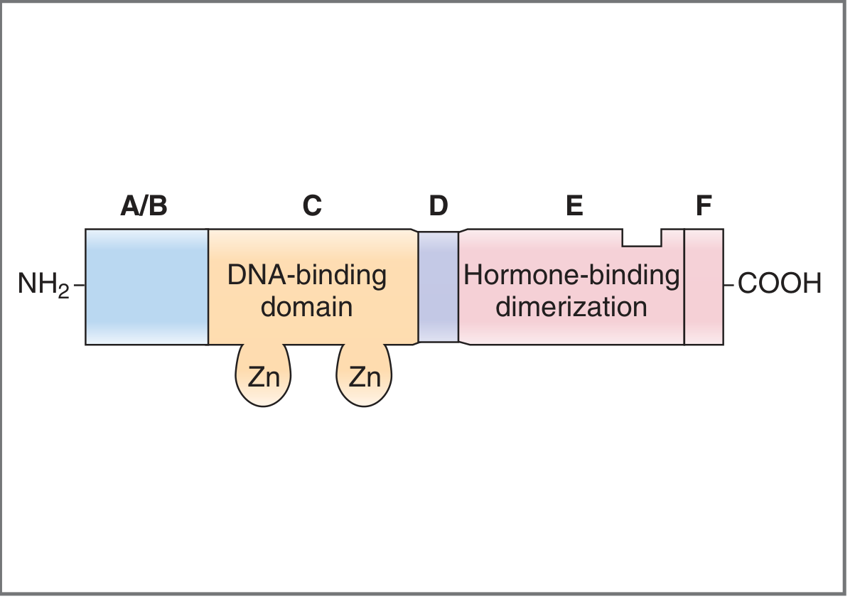
- Domain C (central): highly conserved, contains two zinc fingers, responsible for DNA binding
- Domain E (near C-terminus): the hormone-binding and dimerization site
- Receptor may be located in the cytosol (most steroid hormones) or already in the nucleus (thyroid hormones, retinoids)
Step-by-Step Mechanism
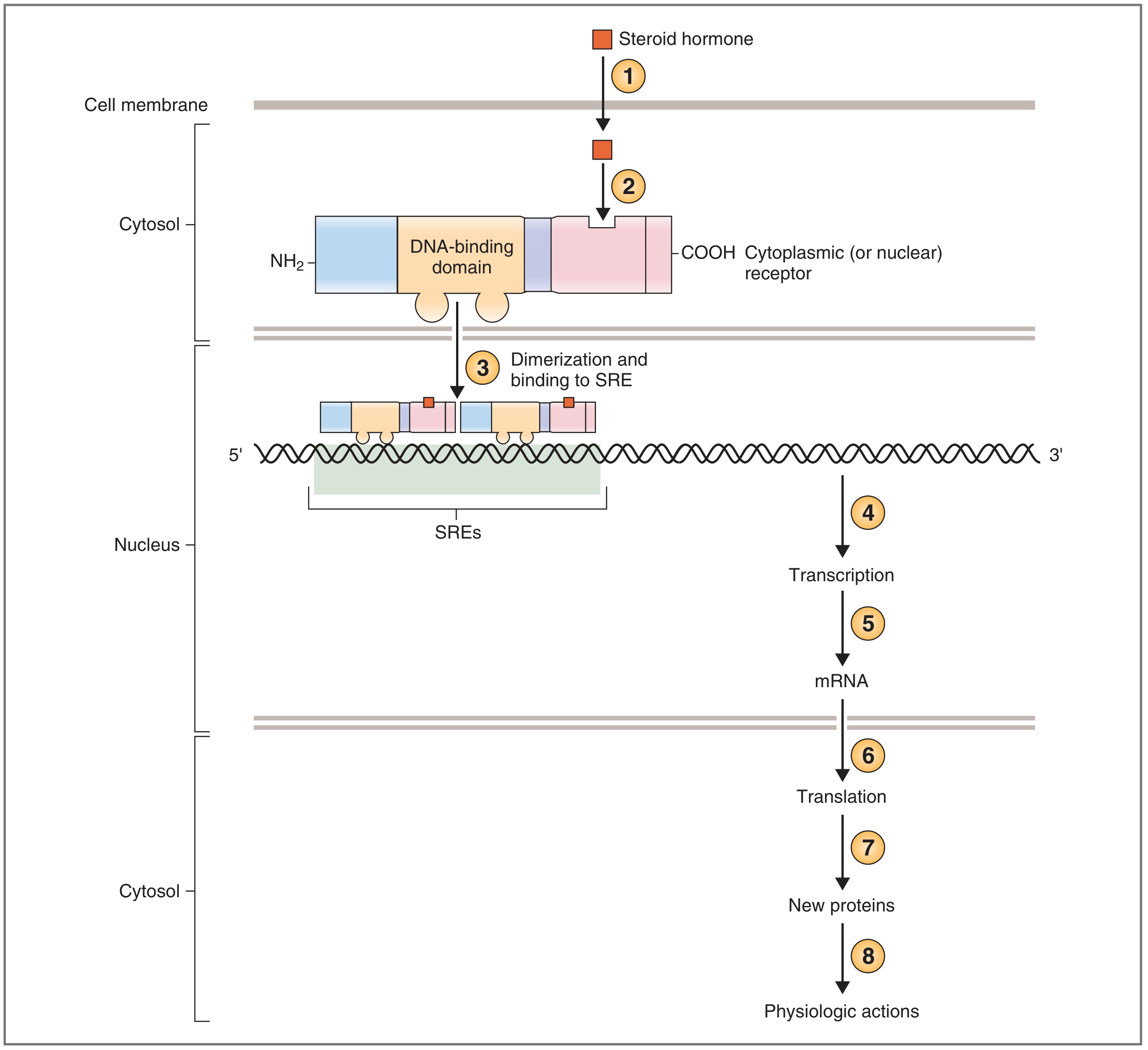
Step 1 - Diffusion across the cell membrane
Because the hormone is lipophilic, it freely diffuses across the plasma membrane into the target cell.
Step 2 - Receptor binding
Inside the cell, the hormone binds to a specific receptor protein located in the cytosol or nucleus. This binding causes a conformational change in the receptor, activating it.
Step 3 - Dimerization and nuclear entry
The activated hormone-receptor complex dimerizes (pairs with another complex) and translocates into the nucleus if it was in the cytosol.
Step 4 - Binding to DNA (Hormone Response Element)
The dimer binds via its zinc fingers (C domain) to specific DNA sequences called Steroid-Responsive Elements (SREs) or Hormone Response Elements (HREs) located in the 5' regulatory region of target genes.
Step 5 - Transcription
The hormone-receptor complex now acts as a transcription factor, either activating or repressing gene transcription.
Step 6 - mRNA formation and export
New mRNA is transcribed, leaves the nucleus through nuclear pores.
Step 7 - Translation
mRNA is translated at ribosomes into new specific proteins.
Step 8 - Physiologic effects
The new proteins produce the hormone's physiologic actions.
The Pathway Illustrated (Guyton & Hall)
Key Examples of Proteins Induced
| Hormone | New Protein Synthesized | Effect |
|---|---|---|
| 1,25-(OH)2 Vitamin D | Ca2+-binding protein (calbindin) | Promotes Ca2+ absorption from intestine |
| Aldosterone | Na+ channels (ENaC) | Promotes Na+ reabsorption in kidney |
| Testosterone | Skeletal muscle proteins | Muscle growth |
| Cortisol | Gluconeogenic enzymes | Increases blood glucose |
Key Characteristics - Comparison with Membrane Receptor Pathway
| Feature | Intracellular Receptor Pathway | Membrane Receptor (e.g., cAMP) Pathway |
|---|---|---|
| Hormone type | Lipophilic (steroids, thyroid, vitamin D) | Hydrophilic (peptides, catecholamines) |
| Receptor location | Cytosol or nucleus | Cell surface |
| Second messenger | None - acts directly on DNA | Yes (cAMP, IP3, Ca2+) |
| Speed of action | Slow (hours to days) | Rapid (seconds to minutes) |
| Effect | New protein synthesis | Enzyme activation/inhibition |
| Duration | Long-lasting | Short-lived |
Tissue Specificity
An important point: many tissues have identical intracellular receptors, but the genes regulated differ between tissues. The response depends not just on the receptor but on which gene regulatory proteins are expressed in that particular tissue. This explains why the same hormone (e.g., cortisol) can have different effects in liver vs. immune cells.
- Guyton and Hall Textbook of Medical Physiology, p. 910-912
- Costanzo Physiology 7th Edition, p. 410
This is a shared conversation. Sign in to Orris to start your own chat.