
Give me a clear explanation of physiology of cardiovascular system
Reading File
Now let me search the library for cardiovascular physiology content across multiple topics in parallel.
Finding Sources
Finding Sources
Finding Sources
Excellent. Now let me read the core physiology sections in parallel from Ganong's and Guyton & Hall (the gold-standard physiology textbooks), plus the cardiac cycle from Costanzo.
Reading File
Reading File
Reading File
Now let me search for more key cardiovascular topics in parallel - blood pressure regulation, Frank-Starling law, and vascular physiology.
Finding Sources
Finding Sources
Finding Sources
Finding Sources
Finding Sources
Reading File
Reading File
I now have comprehensive content from multiple authoritative textbooks. Let me also fetch the key diagrams.
Loading Image
Loading Image
I have comprehensive material from authoritative textbooks. Let me now compose the full answer.
Physiology of the Cardiovascular System
The cardiovascular system is a closed-loop circuit whose primary job is to transport oxygen, nutrients, hormones, and metabolic waste between the tissues and the lungs/organs of elimination. It maintains homeostasis by ensuring cells always operate within a narrow range of pH, oxygen saturation, and osmolality.
1. Overview and Components
The system consists of three main elements:
- The heart - a four-chambered pump
- Blood vessels - arteries, arterioles, capillaries, venules, veins
- Blood - the transport medium
The right heart receives deoxygenated blood from the body and pumps it to the lungs (pulmonary circulation). The left heart receives oxygenated blood from the lungs and pumps it to the body (systemic circulation). Both circuits run in series, so cardiac output is the same through each.
2. The Cardiac Conduction System
The heart generates its own electrical impulses - a property called automaticity.
| Structure | Role |
|---|---|
| SA node (sinoatrial) | Primary pacemaker; fires 60-100 bpm spontaneously |
| Internodal pathways | Conduct impulse across atria |
| AV node | Delays conduction by >0.1 sec; allows atrial emptying before ventricular contraction |
| Bundle of His | Passes impulse from AV node to ventricles |
| Left and right bundle branches | Distribute impulse to each ventricle |
| Purkinje fibers | Rapidly activate ventricular myocardium from apex upward |
The SA node fires fastest and therefore dominates as the pacemaker. Lower-order pacemakers (AV node: 40-60 bpm, ventricles: 20-40 bpm) only take over if the SA node fails.
"The parts of the heart normally beat in orderly sequence: contraction of the atria (atrial systole) is followed by contraction of the ventricles (ventricular systole), and during diastole all four chambers are relaxed." - Ganong's Review of Medical Physiology, 26th Ed.
3. The Cardiac Cycle
Each heartbeat is one complete cardiac cycle (~0.83 sec at 72 bpm). It has 7 phases:
Phase A - Atrial Systole
- Triggered by the P wave on ECG
- Atria contract, pushing the final ~30% of blood into the ventricles
- Mitral and tricuspid valves are open
- Contributes to the a wave in venous pulse
Phase B - Isovolumetric Ventricular Contraction
- Triggered by the QRS complex on ECG
- All four valves are closed; ventricular pressure rises rapidly with no change in volume
- S1 heart sound = mitral valve closing
Phase C - Rapid Ventricular Ejection
- Ventricular pressure exceeds aortic pressure (~80 mmHg on left) → aortic valve opens
- ~70% of stroke volume is ejected rapidly
- Aortic pressure rises to ~120 mmHg
Phase D - Reduced Ventricular Ejection
- Ventricle continues ejecting but at slower rate
- Coincides with the T wave (ventricular repolarization)
- Ventricular volume reaches its minimum (end-systolic volume, ~50 mL)
Phase E - Isovolumetric Ventricular Relaxation
- Aortic valve closes → S2 heart sound
- Ventricular pressure falls rapidly, all valves closed, volume unchanged
- The dicrotic notch on aortic pressure trace = aortic valve closure
Phase F - Rapid Ventricular Filling
- Ventricular pressure falls below atrial pressure → mitral valve opens
- ~70% of ventricular filling occurs passively here
- S3 sound (if present) marks this phase; normal in children, pathological in adults
Phase G - Reduced Ventricular Filling (Diastasis)
- Slow filling continues as pressure gradients equalize
- Cycle then repeats with the next P wave
(Costanzo Physiology, 7th Ed.)
4. Cardiac Output and Its Determinants
Cardiac Output (CO) = Heart Rate (HR) × Stroke Volume (SV)
Normal CO at rest: ~5 L/min (can rise to ~20-25 L/min during intense exercise).
CO depends on four key factors:
4a. Preload
- The degree of ventricular stretch at end-diastole (end-diastolic volume/pressure)
- More filling → more stretch → more force on contraction
- This is the Frank-Starling Law of the Heart
4b. Frank-Starling Law
The greater the initial sarcomere stretch during diastole (preload), the more force generated during systole. At the cellular level, stretching the sarcomere increases the sensitivity of troponin C to calcium and improves actin-myosin overlap, producing a stronger contraction without needing extra calcium or catecholamines.
"This relationship of force to length is referred to as the Frank-Starling law of the heart." - Goldman-Cecil Medicine
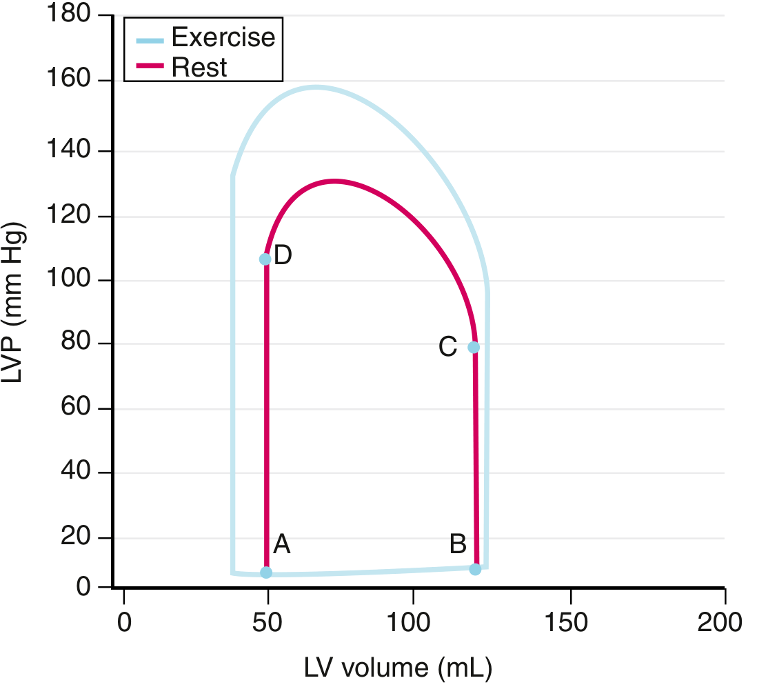
4c. Afterload
- The resistance the ventricle must overcome to eject blood (determined mainly by aortic pressure and systemic vascular resistance)
- High afterload reduces stroke volume; low afterload increases it
- Described by Laplace's Law: wall tension = (Pressure × Radius) / (2 × wall thickness)
4d. Contractility (Inotropy)
- The intrinsic ability of the myocardium to generate force at a given preload/afterload
- Increased by: sympathetic stimulation, catecholamines, digoxin
- Decreased by: acidosis, hypoxia, beta-blockers, heart failure
4e. Heart Rate (Chronotropy)
- Higher HR → higher CO, up to a limit (~150-180 bpm in adults)
- At very high rates, diastolic filling time shortens and SV falls, limiting the CO benefit
5. Pressures in the Circulation
| Location | Pressure |
|---|---|
| Aorta (systolic/diastolic) | 120 / 80 mmHg |
| Arterioles | 40-80 mmHg (major resistance vessels) |
| Capillaries | 17-35 mmHg |
| Venules/veins | 5-10 mmHg |
| Right atrium (CVP) | 0-5 mmHg |
| Pulmonary artery | 25 / 8 mmHg |
| Normal SVR | 10-20 mmHg·min/L |
| Normal PVR | 0.5-1.5 mmHg·min/L |
(Goldman-Cecil Medicine)
Mean arterial pressure (MAP) = Diastolic BP + 1/3 (Pulse Pressure) ~ 93 mmHg at rest.
MAP = CO × Systemic Vascular Resistance (SVR)
6. Vascular Physiology
Arteries
- Elastic (large arteries: aorta) - act as pressure reservoirs, dampening systolic peaks
- Muscular (medium arteries) - regulate distribution of blood
- Arterioles - the main resistance vessels; small changes in diameter produce large changes in resistance (resistance ∝ 1/r⁴ by Poiseuille's Law)
Capillaries
- Site of nutrient/gas/waste exchange via diffusion and filtration
- Starling forces govern fluid movement:
- Outward: capillary hydrostatic pressure, interstitial osmotic pressure
- Inward: plasma oncotic pressure (colloid osmotic pressure), interstitial hydrostatic pressure
- Net filtration is small; excess fluid is returned by lymphatics
Veins
- Low-pressure, high-capacitance vessels; hold ~65% of blood volume
- Act as volume reservoirs; venoconstriction mobilizes blood to the heart (increases preload)
7. Regulation of Blood Pressure
Blood pressure is regulated at multiple timescales:
Short-term: Baroreceptor Reflex (seconds)
- Baroreceptors (stretch receptors) in the carotid sinus and aortic arch detect wall distension
- Afferents travel via CN IX (carotid sinus nerve/Hering's nerve) and CN X (vagus/depressor nerve) to the medullary cardiovascular centers
- A rise in BP → increased baroreceptor firing → medullary inhibition of sympathetic outflow + vagal activation → decreased HR, decreased SV, vasodilation → BP falls back to normal
- A fall in BP → opposite response
- Secondary sensors: chemoreceptors (peripheral: carotid/aortic bodies; central: medulla) - respond to low O₂, high CO₂, low pH → increase CO and ventilation
"A dual system of sensors and neural reflexes controls mean arterial pressure. The primary sensors are baroreceptors... The secondary sensors are chemoreceptors that detect changes in blood PO₂, PCO₂, and pH." - Medical Physiology (Boron & Boulpaep)
Medium-term: Hormonal (minutes to hours)
- Renin-Angiotensin-Aldosterone System (RAAS): low renal perfusion → renin → angiotensin II (vasoconstriction, aldosterone release) → Na⁺/water retention → increased blood volume and BP
- ADH (vasopressin): released from posterior pituitary in response to low BP or high osmolality → water reabsorption, vasoconstriction
- Atrial Natriuretic Peptide (ANP): released when atria are overstretched → natriuresis, vasodilation → lowers BP
Long-term: Renal Pressure-Natriuresis
- The kidney regulates blood volume over days-weeks; increased BP → increased urinary Na⁺ and water excretion → reduced volume → BP normalizes
8. The Autonomic Nervous System and the Heart
| Effect | Sympathetic (β₁) | Parasympathetic (M₂) |
|---|---|---|
| Heart rate | Increases (positive chronotropy) | Decreases (negative chronotropy) |
| Contractility | Increases (positive inotropy) | Minimal effect on ventricles |
| Conduction velocity | Increases (positive dromotropy) | Decreases (AV node) |
| Relaxation speed | Increases (positive lusitropy) | - |
Sympathetic stimulation acts via norepinephrine on β₁ receptors, increasing cAMP → PKA activation → phosphorylation of L-type Ca²⁺ channels and phospholamban → more calcium entry and faster re-uptake → stronger, faster contractions.
9. The Microcirculation and Oxygen Delivery
Oxygen delivery (DO₂) = CO × Arterial O₂ content (CaO₂)
CaO₂ = (Hb × 1.34 × SaO₂) + (0.003 × PaO₂)
At rest, tissues extract ~25% of delivered O₂ (oxygen extraction fraction). During exercise or shock, extraction increases up to ~75%. The P₅₀ (PO₂ at which Hb is 50% saturated) and the Bohr effect (CO₂, H⁺, 2,3-DPG shift the curve rightward) fine-tune O₂ unloading at the tissues.
10. Special Circulations
| Circulation | Key Features |
|---|---|
| Coronary | Perfused mainly in diastole (left coronary); highest O₂ extraction at rest |
| Cerebral | Autoregulated (MAP 60-150 mmHg); CO₂ is the dominant vasodilator |
| Pulmonary | Low resistance, low pressure; hypoxic vasoconstriction (unique - opposite of systemic) |
| Splanchnic | Large reservoir; vasoconstricts markedly during exercise/hemorrhage |
| Renal | Autoregulated; receives ~25% of CO despite being 0.5% of body weight |
| Skin | Primary thermoregulatory organ; AVAs (arteriovenous anastomoses) shunt flow |
Summary
The cardiovascular system operates as an integrated pump-pipe-fluid system controlled by electrical, mechanical, hormonal, and neural signals:
- Electrical - SA node generates rhythmic action potentials; conducted in sequence through the conduction system
- Mechanical - The cardiac cycle converts electrical signals into coordinated pressure-volume changes that eject blood
- Frank-Starling - The heart automatically adjusts output to match venous return
- Neural - Baroreceptors provide second-by-second BP corrections via autonomic reflexes
- Hormonal - RAAS, ADH, and ANP tune volume and resistance over longer time periods
Sources: Ganong's Review of Medical Physiology 26th Ed., Guyton and Hall Textbook of Medical Physiology, Costanzo Physiology 7th Ed., Goldman-Cecil Medicine, Medical Physiology (Boron & Boulpaep)
This is a shared conversation. Sign in to Orris to start your own chat.