
AV valve includes papillary muscle
AV valve mitral tricuspid papillary muscle chordae tendineae anatomy diagram
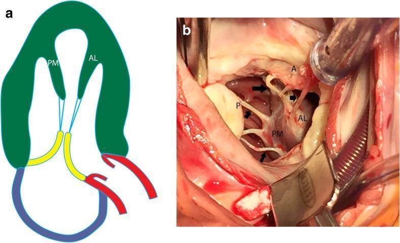
This dual-panel educational material illustrates the normal anatomy of the mitral valve apparatus within the left ventricle. Panel A is an anatomical diagram in a vertical long-axis projection showing the anterolateral (AL) and posteromedial (PM) papillary muscles (green) originating from the ventricular wall. Blue lines represent chordae tendineae connecting the papillary muscles to the mitral valve leaflets (yellow). Panel B is an intraoperative clinical photograph providing a direct surgical view of these structures. It clearly demonstrates the fleshy heads of the anterolateral (AL) and posteromedial (PM) papillary muscles. Multiple thin, white fibrous strands—the chordae tendineae (indicated by black arrows)—extend from the papillary muscle heads to insert into the anterior (A) and posterior (P) mitral valve leaflets. The image highlights the mechanical tethering system essential for valvular competence during the cardiac cycle, specifically demonstrating how both papillary muscles contribute chordae to both leaflets to prevent valve prolapse into the left atrium during systole.
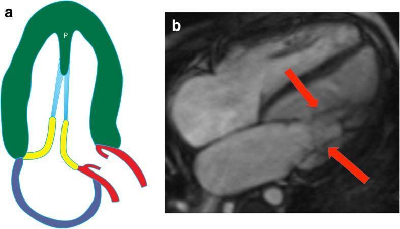
This composite image illustrates the anatomy of a parachute mitral valve (PMV) through a schematic diagram and a diagnostic cardiac MRI. Panel A is an anatomical diagram showing the characteristic single papillary muscle (labeled 'P') within the left ventricle, from which all chordae tendineae (blue) originate. This configuration forces the mitral valve leaflets (yellow) into a restricted, funnel-like shape resembling a parachute. Panel B shows a four-chamber steady-state free precession (SSFP) cardiac MRI sequence. Red arrows indicate the convergent attachment of multiple chordae tendineae to a solitary, centrally located papillary muscle, rather than the typical distribution between anterolateral and posteromedial muscles. This rare congenital anomaly is a significant finding in pediatric and adult cardiology, often associated with mitral stenosis due to reduced leaflet mobility and restricted valve orifice area. It may also present as part of the Shone complex. The image serves as an educational tool for identifying atypical subvalvular apparatus morphology in congenital heart disease.

A three-panel anatomical diagram (A-C) illustrating the step-by-step surgical implantation of artificial chordae using the CardioMech system for mitral valve repair. (A) Shows a transcatheter delivery system (blue catheter with a silver metallic distal tip) approaching the posterior mitral leaflet (PML) to perform a puncture. (B) Illustrates the deployment and anchoring of a blue ePTFE artificial chord into the papillary muscle within the left ventricle. (C) Depicts the final stage where the neochorda is tensioned to adjust the leaflet's position and ensure proper coaptation. The diagram highlights the interaction between the surgical instruments, the thin blue artificial chordae, and the cardiac anatomy, including the mitral leaflets and papillary muscles. This educational visual is designed to demonstrate minimally invasive, beating-heart techniques for correcting mitral regurgitation by replacing ruptured or elongated natural chordae tendineae.
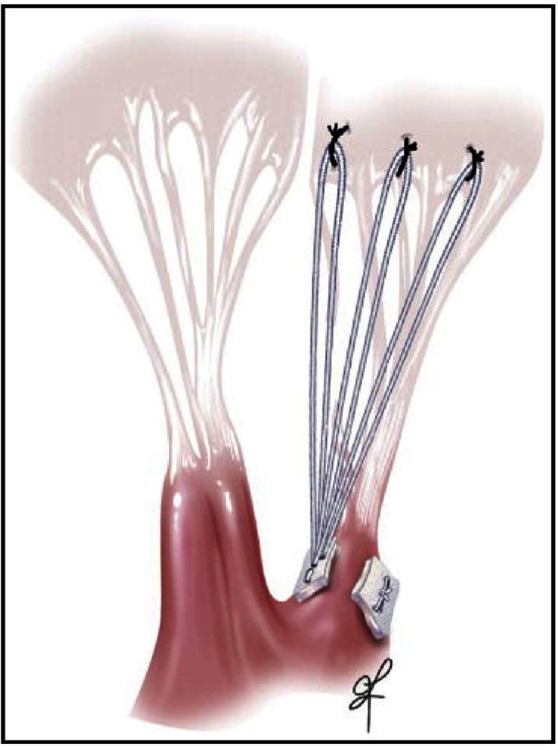
This medical illustration depicts a mitral valve repair utilizing artificial chordae tendineae (neochordae). The diagram shows a surgical view of the subvalvular apparatus, featuring the papillary muscles and mitral valve leaflets. Three sets of artificial chordal loops, represented as thin blue-grey lines made of PTFE, are anchored to the papillary muscle using white felt pledgets. The distal ends of these loops are sutured to the atrial aspect of the posterior mitral valve leaflet at the line of coaptation, visible as distinct black knots. Adjacent to the artificial cords, natural chordae tendineae are shown as thin, translucent string-like structures connecting the opposite leaflet to the papillary muscle. This illustration demonstrates a chordal replacement technique designed to treat mitral regurgitation—specifically for conditions like Barlow’s disease or fibroelastic deficiency—by restoring leaflet support and proper coaptation depth without the need for tissue resection.
AV Valve Apparatus - Components
| Component | Mitral Valve | Tricuspid Valve |
|---|---|---|
| Leaflets | Anterior + Posterior (2) | Anterior + Posterior + Septal (3) |
| Annulus | Oval, saddle-shaped fibrous ring | Larger, more oval ring |
| Chordae tendineae | Primary, secondary, tertiary | Same classification |
| Papillary muscles | Anterolateral + Posteromedial (2) | Anterior + Posterior + Septal (3) |
Papillary Muscles - Key Facts
- Each papillary muscle is an outpouching of subendocardial myocardium
- They contract synchronously with the ventricle
- Papillary muscle contraction tightens the chordae tendineae, preventing excessive leaflet motion beyond the coaptation zone
- This prevents leaflet inversion (prolapse) into the atrium during systole
- Both papillary muscles send chordae to both leaflets of the mitral valve (not just the ipsilateral one)
- Chordal rupture - loss of papillary muscle support causes severe mitral regurgitation (unobstructed retrograde flow from high-pressure LV into low-pressure LA)
- Papillary muscle ischemia/infarction - causes acute mitral regurgitation AND LV systolic dysfunction (papillary muscles contribute to LV apical contraction)
- Surgical implication - removing chordae tendineae-papillary muscle attachments during mitral valve replacement reduces LV ejection fraction
- The septal papillary muscle is a key landmark - its presence helps identify the morphologic RV vs. LV in complex congenital heart disease
Visual - Mitral Valve Papillary Muscle Anatomy
Summary
- Barash's Clinical Anesthesia, 9e, p. 847-848