

IMG20260807171700.jpg
Patient is having itching and pain here
analyse_media
Reading File
Now let me search the medical library for relevant content on cherry angiomas and the surrounding erythema, and also search for a reference image.

Based on the image analysis and textbook data, here is a thorough clinical assessment:
Clinical Assessment - Central Chest Lesion
What the Image Shows
The image shows the central chest (sternum/xiphoid area) with:
- Primary lesion - A small (~3-5 mm), bright red to violaceous, dome-shaped papule just left of the midline
- Surrounding erythema - A faint, ill-defined pink patch centered around the lesion, overlying what appears to be a well-healed midline scar (consistent with a prior sternotomy)
- Satellite lesion - A smaller pinpoint dark red spot in the lower left of the frame
Most Likely Diagnosis: Cherry Angioma (with local irritation)
The primary lesion has the classic appearance of a cherry angioma (also called cherry hemangioma or Campbell-De Morgan spot).
Per Dermatology: 2-Volume Set 5e:
"Bright red, dome-shaped to polypoid papules up to several millimeters in diameter that begin appearing during adult life, most commonly on the trunk and upper extremities. A common, benign lesion found in most individuals by the age of 60 years... Consist of dilated, congested capillaries and postcapillary venules within the papillary dermis."
Here is what multiple cherry angiomas look like for reference:
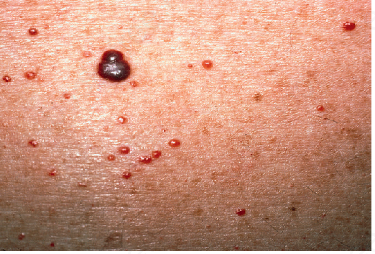
The surrounding pinkish erythema likely represents local skin irritation - possibly from:
- Friction against clothing/undergarment
- Mild contact dermatitis
- Trauma from scratching (given the patient's complaint of itching)
The pain and itch are not typical of uncomplicated cherry angiomas alone. The erythema around the lesion suggests the angioma may have been traumatized (scratched/rubbed), causing local inflammation.
Differential Diagnoses to Consider
| Condition | Features |
|---|---|
| Cherry angioma (most likely) | Bright red dome-shaped papule, blanches with pressure, benign, common on trunk |
| Pyogenic granuloma | Similar red vascular papule but grows rapidly, bleeds easily with minor trauma |
| Angiokeratoma | Dark red/purple, warty surface, doesn't blanch |
| Insect bite reaction | Papule + surrounding erythema + itch - fits well if onset was sudden |
| Traumatized/inflamed angioma | Existing cherry angioma + scratch-induced perilesional dermatitis |
Key Clinical Questions to Ask
- How long has this been present? (Cherry angiomas are long-standing; insect bites are acute)
- Did it appear suddenly? (Pyogenic granulomas grow fast, over days to weeks)
- Does it bleed easily? (Pyogenic granulomas bleed profusely with minor contact)
- Is the pink area spreading, warm, or tender? (Rules out cellulitis/secondary infection)
- Any similar lesions elsewhere on the body?
- Previous cardiac surgery? (The midline scar noted is relevant to context)
Management
For Cherry Angioma with Local Irritation:
- Reassure the patient - cherry angiomas are entirely benign
- Avoid scratching to prevent trauma and secondary infection
- Short course of mild topical steroid (e.g., hydrocortisone 1%) for the perilesional erythema/itch
- Oral antihistamine (e.g., cetirizine) if itch is significant
- If the lesion is cosmetically bothersome or symptomatic: removal options include electrodessication, laser (pulsed dye laser), or cryotherapy - all effective per Dermatology 2-Volume Set 5e
Red flags requiring urgent evaluation:
- Rapid growth over days
- Spontaneous or easy bleeding
- Spreading redness, warmth, or fever (suggests cellulitis)
- Any change in color/border (to exclude rare vascular malignancy)
Disclaimer: This assessment is based on photographic analysis and should not replace an in-person clinical examination. A dermatologist can use dermoscopy to confirm the diagnosis definitively.
This is a shared conversation. Sign in to Orris to start your own chat.