
Short note on Submandibular ganglion for University exam
Reading File
The submandibular ganglion is a parasympathetic ganglion in the floor of the mouth that supplies the submandibular and sublingual salivary glands. It receives preganglionic fibres via the chorda tympani (branch of CN VII) and sends postganglionic fibres to the glands. Let me pull the full textbook content now.
Searching sources...
Finding Sources
Finding Sources
Excellent results. Let me read the dedicated section from Scott-Brown's and the Gray's Anatomy sections for complete detail.
Reading File
Reading File
Reading File
I have all the information I need. Let me also fetch the anatomical diagram showing the submandibular ganglion.
Loading Image
Loading Image
I now have everything needed for a comprehensive exam note. Here is the complete short note:
Submandibular Ganglion
Definition and Type
The submandibular ganglion is one of the four parasympathetic ganglia of the head and neck. It is a small peripheral ganglion of the autonomic nervous system, functionally classified as a secretomotor (parasympathetic) ganglion that serves the submandibular and sublingual salivary glands.
Location
- Situated in the floor of the oral cavity, on the lateral surface of the hyoglossus muscle
- It appears to "hang off" the inferior surface of the lingual nerve by two small nerve roots (a useful anatomical landmark)
- It lies above the deep part of the submandibular gland, medial to the mandible
Roots (Fibres Entering the Ganglion)
The ganglion has three functional roots:
| Root | Fibres | Origin | Fate |
|---|---|---|---|
| Parasympathetic (secretomotor) | Preganglionic GVE | Superior salivatory nucleus (pons) → Facial nerve [VII] → Chorda tympani → Lingual nerve [V3] → ganglion | Synapse here; postganglionic fibres go to glands |
| Sympathetic | Postganglionic (already relayed) | Superior cervical ganglion → sympathetic plexus on facial artery | Pass through ganglion WITHOUT synapsing; supply blood vessels of the glands (vasoconstriction) |
| Sensory | Afferent (GSA) | Lingual nerve [V3] (mandibular division of trigeminal) | Pass through without synapsing; carry sensation from the glands |
Key exam point: Only the parasympathetic fibres synapse in the ganglion. Sympathetic and sensory fibres merely pass through it.
Preganglionic Parasympathetic Pathway (in detail)
- Superior salivatory nucleus (pons, CN VII)
- → Facial nerve [VII] in the petrous temporal bone
- → Chorda tympani (branch of facial nerve)
- Exits via the petrotympanic fissure
- Crosses the middle ear medial to the handle of the malleus
- Descends into the infratemporal fossa
- → Joins the lingual nerve [V3] in the infratemporal fossa
- → Travels with the lingual nerve to the floor of the oral cavity
- → Preganglionic fibres peel off the inferior aspect of the lingual nerve on the surface of hyoglossus
- → Synapse in the submandibular ganglion
Postganglionic Parasympathetic Distribution
After synapsing in the ganglion, postganglionic fibres take two routes:
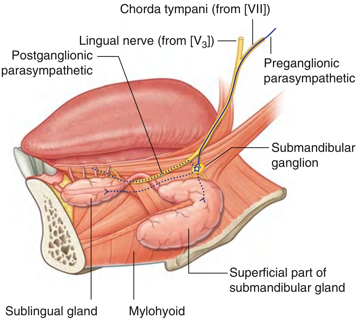
- Pass directly from the ganglion to the submandibular gland (the larger gland immediately below)
- Re-enter the lingual nerve and travel with its terminal branches to reach the sublingual gland and small mucous glands in the floor of the mouth and tongue
The net effect is increased secretion (watery, serous saliva) from both the submandibular and sublingual glands.
Sympathetic Supply
- Preganglionic sympathetic fibres originate at T1 spinal level
- Relay in the superior cervical sympathetic ganglion
- Postganglionic fibres travel along the plexus on the facial artery to reach the ganglion and pass through it to the glands
- Effect: vasoconstriction of glandular blood vessels, producing a small amount of thick, viscous secretion
Summary Diagram
Course of parasympathetic fibres in the chorda tympani to the submandibular ganglion (Gray's Anatomy for Students):
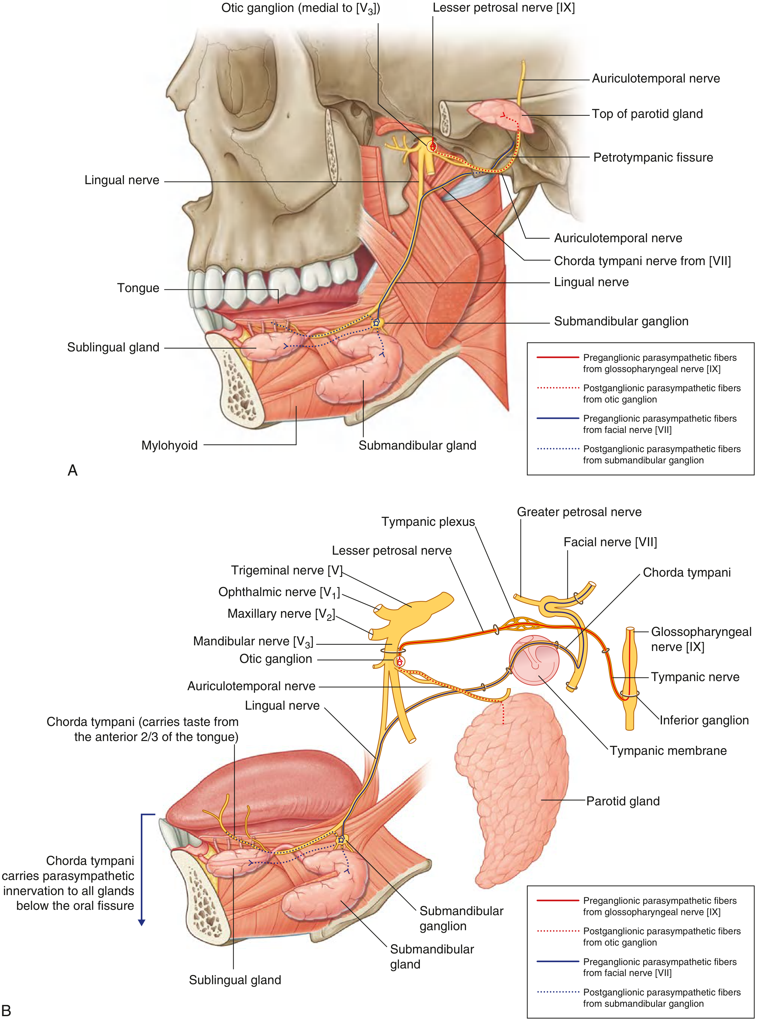
Comparison of submandibular ganglion with the otic ganglion, showing the chorda tympani pathway and target glands:
Clinical Relevance
- Lingual nerve injury proximal to the chorda tympani junction: Loss of general sensation from the anterior 2/3 of the tongue + loss of salivation from submandibular/sublingual glands + loss of taste from anterior 2/3 of tongue
- Lingual nerve injury distal to the chorda tympani junction: Loss of general sensation from anterior 2/3 of tongue only (taste and secretion are preserved)
- The ganglion and lingual nerve are important surgical landmarks during Level I neck dissection and excision of parapharyngeal salivary gland tumours
Comparison with Other Parasympathetic Ganglia of the Head
| Ganglion | CN carrying preganglionic fibre | Target |
|---|---|---|
| Ciliary | CN III (oculomotor) | Sphincter pupillae, ciliary muscle |
| Pterygopalatine | CN VII (greater petrosal) | Lacrimal gland, nasal/palatal glands |
| Submandibular | CN VII (chorda tympani) | Submandibular & sublingual glands |
| Otic | CN IX (lesser petrosal) | Parotid gland |
Sources: Scott-Brown's Otorhinolaryngology Head & Neck Surgery Vol. 2; Gray's Anatomy for Students; Medical Physiology (Boron & Boulpaep)
Short note on Submandibular ganglion for University exam
The submandibular ganglion is a parasympathetic ganglion suspended from the lingual nerve in the floor of the mouth. It relays secretomotor fibres to the submandibular and sublingual salivary glands via the chorda tympani branch of CN VII.
Searching sources...
Finding Sources
Finding Sources
Reading File
Reading File
Reading File
Loading Image
Loading Image
Here is the complete short note, exam-ready:
Submandibular Ganglion
Introduction
The submandibular ganglion is one of the four parasympathetic ganglia of the head (the others being the ciliary, pterygopalatine, and otic ganglia). It is a secretomotor (parasympathetic) ganglion that controls salivary secretion from the submandibular and sublingual glands.
Location
- Situated in the floor of the oral cavity, on the lateral surface of the hyoglossus muscle
- It characteristically "hangs off" the inferior surface of the lingual nerve by two small nerve roots - this is a classic anatomical description
- It lies immediately above the deep part of the submandibular gland, medial to the body of the mandible
Roots (Fibres)
The ganglion receives three types of fibres:
| Root | Fibre Type | Origin | Fate in Ganglion |
|---|---|---|---|
| Parasympathetic (secretomotor) | Preganglionic GVE | Superior salivatory nucleus (pons) → CN VII → Chorda tympani → Lingual nerve | Synapse here |
| Sympathetic | Postganglionic (already relayed) | T1 spinal cord → superior cervical ganglion → plexus on facial artery | Pass through without synapsing |
| Sensory | GSA | Lingual nerve (V3) | Pass through without synapsing |
Key exam point: Only parasympathetic fibres synapse in the submandibular ganglion. Sympathetic and sensory fibres are merely hitchhikers passing through.
Preganglionic Parasympathetic Pathway (Step-by-Step)
- Superior salivatory nucleus (lower pons) - origin of preganglionic fibres
- → Facial nerve [CN VII] in the internal acoustic meatus and petrous temporal bone
- → Chorda tympani (branch of facial nerve)
- Exits via the petrotympanic fissure into the infratemporal fossa
- Crosses the middle ear cavity, lying medial to the handle of the malleus
- → Joins the lingual nerve [V3] in the infratemporal fossa
- → Travels with the lingual nerve into the floor of the oral cavity
- → Preganglionic fibres peel off the inferior aspect of the lingual nerve on the surface of the hyoglossus muscle
- → Synapse in the submandibular ganglion
Postganglionic Distribution
After synapsing, postganglionic parasympathetic fibres take two routes:
- Pass directly from the ganglion into the submandibular gland (immediately below)
- Re-enter the lingual nerve and travel with its terminal branches to reach the sublingual gland and the small mucous glands of the floor of the mouth, lower lip, and tongue
Effect: Increased watery (serous) secretion from both glands.
Note: Taste fibres (special afferent) carried in the chorda tympani do not pass through the ganglion - they continue with the lingual nerve directly to the tongue.
Sympathetic Supply
- Preganglionic: T1 spinal level → sympathetic trunk
- Relay: Superior cervical sympathetic ganglion
- Postganglionic fibres travel along the plexus on the facial artery → pass through the ganglion without synapsing → reach blood vessels of both glands
- Effect: Vasoconstriction; produces small amounts of thick viscous secretion
Diagrams
Parasympathetic pathway via chorda tympani to the submandibular ganglion (Gray's Anatomy for Students, Fig. 8.270):
Comparison of the submandibular ganglion with the otic ganglion, showing the full pathway (Gray's Anatomy for Students, Fig. 8.153):
Clinical Relevance
Lingual nerve injury has different effects depending on the level:
| Level of Injury | Features Lost |
|---|---|
| Proximal (above where chorda tympani joins) | General sensation from anterior 2/3 tongue + loss of taste + loss of salivation from submandibular & sublingual glands |
| Distal (below where chorda tympani joins) | General sensation from anterior 2/3 tongue only (taste and salivation preserved) |
The ganglion and lingual nerve are important surgical landmarks during Level I neck dissection and parapharyngeal tumour excision - the ganglion is exposed alongside Wharton's duct during these procedures.
Comparison with Other Head Parasympathetic Ganglia
| Ganglion | Preganglionic Nerve | Target Organ |
|---|---|---|
| Ciliary | CN III (oculomotor) | Sphincter pupillae, ciliary muscle |
| Pterygopalatine | CN VII (greater petrosal) | Lacrimal gland, nasal & palatal glands |
| Submandibular | CN VII (chorda tympani) | Submandibular & sublingual glands |
| Otic | CN IX (lesser petrosal) | Parotid gland |
Sources: Scott-Brown's Otorhinolaryngology Head & Neck Surgery Vol. 2; Gray's Anatomy for Students; Medical Physiology (Boron & Boulpaep)
This is a shared conversation. Sign in to Orris to start your own chat.