
Zones of respiratory auscultation
Reading File
Reading File
Finding Sources
Searching Images
lung auscultation sites anterior posterior chest
Reading File
Reading File
Finding Sources
query_images_db
Searching PubMed
"Auscultation"[MeSH Terms] AND "Respiratory Sounds"[MeSH Terms]
Lung auscultation is done in a fixed, symmetrical pattern. You listen at the apices and over the upper, middle and lower lung fields, front and back, and compare left with right at each level. This is the standard approach. The textbooks I found describe it only briefly, so the specific landmarks below come from standard clinical teaching.
Principles
- Listen over bare skin in a quiet room, with the patient sitting upright if possible. Use the diaphragm of the stethoscope.
- Ask the patient to breathe deeply through the mouth. Listen for at least one full inspiration and expiration at each site.
- Move side to side at the same level so the two sides can be compared.
- Goldman-Cecil Medicine says to listen "at both apices and over both upper and lower lobes, anteriorly and posteriorly."
- Textbook of Family Medicine 9e says to listen over the upper, middle and lower fields of each lung posteriorly, and over the apices and mid-lung fields anteriorly. It adds that this includes the right middle lobe and the lingula of the left lung.
Anterior chest
| Zone | Landmark | Underlying lung |
|---|---|---|
| Apical / supraclavicular | Above the clavicle, in the supraclavicular fossa | Apex (upper lobe) |
| Upper | 2nd intercostal space (ICS), midclavicular line | Upper lobe |
| Middle | 4th-5th ICS, midclavicular line | Right middle lobe on the right; lingula of the upper lobe on the left |
| Lower | 6th ICS, midclavicular line | Lower lobe (anterior basal) |
- The trachea and main bronchi can be heard over the manubrium and the parasternal 1st-2nd ICS. Sounds here are normally bronchial or tracheal.
- Over the rest of the chest wall, normal sounds are vesicular.
Lateral (axillary) zones
- Upper axilla: 4th-5th ICS, midaxillary line. This is over the right middle lobe and the lingula. It is the best place to hear the middle lobe, because the fissure runs obliquely.
- Lower axilla: 6th-8th ICS, midaxillary line. This is over the lower lobes.
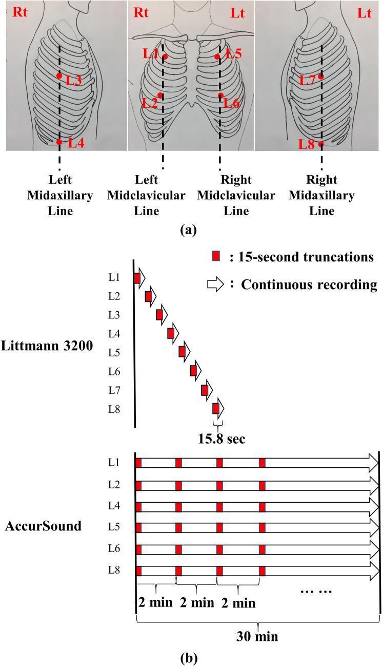
The paired points in the figure below follow this pattern. On the right they are the 2nd ICS midclavicular line (L1), the 5th ICS midclavicular line (L2), the 4th ICS midaxillary line (L3) and the 10th ICS midaxillary line (L4). The left side (L5-L8) mirrors these.
Posterior chest
| Zone | Landmark | Underlying lung |
|---|---|---|
| Apical (supraspinous) | Above the scapular spine, medial to the scapula | Apex of the upper lobe |
| Upper interscapular | Between the scapulae, T3-T4 level | Upper lobe |
| Middle interscapular | Between the scapulae, around T5-T6 | Upper and lower lobe junction (the oblique fissure begins at T3-T4) |
| Lower (infrascapular) | Below the scapula, T8-T10, extending laterally | Lower lobes |
- The posterior chest is the best place to assess the lower lobes, because they lie mostly posteriorly.
- The apex can also be heard in the supraclavicular fossa. The Color Atlas of Human Anatomy notes that the apex "can be percussed and auscultated in the supraclavicular fossa," although it is not highly ventilated.
- Posteriorly, the lower margin of the lung lies at about the 10th rib at the midscapular line (about T10 at the paravertebral line) on quiet breathing. It moves down about 5 cm on deep inspiration.
Lobar correlation
- Right lung: the upper lobe is heard anteriorly above the 4th rib, and also at the posterior apex. The middle lobe is heard anteriorly and in the axilla, between the 4th and 6th ribs. The lower lobe is heard posteriorly and laterally, below the scapular spine.
- Left lung: the upper lobe is heard anteriorly, including the lingula (which corresponds to the middle lobe), and at the posterior apex. The lower lobe is heard posteriorly and laterally.
- The oblique fissure runs from T3-T4 posteriorly to the 6th costal cartilage anteriorly. The horizontal fissure (right only) follows the 4th rib from the sternum to the midaxillary line.
Normal breath sounds by zone
- Tracheal: loud and harsh, heard over the trachea in the neck.
- Bronchovesicular: moderate intensity, heard over the main bronchi (parasternal 1st-2nd ICS anteriorly, and between the scapulae posteriorly).
- Vesicular: soft and low-pitched, with a short expiratory phase, heard over most of the peripheral lung fields.
- Bronchial sounds heard over the peripheral lung (for example, in lobar consolidation) are abnormal.
Clinical points
- Upper-zone findings point to conditions such as tuberculosis, silicosis and chronic hypersensitivity pneumonitis.
- Lower-zone fine crackles suggest interstitial fibrosis or pulmonary edema.
- Findings are anatomical clues. For example, right-sided crackles in the axilla suggest middle-lobe disease.
- Do not forget the posterior bases. A pleural effusion shows as reduced breath sounds with dullness to percussion, and consolidation shows as bronchial breathing.
- Murray & Nadel's Textbook of Respiratory Medicine uses the standardized American Thoracic Society terms for the sounds you hear: crackles, wheezes, rhonchi and stridor.
Recent evidence check: a quick PubMed search of the last two years found only AI and digital lung-sound analysis reviews (PMID 40249944, PMID 38400330). Neither contradicts the standard zone-based approach.
Sources:
- Color Atlas of Human Anatomy, Vol 2, Internal Organs.
- Textbook of Family Medicine 9e, History and Physical Examination.
- Goldman-Cecil Medicine, Approach to the Patient with Respiratory Disease.
- Murray & Nadel's Textbook of Respiratory Medicine, Auscultation.
This is a shared conversation. Sign in to Orris to start your own chat.