
The central canal of medulla at sensory trans-- sensory decussation like we take a TS. So, central canal contains hypoglossal nucleus, dorsal nucleus of vagus, and nucleus of tractus solitarius, or a centre matter around has grey matter which, अ, अ, which contains that.
Reading File
Now let me search the library for the medulla at sensory decussation level.
Finding Sources
Finding Sources
Finding Sources
Finding Sources
Reading File
Finding Sources
Reading File
Finding Sources
Reading File
Loading Image
Now let me also get the image of the sensory decussation level specifically from Gray's Anatomy:
Loading Image
This second image is exactly what's needed - it is a transverse section (TS) at the level of the sensory decussation (internal arcuate fiber decussation). Now I have everything to give a thorough answer.
Great question - and the key point you're asking about is an important distinction. Let me clarify.
TS of Medulla at the Level of Sensory Decussation
The sensory decussation (also called the decussation of internal arcuate fibers / decussation of the lemniscus) occurs at a more caudal level of the medulla - this is the "closed medulla" (canal is not yet open to the 4th ventricle). At this level the central canal is present.
What is in the grey matter AROUND the central canal at this level?
At the level of sensory decussation, the central grey around the canal contains:
| Structure | Position |
|---|---|
| Nucleus gracilis | Dorsomedial - forms the gracile tubercle |
| Nucleus cuneatus | Dorsolateral to gracilis - forms the cuneate tubercle |
| Nucleus of spinal tract of trigeminal (pars caudalis) | Lateral |
| Central grey substance | Surrounding the canal |
The internal arcuate fibers arise from nucleus gracilis and nucleus cuneatus, sweep ventrally around the grey matter, and cross the midline = sensory decussation / lemniscal decussation.
Your question about Hypoglossal nucleus, Dorsal nucleus of vagus, and NTS
These three nuclei are NOT at the level of sensory decussation. They appear at a higher (more rostral) level of the medulla:
| Nucleus | Level |
|---|---|
| Hypoglossal nucleus (CN XII) | Open medulla / mid-medulla (near floor of 4th ventricle) |
| Dorsal motor nucleus of vagus (CN X) | Open medulla / mid-medulla |
| Nucleus tractus solitarius (NTS) | Open medulla / mid-medulla |
These nuclei cluster in the dorsal grey of the open medulla, near the floor of the 4th ventricle - not near the central canal of the sensory decussation level.
The image above (mid-medulla section with the IV ventricle open) shows exactly this - hypoglossal nucleus, dorsal motor nucleus of vagus (X), vestibular nuclei, etc. are all visible in the open medulla.
The Two Key Levels to Remember
Here is the classic TS of the medulla at the sensory decussation level (closed medulla):
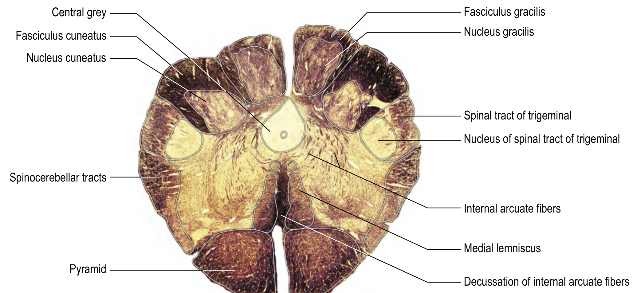
Notice in this section:
- The central canal is present (small circle at the centre)
- Dorsal grey: Nucleus gracilis (medial) and Nucleus cuneatus (lateral)
- Internal arcuate fibers are sweeping ventrally from these nuclei
- They decussate near the midline to form the medial lemniscus
- Pyramids ventral
- Spinal tract of trigeminal and its nucleus lateral
- Spinocerebellar tracts laterally
At this level, you do NOT see hypoglossal nucleus, dorsal vagal nucleus, or NTS because those are at the rostral/open medulla level.
Summary
At sensory decussation level (closed medulla, central canal present):
- Grey matter around canal = nucleus gracilis + nucleus cuneatus
- Key event = internal arcuate fibers decussating → become medial lemniscus
Hypoglossal nucleus + Dorsal nucleus of vagus + NTS = rostral/open medulla (floor of 4th ventricle), NOT at sensory decussation level.
- Localization in Clinical Neurology, 8e, p. 916
- Gray's Anatomy for Students, p. 1321
This is a shared conversation. Sign in to Orris to start your own chat.