
Perthes disease
Reading File
Perthes disease (Legg-Calve-Perthes disease) is avascular necrosis of the femoral head in children, typically ages 4-10. Let me search the medical library for comprehensive coverage.
Searching sources...
Finding Sources
Searching PubMed
Legg-Calve-Perthes disease management
Excellent - Campbell's has a very detailed section. Let me read the main block from Campbell's and also Bailey & Love's.
Reading File
Reading File
I hit the references section of Campbell's. Let me read the main content section (the block13 section that starts at line 1304) and continue Bailey & Love's:
Reading File
Reading File
I have good foundational content. Now let me read more of the Campbell's detailed content on classification, imaging, and management:
Reading File
Reading File
I have enough content now. Let me also fetch the key radiographic X-ray image from Bailey & Love's and the Herring classification image from Campbell's:
Loading Image
Reading File
I now have comprehensive information. Let me compile the full answer.
Legg-Calvé-Perthes (LCP) Disease
Definition & Epidemiology
Legg-Calvé-Perthes (LCP) disease is idiopathic avascular necrosis (AVN) of the proximal femoral epiphysis in children. Key demographic features:
- Age: Typically 4-10 years; average age at diagnosis ~7 years
- Sex: Boys affected 3-4x more than girls (but girls have worse prognosis)
- Bilaterality: ~10% of cases are bilateral (though usually at different stages)
- Laterality: Rarely truly simultaneous - bilateral presentation should prompt search for alternative diagnoses
Aetiology & Pathogenesis
The exact cause remains unknown. The leading hypothesis involves coagulation disorder promoting thrombotic occlusion of the vascular supply to the femoral head, leading to bone death. Contributing factors include:
- Socioeconomic deprivation
- Passive smoking
- Delayed bone age (skeletal maturity is typically delayed in these children)
- Thrombophilia (some evidence of inherited risk factors)
The disease is self-limiting and follows a predictable sequence:
- Avascular/Necrotic phase: Ischaemia causes bone death in all or part of the femoral epiphysis
- Revascularisation/Fragmentation phase: Blood supply returns; dead bone is resorbed and the ossific nucleus fragments within the cartilaginous femoral head
- Reossification/Healing phase: Bony epiphysis regenerates ("heals")
- Residual phase: Final shape of the head is established
During the collapse and fragmentation phases, the cartilage follows the shape of the reossifying epiphysis - leading to irreversible femoral head deformity if the head is not protected.
Differential Diagnosis
Other causes of femoral head AVN must be excluded, especially in bilateral cases:
| Condition | Key Distinguishing Feature |
|---|---|
| Steroid-induced AVN | History of steroid use |
| Septic arthritis sequelae | Preceding infection |
| Sickle cell disease | Haemoglobinopathy |
| Hypothyroidism | TFTs, systemic features |
| Multiple epiphyseal dysplasia | Bilateral, other joints involved |
| Meyer dysplasia | Age <4 yrs, bilateral, NO subchondral fracture or subluxation |
Meyer dysplasia in particular can mimic LCP and lead to unnecessary treatment - it shows delayed/smaller ossification centres and cystic changes but lacks condensation, subchondral fractures, and fragmentation.
Clinical Features
- Limp (antalgic or Trendelenburg)
- Pain in hip or referred to knee (intermittent)
- Restricted hip movement - especially abduction and internal rotation
- Symptoms typically present for ~6 weeks before diagnosis is made
- Irritability of the hip joint
Diagnosis & Imaging
Plain Radiography
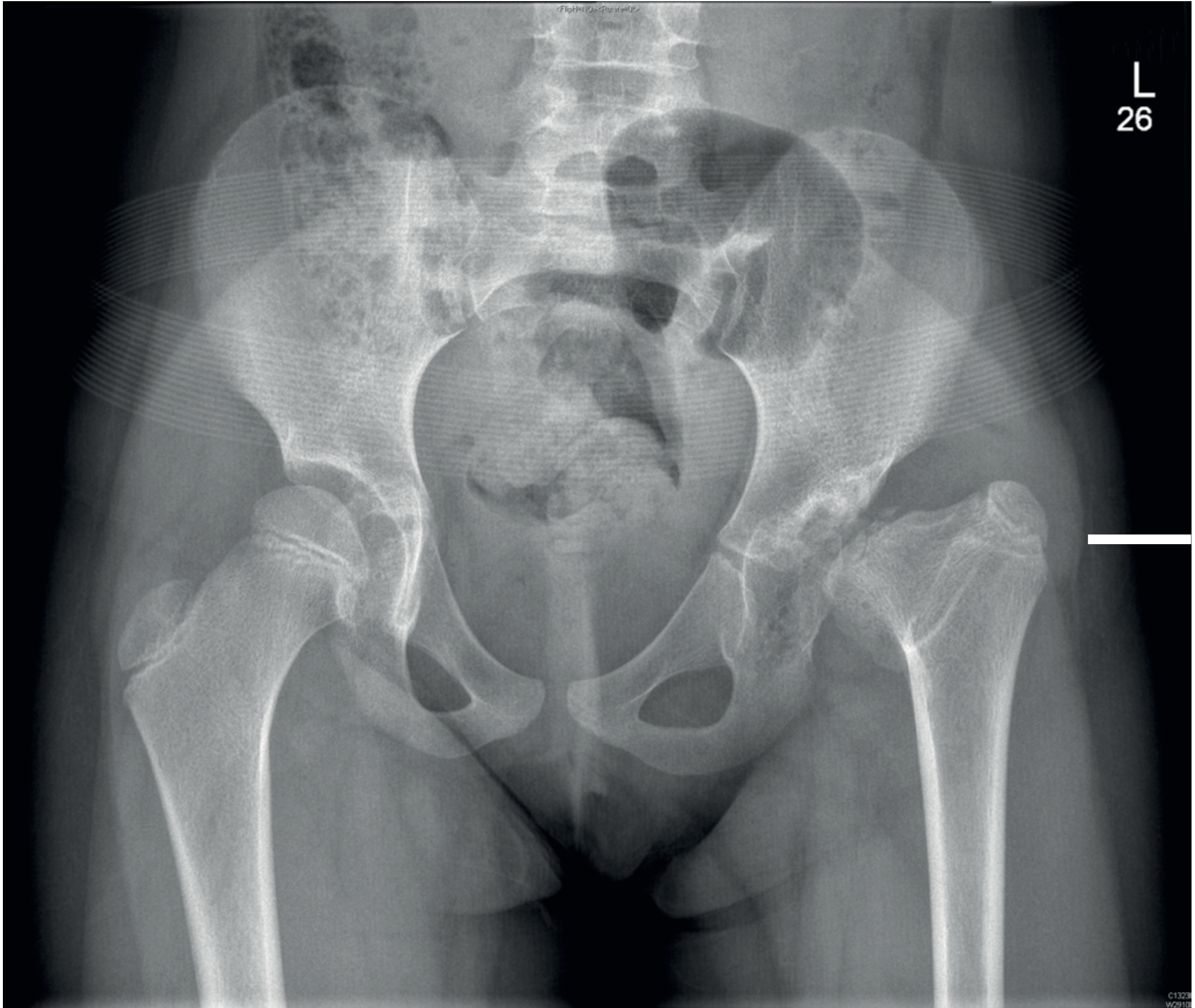
AP pelvis + "frog-leg" lateral views are the first-line investigation. Radiographic changes lag 6 weeks behind clinical onset. If early presentation with normal X-rays - repeat at 6 weeks if still symptomatic.
Waldenström radiographic stages (modified by International Perthes Study Group - IPSG):
- Initial: Slight widening of joint space, smaller ossific nucleus
- Fragmentation: Dense, fragmented femoral head epiphysis
- Reossification: New bone formation within the epiphysis
- Healed/Residual: Final shape established
MRI
Superior to scintigraphy for early detection and defining extent of involvement. Gadolinium-enhanced perfusion MRI can show lateral pillar involvement earlier than plain films (at initial fragmentation stage rather than mid-fragmentation), potentially allowing earlier treatment decisions. ~50% of children require sedation/GA. Reperfusion progresses in a horseshoe pattern - posterior to anterior.
Bone Scintigraphy
Can establish early diagnosis but now largely superseded by MRI.
Classification Systems
Herring Lateral Pillar Classification (most commonly used)
Applied during the fragmentation phase - based on height of the lateral pillar of the femoral head on AP radiograph:
| Group | Lateral Pillar Height | Prognosis |
|---|---|---|
| A | Fully maintained (no involvement) | Good |
| B | >50% of original height preserved | Intermediate |
| B/C border | Exactly 50% height or narrow pillar | Poorer |
| C | <50% of original height | Poor |
If the anterolateral portion of the head is preserved, prognosis is good.
Catterall Classification (older)
Divides into groups I-IV based on proportion of femoral head involved (25%, 50%, 75%, total).
Salter-Thompson Classification
Groups A and B - based on extent of subchondral fracture.
Stulberg Classification (outcome)
Used at skeletal maturity to grade residual femoral head sphericity (I-V); higher grades predict early osteoarthritis.
Prognosis
Key prognostic factors:
- Age at onset: Younger children (<6 yrs) have better prognosis due to greater remodelling potential
- Sex: Girls have worse prognosis
- Herring grade: Group A > B > C
- Extent of head involvement: Partial involvement better than total
- Maintenance of motion: Loss of hip movement is a poor sign
Management
Principles
The goal is to minimise femoral head deformity and prevent secondary acetabular dysplasia by maintaining a good range of joint movement and ensuring femoral head containment within the acetabulum ("containment principle").
Non-operative
- Analgesia and physiotherapy to maintain ROM
- Avoid crutches/wheelchairs (promote flexion/adduction deformity)
- Bracing: Scottish Rite (Atlanta) orthosis or Petrie casts for abduction - but evidence shows brace management does not alter natural history; mainly used in younger children
- Activity modification, anti-inflammatories
- Observation in: children <6 years, Herring Group A, Herring Group B <8 years
Operative (Containment Surgery)
Indicated in higher-risk cases - older children, Herring B/C or C, loss of containment.
A. Proximal Femoral Varus Derotational Osteotomy (VDRO)
- Procedure of choice when containment needed but bracing not feasible
- Preferred in children 8-10 years, with significant femoral anteversion
- Achieves 0-15° of varus correction
- Advantages: Maximal femoral head coverage, corrects anteversion
- Disadvantages: Limb shortening, risk of gluteal lurch, premature physeal closure, poor results in Group C aged >9 years
B. Innominate (Salter) Osteotomy
- Provides anterolateral femoral head coverage
- Advantages: May slightly lengthen extremity, avoids second operation for plate removal
- Disadvantages: Risk of over-correction, can compress already compromised femoral head, does not accelerate healing
Note: Neither osteotomy has been conclusively shown to be superior to the other - choice depends on surgeon's expertise and individual patient factors.
C. Shelf Acetabuloplasty / Chiari Osteotomy
- Late "salvage" procedures for established deformity limiting movement
D. Arthrodiastasis (Joint Distraction)
- Used in late-onset or severe cases
- External fixator maintains joint space during revascularisation
Late/Salvage Surgery
When deformity is established:
- Surgical hip dislocation with femoral head reshaping
- Relative femoral neck lengthening
- Joint-preserving surgery (improves pain, ROM, and abductor strength)
- Total hip replacement in adults with end-stage osteoarthritis
Treatment Algorithm (Summary)
| Patient Group | Recommended Management |
|---|---|
| Age <6 years, any Herring group | Observation ± physio |
| Age 6-8 years, Herring A | Observation |
| Age 6-8 years, Herring B | Consider containment (brace or surgery) |
| Age 6-8 years, Herring C | Surgery (osteotomy) |
| Age >8 years, Herring B or C | Surgery (osteotomy) |
| Any age, Herring B/C border | Surgery generally favoured |
Sources:
- Campbell's Operative Orthopaedics 15th Ed 2026, Ch. 34 (Legg-Calvé-Perthes Disease)
- Bailey and Love's Short Practice of Surgery 28th Ed, Ch. 44
Recent evidence note: A 2026 systematic review and meta-analysis (PMID 41308557) evaluated the diagnostic performance of diffusion-weighted MRI (DWI) in LCP disease, supporting the growing role of advanced MRI techniques in early diagnosis.
Osteochondritis
Searching sources...
Finding Sources
Searching PubMed
osteochondritis dissecans treatment
Good - multiple rich sources found. Let me read the main sections in parallel:
Reading File
Reading File
Reading File
Good - let me now get the detailed classification/staging and treatment content from Campbell's, and also the Rheumatology textbook section:
Reading File
Reading File
Let me get the rest of the rheumatology section and the OCD at other joints content from Campbell's:
Reading File
Reading File
Loading Image
I have comprehensive content. Now let me also fetch the knee X-ray/MRI image showing OCD lesion:
Loading Image
Loading Image
Excellent - I have comprehensive content and great images. Let me now compile the full answer.
Osteochondritis Dissecans (OCD)
Definition
Osteochondritis dissecans is a condition where an area of subchondral bone undergoes avascular necrosis, with degenerative changes then developing in the overlying articular cartilage. The necrotic bone and overlying cartilage gradually separate from adjacent tissue, and if the process is not interrupted, become a loose body within the joint. It is the most common source of loose bodies in the knee.
Epidemiology
- Most common in individuals aged 12-19 years (peak adolescence), though it occurs in adults
- Adolescent form typically ages 10-15 years
- Associated with sports participation and repetitive loading
- Childhood obesity is an associated risk factor
- Bilateral in 20-30% of knee OCD cases
Aetiology
The exact cause is multifactorial and not fully understood:
- Genetic predisposition
- Mechanical stress and repetitive microtrauma
- Microvascular insufficiency to subchondral bone
- Occult trauma (repetitive rather than single episode)
The condition can occur in any joint but has characteristic sites at each location.
Sites of Involvement
Knee (most common):
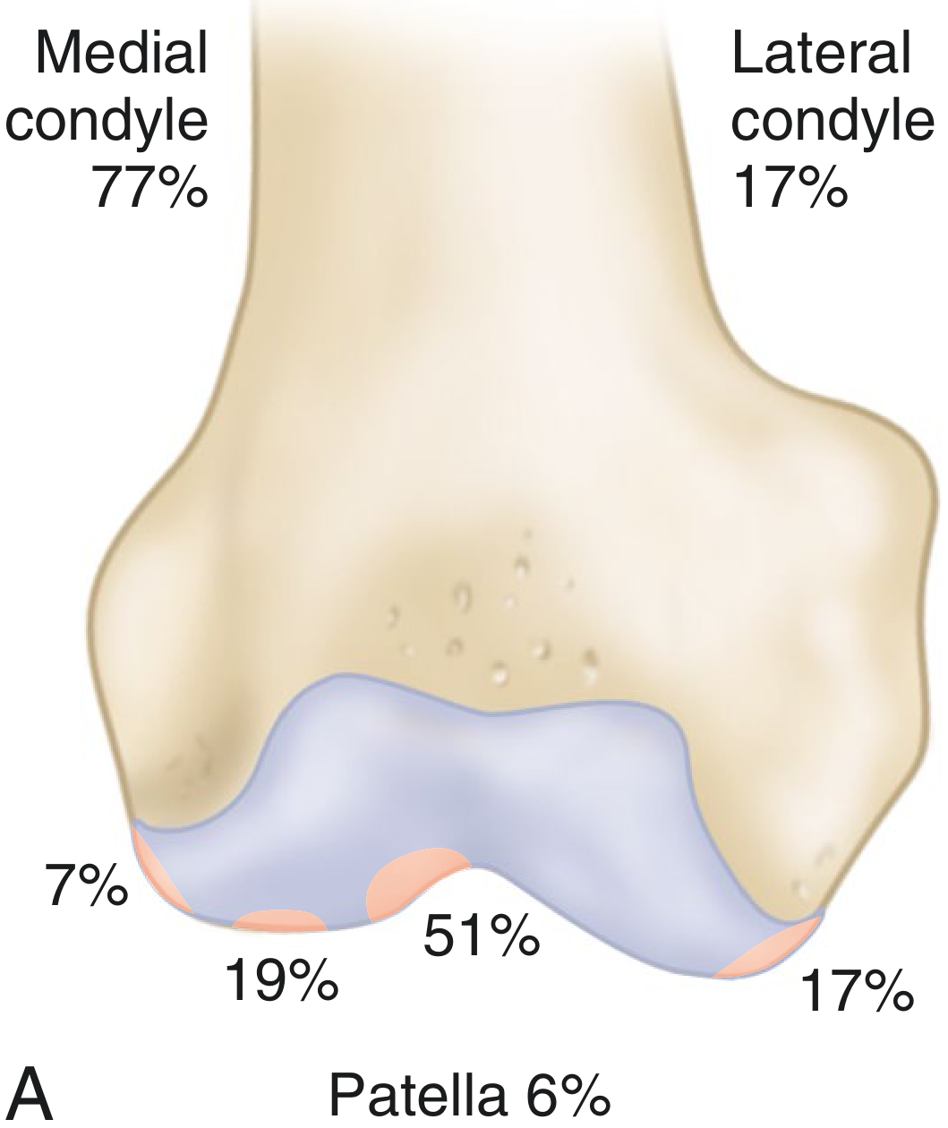
- Lateral aspect of the medial femoral condyle near PCL attachment: ~66-80% (classic site)
- Posterolateral femoral condyle: ~15-18%
- Inferomedial patella: ~5-6%
Other joints:
- Talus - second most common; posteromedial or anterolateral talar dome
- Elbow - capitellum (common in throwing athletes and gymnasts)
- Hip - femoral head
- Shoulder - humeral head
Clinical Features
- Vague, poorly localised joint pain - often described as dull or aching
- Swelling/effusion of the joint
- Limp (if lower limb)
- Mechanical symptoms (locking, catching, giving way) - when the fragment is unstable or loose
- Local tenderness over the OCD lesion on palpation
- Wilson's sign (knee): pain on internal rotation at 30° flexion, relieved by external rotation - positive in medial femoral condyle OCD
Diagnosis & Imaging
Plain Radiography
- First-line investigation
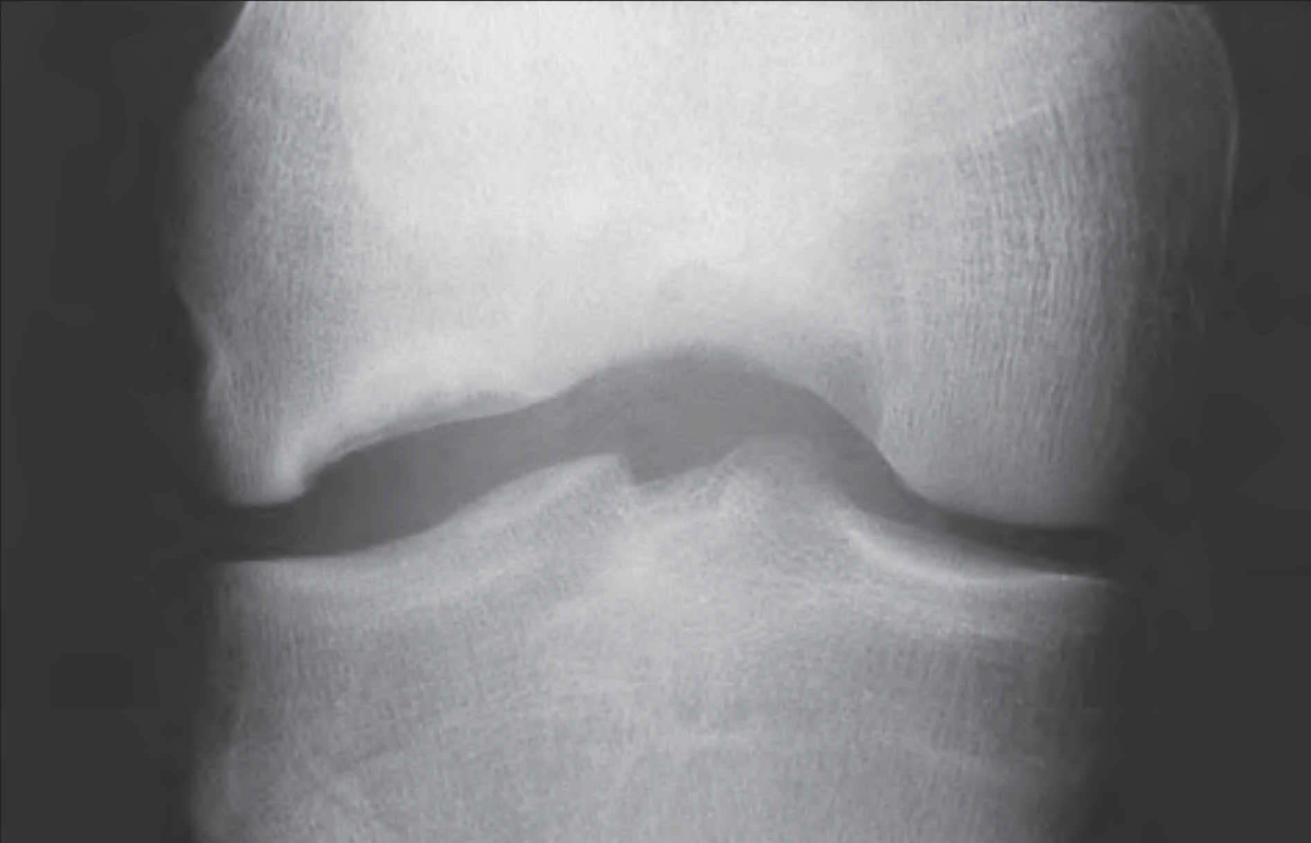
- Shows a well-defined lucent defect in subchondral bone, sometimes with a sclerotic margin
- Displaced fragment seen as loose body
- Tunnel (notch) view - especially useful for classic OCD of the lateral aspect of the medial femoral condyle
MRI (Investigation of Choice)
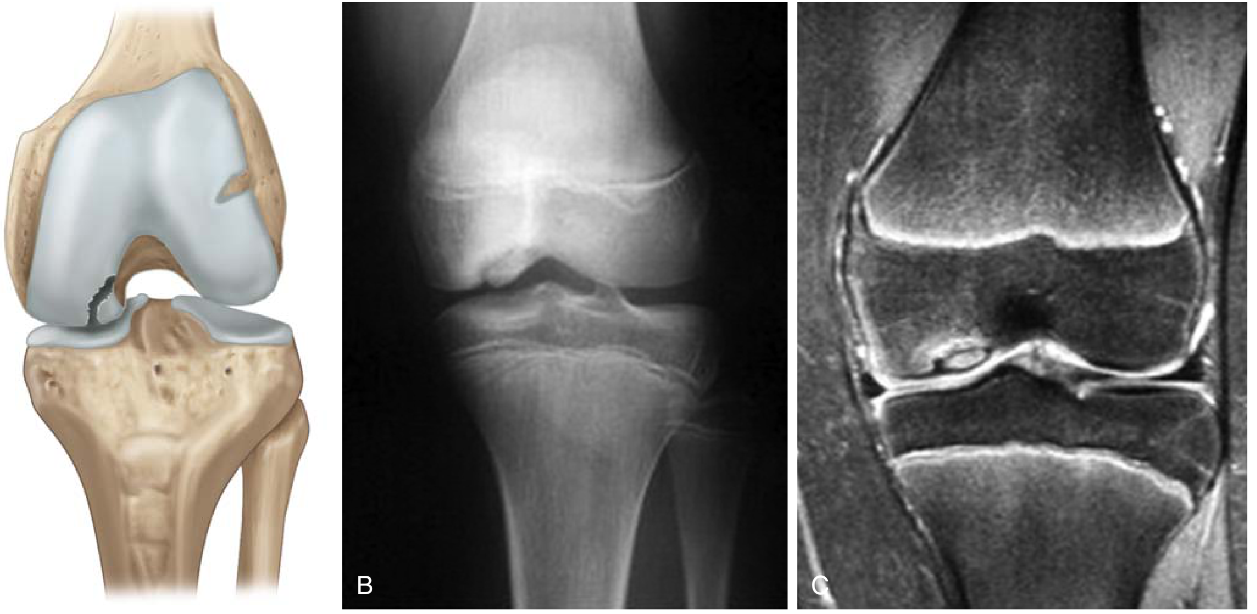
- Best method for demonstrating site, extent, and stability of the lesion
- Differentiates stable from unstable lesions (critical for management)
- MR arthrography especially useful for assessing cartilage integrity
- Note: MRI underestimates the size of articular cartilage defects in ~75% of cases
CT
- Helpful for defining the precise osseous extent of the lesion
- Less useful for cartilage assessment
Staging (DiPaola Classification)
| Stage | Arthroscopy | MRI | Radiography |
|---|---|---|---|
| I | Articular cartilage softening, intact; no fragment | Low signal changes; thickened cartilage | Compression lesion; no visible fragment |
| II | Cartilage breached; fragment not displaceable | Low signal rim behind fragment (fibrous attachment) | Fragment attached |
| III | Cartilage breached; fragment displaceable but attached by overlying cartilage | High signal behind fragment (synovial fluid interposed) | Non-displaced fragment without attachment |
| IV | Loose body | Loose body | Displaced fragment |
Stages I-II = stable lesions | Stages III-IV = unstable lesions
Treatment
Treatment depends on patient age (skeletally mature vs. immature) and lesion stability.
Non-operative (Stable lesions, skeletally immature)
- Observation - most juvenile OCD (open physes) will resolve spontaneously
- Activity modification - avoid high-impact sports
- Protected weight bearing ± crutches
- Gentle physiotherapy
- Short-term immobilisation in a cast (avoid prolonged - impairs cartilage nutrition)
- Most juvenile cases resolve; ~50% of adolescent cases resolve; adult OCD rarely heals spontaneously
Operative
Stable in-situ lesions:
- Retrograde drilling (transarticular or retroarticular) - stimulates revascularisation and fibrocartilage formation through the intact overlying cartilage without breaching it; preferred in skeletally immature with open physes
- Transarticular drilling through the cartilage - done arthroscopically
Unstable lesions (Stage III-IV):
- Fragment fixation - for large, viable fragments; using cannulated screws or bioabsorbable devices
- Removal of loose bodies - for small, non-viable fragments
- Arthroscopic debridement of the crater bed
Large cartilage defects (salvage/restoration):
| Technique | Principle | Notes |
|---|---|---|
| Microfracture | Drill/awl holes in subchondral bone to release marrow cells → fibrocartilage | Produces fibrocartilage (inferior to hyaline); degenerates over time; best for smaller defects |
| Abrasion arthroplasty | Debride and abrade the bony bed | Similar fibrocartilage result |
| Osteochondral autograft transfer (OATS / mosaicplasty) | Osteochondral plugs from non-weight-bearing surface transplanted to defect | Limited by donor site availability and morbidity |
| Osteochondral allograft | Cadaveric osteochondral graft | Larger defects; small risk of disease transmission |
| Autologous Chondrocyte Implantation (ACI) | Two-stage: 1) harvest + culture chondrocytes ex vivo; 2) implant under periosteal patch or membrane | Produces more hyaline-like repair tissue; best in younger, active males with smaller lesions |
Prognosis
Good prognostic factors:
- Skeletally immature (open physes)
- Smaller lesion size
- Stable lesion (Stage I-II)
- Younger age at diagnosis
- Male sex (for ACI outcomes)
Poor prognostic factors:
- Skeletal maturity at presentation
- Larger, more severe lesions
- Unstable lesion (Stage III-IV)
- Patellar or elbow location
- BMI >25 kg/m² at diagnosis
- Presence of discoid meniscus (knee OCD)
- Mechanical symptoms
Adult OCD is usually symptomatic and leads to osteoarthritis if left untreated. A cumulative incidence of arthritis of 30% at 35 years after diagnosis has been reported after nonoperative treatment.
OCD vs. Related Conditions (Quick Distinctions)
| OCD | Perthes Disease | Osgood-Schlatter | |
|---|---|---|---|
| Site | Any joint articular surface | Femoral head epiphysis | Tibial tubercle apophysis |
| Age | 12-19 yrs (peak) | 4-10 yrs | Adolescence |
| Pathology | Subchondral AVN → fragment | Femoral head AVN | Traction apophysitis |
| Loose bodies | Yes (Stage IV) | No | No |
Sources:
- Campbell's Operative Orthopaedics 15th Ed 2026, Ch. 50 (Osteochondritis Dissecans)
- Rheumatology 2-Volume Set (Elsevier 2022), Ch. 83
- Miller's Review of Orthopaedics 9th Ed, Section 4
- Bailey and Love's Short Practice of Surgery 28th Ed, Ch. 44
Recent evidence: A 2024 systematic review (PMID 38340961) on retroarticular drilling for OCD of the talus supports this technique as effective. A 2026 meta-analysis (PMID 41979334) on fresh osteochondral allograft transplantation of the femoral head found clinical improvement but noted a substantial arthroplasty conversion risk - underscoring the importance of early intervention.
This is a shared conversation. Sign in to Orris to start your own chat.