
i am md pediatrics currently doing fellowship in pediatric rheuematology. tell me in detail about diagnosis criteria classification and clinical feature of sle in detail based on standard textbooks and latest guidelines
Reading File
Now let me run comprehensive parallel searches across the medical library for SLE in pediatrics.
Finding Sources
Finding Sources
Finding Sources
Searching PubMed
"systemic lupus erythematosus"[MeSH Terms] AND "classification criteria"
Good, I found key sources. Now let me read the most authoritative content - Harrison's 22E, Textbook of Pediatric Rheumatology, Tietz, and run PubMed search in parallel.
Finding Sources
Reading File
Searching PubMed
"lupus erythematosus" AND "pediatric" AND "classification"
Harrison's 22E has excellent SLE content. Now let me read the full SLE section and find pediatric rheumatology textbook content.
Reading File
Finding Sources
Finding Sources
Finding Sources
Reading File
I have the Harrison's content. Now let me get the full clinical features and criteria tables from Harrison's, and also search for pediatric-specific SLE content.
Reading File
Finding Sources
Searching PubMed
"childhood-onset systemic lupus erythematosus" AND "clinical features"
Excellent. Now let me get the clinical features section and the EULAR/ACR criteria table from Harrison's, plus the Tietz book content.
Reading File
Reading File
Excellent content. I have the 2019 EULAR/ACR criteria table. Let me read more from both files to get the full clinical features and criteria.
Reading File
Reading File
I now have extensive content from Harrison's 22E and Tietz. Let me get a few more key sections - particularly the pediatric-specific features and the SLICC criteria table.
Finding Sources
Reading File
I now have all the key content I need. Let me also fetch the clinical image from Harrison's and get the Tietz criteria table content.
Loading Image
Reading File
I now have comprehensive content. Let me update todos and compose the final detailed answer.Here is the comprehensive, fellowship-level guide to SLE - covering classification, criteria, and clinical features in full detail.
Systemic Lupus Erythematosus (SLE): Classification, Criteria & Clinical Features
Based on Harrison's Principles of Internal Medicine 22E (2025), Tietz Textbook of Laboratory Medicine 7th Ed, Braunwald's Heart Disease, Bradley & Daroff's Neurology, and current EULAR/ACR guidelines.
1. DEFINITION AND EPIDEMIOLOGY
SLE is a chronic, multisystem autoimmune disease driven by overactive innate and adaptive immunity, leading to tissue damage via autoantibodies and immune complex deposition. Multiple organ systems are affected, with cutaneous, musculoskeletal, and renal being most prevalent, followed by pulmonary, hematologic, cardiovascular, serosal, and CNS involvement.
Epidemiology:
- ~90% of patients are women, predominantly of childbearing age
- Female:male ratio is 13:1 in the 15-44 years age group but drops to 2:1 in children and the elderly - this makes pediatric SLE distinctive, as boys are relatively more affected than in adults
- Higher prevalence and severity in non-Caucasian populations (Black > Hispanic > White > Asian)
- CDC National Lupus Registry prevalence in the US: ~204,295 cases
- Autoantibodies can precede clinical diagnosis by years
2. CLASSIFICATION CRITERIA
Critical distinction: Classification criteria are developed for research/clinical trial enrollment, NOT for clinical diagnosis. Diagnosis remains a clinical judgment based on the totality of clinical and laboratory findings. Approximately 5% of SLE patients are ANA-negative and would be excluded from trials using EULAR/ACR 2019 criteria.
A. 2019 EULAR/ACR Classification Criteria (Current Standard)
Published: Aringer M et al, Ann Rheum Dis 2019;78:1151-9
Entry Criterion (mandatory):
ANA titer ≥1:80 on HEp-2 cells by indirect immunofluorescence (IIF), or an equivalent positive test at any time. If ANA is absent, SLE classification cannot be made.
Scoring Domains (additive, weighted):
Only the highest-weighted criterion within each domain counts. Criteria need not occur simultaneously - occurrence on at least one occasion is sufficient.
| Domain | Criterion | Points |
|---|---|---|
| Constitutional | Fever | 2 |
| Hematologic | Leukopenia | 3 |
| Thrombocytopenia (<100 × 10⁹/L) | 4 | |
| Autoimmune hemolysis | 4 | |
| Neuropsychiatric | Delirium | 2 |
| Psychosis | 3 | |
| Seizure | 5 | |
| Mucocutaneous | Non-scarring alopecia | 2 |
| Oral ulcers | 2 | |
| Subacute cutaneous or discoid lupus | 4 | |
| Acute cutaneous lupus (malar rash) | 6 | |
| Serosal | Pleural or pericardial effusion | 5 |
| Acute pericarditis | 6 | |
| Musculoskeletal | Joint involvement (synovitis ≥2 joints OR morning stiffness ≥30 min) | 6 |
| Renal | Proteinuria >0.5 g/24h | 4 |
| Biopsy-proven class II or V lupus nephritis | 8 | |
| Biopsy-proven class III or IV lupus nephritis | 10 | |
| Antiphospholipid antibodies | aCL OR anti-β2GPI OR lupus anticoagulant | 2 |
| Complement | Low C3 OR low C4 | 3 |
| Low C3 AND low C4 | 4 | |
| SLE-specific antibodies | Anti-dsDNA OR anti-Sm | 6 |
Classification requires: ≥1 clinical criterion AND total score ≥10 points
Notable: A renal biopsy showing Class III or IV lupus nephritis alone yields 10 points - the only single criterion sufficient to classify as SLE (plus the mandatory ANA entry criterion).
- Harrison's Principles of Internal Medicine 22E, p. 2871; Tietz Textbook of Laboratory Medicine 7th Ed, p. 3787
B. 2012 SLICC Classification Criteria
Petri M et al, Arthritis Rheum 2012;64:2677-86
The SLICC criteria improved sensitivity over the older 1997 ACR criteria (sensitivity 97%, specificity 84%).
Rules:
- Satisfy at least 4 of 17 criteria (at least 1 clinical + 1 immunologic), OR
- Biopsy-proven lupus nephritis in the presence of ANA or anti-dsDNA antibodies
11 Clinical Criteria:
| # | Criterion | Key Details |
|---|---|---|
| 1 | Acute cutaneous lupus | Malar rash, maculopapular rash, bullous lupus, TEN variant, SCLE |
| 2 | Chronic cutaneous lupus | Discoid rash, verrucous lupus, lupus panniculitis, mucosal, chilblains |
| 3 | Oral/nasal ulcers | Palate, buccal, tongue, or nasal ulcers (painless or painful) |
| 4 | Non-scarring alopecia | Diffuse hair thinning or fragility (lupus hair) |
| 5 | Synovitis | ≥2 joints with synovitis, OR tender + morning stiffness ≥30 min |
| 6 | Serositis | Pleuritis (≥1 day) or pericarditis (≥1 day) |
| 7 | Renal | Urine protein/creatinine ratio >500 mg/g, or RBC casts in urine |
| 8 | Neurologic | Seizures, psychosis, mononeuritis multiplex, myelitis, peripheral or cranial neuropathy, acute confusional state |
| 9 | Hemolytic anemia | Direct Coombs positive, falling hemoglobin |
| 10 | Leukopenia (<4000/mm³) or Lymphopenia (<1000/mm³) | At least once; exclude other causes |
| 11 | Thrombocytopenia (<100,000/mm³) | At least once; exclude other causes |
6 Immunologic Criteria:
| # | Criterion |
|---|---|
| 1 | ANA above laboratory reference range |
| 2 | Anti-dsDNA above lab reference (or ≥2× ELISA) |
| 3 | Anti-Sm |
| 4 | Antiphospholipid antibodies (LA, aCL IgG/IgM, anti-β2GPI IgG/IgM, false-positive VDRL) |
| 5 | Low complement (C3, C4, or CH50) |
| 6 | Direct Coombs test positive (without hemolytic anemia) |
- Tietz Textbook of Laboratory Medicine 7th Ed; Henry's Clinical Diagnosis 23E
C. Historical: 1997 ACR Criteria (Rarely Used Now)
The original 11-criterion system (≥4 required) is rarely used for modern classification but historically important. It included malar rash, discoid rash, photosensitivity, oral ulcers, arthritis, serositis, renal disorder, neurologic disorder, hematologic disorder, immunologic disorder (anti-dsDNA, anti-Sm, aPL, false-positive VDRL), and ANA.
D. Type 1 vs Type 2 SLE (Emerging Classification - Harrison's 22E)
Harrison's 22E introduces a newer conceptual classification:
- Type 1 SLE: Predominant classic immune-inflammatory manifestations (nephritis, arthritis, vasculitis) - responds to immunosuppression
- Type 2 SLE: Predominant fibromyalgia-like symptoms (fatigue, diffuse pain, depression, cognitive dysfunction, sleep disturbance, brain fog) - less responsive to immunosuppressives
This has implications for treatment approach and trial design.
3. CLINICAL FEATURES
A. Constitutional Symptoms
- Fatigue - most common symptom, often the presenting complaint, present in >80%
- Fever (may be low-grade or high-grade, signaling disease flare or infection)
- Anorexia and unintentional weight loss
- Malaise
- ~15% have relatively mild disease with predominant fatigue/arthralgia
B. Cutaneous Manifestations (~90% of patients)
Three major categories of lupus skin disease:
1. Acute Cutaneous Lupus Erythematosus (ACLE)
- Occurs with systemic disease activity
- Classic form: Malar (butterfly) rash - erythematous, slightly raised, photosensitive rash over cheeks and nasal bridge with nasolabial fold sparing (this distinguishes it from rosacea, which involves nasolabial folds)
- Lasts >3 weeks; associated with arthralgias and fatigue
- Generalized ACLE: Maculopapular rash in sun-exposed areas
- Rare variants: Bullous lupus, toxic epidermal necrolysis (TEN)-like variant
2. Subacute Cutaneous Lupus Erythematosus (SCLE)
- Photosensitive, may occur without systemic disease
- Associated with anti-Ro/SSA antibodies
- Flat, red-rimmed annular or psoriasiform lesions
- Non-scarring, widespread
3. Chronic Cutaneous Lupus
- Discoid lupus erythematosus (DLE): Rough, circular, slightly raised, dyskeratotic and hyperpigmented with depigmented atrophic centers and erythematous rims. Most common chronic form. Scarring alopecia if scalp involved.
- Other forms: Hypertrophic/verrucous lupus, lupus panniculitis (lupus profundus), tumid lupus, chilblains lupus, discoid/lichen planus overlap
Non-specific cutaneous features:
- Non-scarring alopecia (diffuse or patchy "lupus hair")
- Oral and nasal mucosal ulcers (painful or painless)
- Raynaud's phenomenon (~1/3 of patients)
- Leukocytoclastic vasculitis, urticarial vasculitis
- Periungual telangiectasias, livedo reticularis
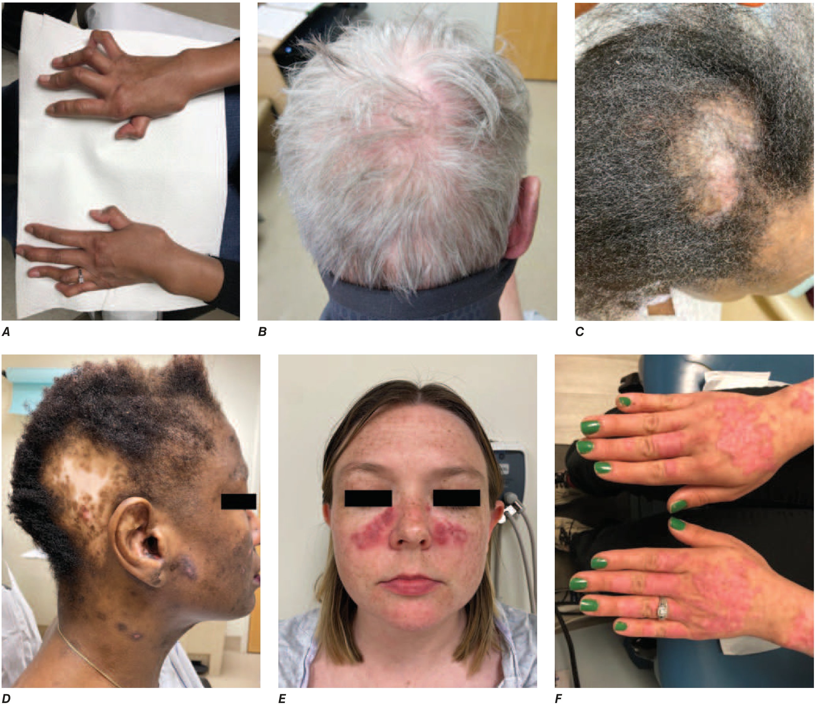
C. Musculoskeletal Manifestations (~90% of patients)
- Arthralgia and myalgia: Extremely common, often the presenting complaint
- Lupus arthritis: Symmetric polyarthritis most commonly involving:
- Wrists
- Metacarpophalangeal (MCP) joints
- Proximal interphalangeal (PIP) joints of the hands
- Knees
- Non-erosive by plain radiograph (unlike RA), but emerging MRI/US data show possible mild erosive changes
- Jaccoud arthropathy: Chronic nonerosive, reversible deviations from periarticular ligament damage leading to MCP subluxation and ulnar deviation - a distinctive feature
- Rhupus: Overlap of RA and SLE
- Avascular necrosis (AVN) of bone: Pain in a single hip, shoulder, or knee out of proportion to other joints should prompt consideration, especially with corticosteroid history
- Inflammatory myopathy: Symmetric proximal weakness, elevated CK and aldolase, inflammatory changes on muscle biopsy
D. Renal Manifestations (50% of patients; up to 70% in Black patients)
Lupus nephritis (LN) is a major cause of morbidity and mortality. All SLE patients should be screened with urine protein/creatinine ratio.
WHO/ISN-RPS Classification of Lupus Nephritis:
- Class I: Minimal mesangial LN
- Class II: Mesangial proliferative LN
- Class III: Focal LN (<50% glomeruli involved)
- Class IV: Diffuse LN (≥50% glomeruli; most severe; segmental [IV-S] or global [IV-G])
- Class V: Membranous LN (can coexist with III or IV)
- Class VI: Advanced sclerosing LN (>90% sclerosis)
Clinical clues to LN activity:
- Proteinuria >0.5 g/24h
- Red blood cell casts in urine (pathognomonic)
- Hypocomplementemia (low C3/C4)
- Rising anti-dsDNA titers
- Hypertension, edema, rising creatinine
E. Hematologic Manifestations
Cytopenias are common and often multifactorial (disease activity, medications, infection):
| Manifestation | Frequency/Notes |
|---|---|
| Anemia | >50% of patients; usually anemia of chronic disease; also hemolytic anemia (AIHA) with positive direct Coombs |
| Leukopenia | <4000/mm³; often lymphopenia (<1000/mm³) |
| Thrombocytopenia | <100,000/mm³; can be severe (<20,000) with immune thrombocytopenia |
| Lymphadenopathy | Common in active disease |
| Splenomegaly | Present in active disease |
| Thrombosis | Venous or arterial; markedly increased with antiphospholipid syndrome (APS) |
| Atypical HUS | Rare but occurs |
F. Neuropsychiatric SLE (NPSLE)
The ACR defined 19 neuropsychiatric syndromes attributable to SLE. Key manifestations:
Central nervous system:
- Seizures (most common NPSLE manifestation)
- Cognitive dysfunction/brain fog (very common, often overlooked)
- Cerebrovascular disease/stroke (up to 19% of SLE patients)
- Aseptic meningitis
- Demyelinating syndrome
- Headache (non-specific, very common)
- Myelopathy/transverse myelitis
- Movement disorders (chorea)
- Acute confusional state/delirium
- Psychosis (corticosteroid-induced vs SLE-mediated - important distinction)
- Mood disorders (depression/anxiety very common)
Peripheral nervous system:
- Mononeuritis multiplex
- Cranial neuropathies
- Peripheral neuropathy (sensorimotor)
- Autonomic neuropathy
Pathomechanisms include: autoantibody-mediated injury (anti-ribosomal P antibodies linked to CNS lupus/psychosis), immune complex deposition, antiphospholipid antibody-related thrombosis, and cytokine-mediated inflammation.
G. Cardiovascular Manifestations
Pericardial involvement - most frequent cardiac manifestation:
- Pericarditis: Can be managed with NSAIDs, colchicine, anti-IL-1 agents; rarely tamponade
- Pericardial effusion
Myocarditis:
- Less common but serious
- Left-sided heart failure and/or arrhythmia
Libman-Sacks endocarditis:
- Fibrinous sterile verrucous endocarditis on mitral and aortic valves
- Associated with antiphospholipid antibodies
- Risk of embolic events
Accelerated atherosclerosis and coronary artery disease:
- Significant cause of premature death, especially in young patients without traditional risk factors
- Driven by type 1 IFN, immune complex vascular injury, APS, dyslipidemia, corticosteroid use
Pulmonary arterial hypertension: With or without APS
H. Pulmonary Manifestations
| Manifestation | Notes |
|---|---|
| Pleuritis | Most common pulmonary manifestation; with or without exudative pleural effusion |
| Acute pneumonitis | Pulmonary infiltrates indistinguishable from infection on imaging; urgent workup needed |
| Diffuse alveolar hemorrhage (DAH) | Life-threatening; capillaritis; hemoptysis, falling hemoglobin with pulmonary infiltrates |
| Interstitial lung disease (ILD) | Less common than in Sjögren's or SSc |
| Shrinking lung syndrome | Restrictive defect from reduced lung volumes; diaphragmatic dysfunction |
| Pulmonary embolism | Associated with APS |
I. Gastrointestinal Manifestations
- Nausea, vomiting, diarrhea during flares (nonspecific)
- Lupus peritonitis - serositis of the peritoneum
- Lupus enteritis/vasculitis - abdominal pain, can lead to ischemia, perforation, bleeding; requires high-dose glucocorticoids
- Pancreatitis - rare but recognized association
- Protein-losing enteropathy
- Elevated liver enzymes (transaminitis from active disease or medications)
- Autoimmune hepatitis overlap
- APS-related: Mesenteric thrombosis, Budd-Chiari syndrome, hepatic venoocclusive disease
J. Ocular Manifestations
- Keratoconjunctivitis sicca - most common (secondary Sjögren's, or dry eyes from inflammation)
- Retinal vasculitis - rare but aggressive; risk of blindness
- Optic neuritis
- Uveitis, scleritis, episcleritis
- Peripheral ulcerative keratitis
- Drug-related: HCQ maculopathy (with prolonged use), corticosteroid-induced cataracts and glaucoma
4. AUTOANTIBODIES IN SLE - Clinical Significance
| Autoantibody | Prevalence | Clinical Association |
|---|---|---|
| ANA (IIF) | >95% | Screening test; homogeneous and speckled patterns most common |
| Anti-dsDNA | 60-70% | Specific for SLE; titer correlates with disease activity, especially nephritis; fluctuates with flares |
| Anti-Sm (anti-Smith) | 25-30% | Highly specific for SLE (~99%); does NOT correlate with disease activity |
| Anti-Ro/SSA | 30-40% | SCLE, neonatal lupus, secondary Sjögren's, photosensitivity, congenital heart block in offspring |
| Anti-La/SSB | 10-15% | Associated with anti-Ro; neonatal lupus |
| Anti-histone | 60-70% | Drug-induced lupus (high sensitivity); also SLE |
| Anti-U1 RNP | 25-30% | Mixed connective tissue disease (MCTD) overlap; Raynaud's, myositis |
| Antiphospholipid (aCL, LA, anti-β2GPI) | 30-40% | Thrombosis, pregnancy loss, thrombocytopenia - antiphospholipid syndrome |
| Anti-ribosomal P | 10-20% | CNS lupus, psychosis, hepatitis |
| Anti-C1q | 30-40% | Lupus nephritis activity |
| Direct Coombs | 30% | Hemolytic anemia when positive |
5. PEDIATRIC-ONSET SLE (cSLE / pSLE) - KEY DIFFERENCES
Children (<18 years) represent approximately 15-20% of all SLE cases. Pediatric onset is clinically distinct and generally more severe.
Epidemiology in pSLE:
- Peak onset in pre-adolescent and adolescent age groups (mean onset ~12-13 years)
- Can present before age 5 (consider monogenic forms - C1q, C2, C4 deficiency; TREX1 mutations, etc.)
- Female:male ratio ~4-5:1 (closer than in adult SLE at ~9:1) - boys make up a larger relative proportion
- Higher prevalence in Asian, Black, and Hispanic children
Clinically more severe at onset and overall:
- More frequent renal involvement: LN at onset in 50-75% of cSLE (vs ~50% adult); more class III/IV nephritis
- More frequent hematologic involvement: Cytopenias at presentation more common
- More frequent neuropsychiatric involvement: Seizures, headache, cognitive dysfunction, chorea
- Malar rash is a common presenting feature
- Fever at onset is more common than in adults
- Constitutional symptoms (fever, weight loss, fatigue) very prominent
- Serositis (pleuritis, pericarditis) frequent
Complications specific to or more prominent in pSLE:
- Growth failure and pubertal delay from chronic disease and glucocorticoid use
- Macrophage Activation Syndrome (MAS) as a life-threatening complication - must always be considered in a cSLE patient with persistent fever, pancytopenia, elevated ferritin, elevated LDH, hepatosplenomegaly
- Infection risk from immunosuppression
- Psychosocial impact: Depression, anxiety, school absenteeism, reduced quality of life - longitudinal screening is now recommended
Serologic differences:
- ANA positivity >95%
- Anti-dsDNA positivity is high (~75-80%)
- Anti-Sm positivity slightly higher than in adults
- Complement consumption (low C3/C4) is a reliable activity marker
Monogenic/Early-onset SLE (<5 years):
If SLE presents in infants or very young children, genetic causes should be sought:
- Complement deficiencies (C1q, C2, C4) - most strongly associated with SLE
- TREX1 mutations
- DNASE1L3 mutations
- RNASEH2 defects
- SAMHD1, ADAR mutations (Aicardi-Goutières spectrum) These represent interferonopathies and monogenic lupus-like syndromes.
Disease monitoring in pSLE:
- SLEDAI-2K (SLE Disease Activity Index) adapted for pediatric use (pSLEDAI)
- BILAG (British Isles Lupus Assessment Group) index
- SLICC/ACR Damage Index
- Urine protein:creatinine ratio at every visit
- Complement levels and anti-dsDNA as activity biomarkers
6. LABORATORY EVALUATION - COMPLETE WORKUP
For initial diagnosis:
- ANA (IIF on HEp-2 cells) - screening
- Anti-dsDNA (Crithidia/ELISA/Farr)
- Anti-Sm, anti-Ro/SSA, anti-La/SSB, anti-U1 RNP, anti-histone
- Antiphospholipid panel: aCL (IgG/IgM), anti-β2GPI (IgG/IgM), lupus anticoagulant
- Complement: C3, C4, CH50
- Direct Coombs test
- CBC with differential
- Comprehensive metabolic panel (LFTs, renal function)
- Urinalysis with microscopy + urine protein:creatinine ratio
- ESR, CRP (often markedly elevated ESR with relatively normal CRP in SLE; very high CRP suggests intercurrent infection)
Monitoring (ongoing):
- C3, C4, anti-dsDNA (disease activity markers - fall with flares)
- CBC, CMP, urinalysis at each visit
- Ophthalmology screening for HCQ maculopathy (baseline then yearly after 5 years of use)
- Blood pressure at every visit
7. DIFFERENTIAL DIAGNOSIS
| Condition | Key Distinguishing Features |
|---|---|
| Juvenile idiopathic arthritis (JIA) | ANA usually low titer; no multi-system involvement; anti-dsDNA negative |
| Viral arthritis/SLE-like viral illness | Acute onset, self-limited, viral serology positive |
| Drug-induced lupus | Anti-histone antibodies positive; anti-dsDNA usually negative; resolves with drug withdrawal |
| Mixed connective tissue disease (MCTD) | High titer anti-U1 RNP; overlap features; Raynaud's prominent |
| Dermatomyositis | Heliotrope rash, Gottron's papules, elevated CK, myositis-specific antibodies |
| Antiphospholipid syndrome (primary) | Thrombosis/pregnancy morbidity without other SLE features |
| Sjögren's syndrome | Sicca predominant; anti-Ro/La; less organ involvement |
| Vasculitis (ANCA-associated) | ANCA positive; different organ pattern |
| Autoinflammatory diseases | In young children; periodic fever syndromes; ANA negative |
| Infectious (EBV, CMV, parvovirus) | Acute illness; specific serology; no anti-dsDNA |
8. SUMMARY TABLE: Comparison of Classification Criteria
| Feature | SLICC 2012 | EULAR/ACR 2019 |
|---|---|---|
| ANA required? | No (but included as immunologic criterion) | Yes (entry criterion, ≥1:80) |
| Total criteria | 17 (11 clinical + 6 immunologic) | 10 domains, weighted scoring |
| Classification rule | ≥4 criteria (≥1 clinical + ≥1 immunologic) OR biopsy-proven LN + ANA/anti-dsDNA | ANA entry + ≥10 points + ≥1 clinical domain |
| Sensitivity | ~97% | ~96.1% |
| Specificity | ~84% | ~93.4% |
| Handles ANA-neg SLE? | Yes (can classify without ANA) | No |
| Useful for pSLE? | More applicable | Used but ANA requirement may miss rare cases |
References:
- Harrison's Principles of Internal Medicine 22E (2025, McGraw-Hill), Chapter 368
- Tietz Textbook of Laboratory Medicine 7th Edition, Chapter 94
- Aringer M et al. 2019 EULAR/ACR classification criteria for SLE. Ann Rheum Dis 2019;78:1151-9
- Petri M et al. Derivation and validation of the SLICC classification criteria for SLE. Arthritis Rheum 2012;64:2677-86
- Recent PubMed literature: Genetics of Childhood-Onset SLE (Carlomagno R et al., Arthritis Rheumatol 2025; PMID 40356234)---
A few high-yield clinical pearls specifically for your fellowship practice:
-
EULAR/ACR 2019 vs SLICC 2012 in pSLE: The 2019 criteria are more specific but require ANA ≥1:80 as a mandatory entry criterion - this is rarely a problem in pediatric SLE (>95% ANA positive), but early-onset or monogenic forms may be ANA-negative, making SLICC 2012 more inclusive and practically useful.
-
Anti-dsDNA fluctuates with disease activity - useful for monitoring flares, especially renal. Anti-Sm is highly specific but does NOT track activity.
-
Complement consumption pattern (falling C3/C4 + rising anti-dsDNA) is the most reliable combination for predicting lupus nephritis flare.
-
Seronegative SLE (~5% anti-dsDNA negative, ~5% ANA negative): Do not dismiss SLE on serology alone if the clinical picture is convincing.
-
Macrophage Activation Syndrome must be actively ruled out in any cSLE patient with fever, cytopenias, hyperferritinemia (>500 ng/mL, especially >10,000), and hepatosplenomegaly - it is a disease-defining emergency.
-
Psychosocial screening (anxiety, depression) is now recommended longitudinally in cSLE as per 2025-2026 literature (Valente Soto et al., Lupus 2026; PMID 41665227).
ok now lets move on to treatment and management
Summarising Conversation
Searching sources...
Reading File
Finding Sources
Finding Sources
Searching PubMed
"systemic lupus erythematosus" AND "treatment" AND "guidelines"
I found the 2023 EULAR update systematic review (PMID 38777375) and the Brenner & Rector pediatric LN table. Let me read all these sources in parallel now.
Reading File
Reading File
Reading File
Reading Article
PMID: 38777375
Excellent. Now let me get the Harrison's LN treatment section and the 2023 EULAR update content, plus the pediatric LN table.
Reading File
Reading File
Searching PubMed
"lupus nephritis" AND "voclosporin OR belimumab OR obinutuzumab"
Excellent - I found the 2025 EULAR LN recommendations (PMID 41107121) and the obinutuzumab Phase III RCT (PMID 39927615). Let me get the details on both.
Reading Article
PMID: 41107121
Reading Article
PMID: 39927615
Reading File
I now have comprehensive treatment content. Let me get the remaining biologics/NPSLE/APS sections and the pediatric SLE treatment section.
Reading File
Reading File
I now have comprehensive treatment data from Harrison's 22E, Brenner & Rector (pediatric nephrology), Goldman-Cecil, and the latest 2025 EULAR LN recommendations + obinutuzumab RCT. Let me now compile the complete treatment guide.Here is the comprehensive fellowship-level treatment guide, fully updated through 2025-2026.
SLE: Treatment and Management
Based on Harrison's Principles of Internal Medicine 22E (2025), Brenner & Rector's The Kidney, Goldman-Cecil Medicine, 2023 EULAR SLE Recommendations (Fanouriakis et al., Ann Rheum Dis 2024), 2025 EULAR LN Recommendations (PMID 41107121), and obinutuzumab Phase III data (NEJM 2025, PMID 39927615).
1. TREATMENT GOALS (EULAR Framework)
The overarching principle of SLE management is treat-to-target:
| Target | Definition |
|---|---|
| Remission | SLEDAI-2K = 0, on stable HCQ ± immunosuppressives, prednisone ≤5 mg/day |
| Low Disease Activity (LLDAS) | SLEDAI-2K 0-4, no new activity, on stable therapy, prednisone ≤7.5 mg/day |
| Prevention of damage | Minimise cumulative corticosteroid dose, organ-threatening flares, infections |
| Quality of life | Address fatigue, pain, mood, schooling (in pediatric patients) |
Additional EULAR goals for lupus nephritis:
- 3-month milestone: ≥25% reduction in urine protein:creatinine ratio (UPCR), eGFR not worsened
- 6-month milestone: ≥50% reduction in UPCR to <3 g/day
- 12-24 month target: UPCR <0.5-0.7 g/day with eGFR within 10% of baseline
2. GENERAL PROTECTIVE MEASURES (ALL PATIENTS)
These apply to every SLE patient regardless of disease severity:
- Daily broad-spectrum sunscreen (SPF ≥50) on sun-exposed areas - UV light triggers flares
- Avoid UV light exposure (especially 10am-4pm), wear protective clothing
- Hydroxychloroquine (HCQ) for all patients unless contraindicated (see below)
- Healthy diet, maintain normal weight
- No smoking - tobacco reduces antimalarial efficacy
- Regular aerobic exercise
- Strict BP control (target <130/80 in LN)
- Lipid and glucose monitoring
- Vaccinations: Influenza (annual), pneumococcal (PCV15/20 or PCV13 + PPSV23), COVID-19, HPV, varicella (live vaccines should be given before starting immunosuppression or when off significant immunosuppression)
- Bone protection: Calcium + vitamin D supplementation for all patients on glucocorticoids; bisphosphonates if high fracture risk
- ACE inhibitor or ARB for all patients with proteinuria/hypertension
- SGLT2 inhibitor (e.g., dapagliflozin) for patients with LN + reduced eGFR - reduces glomerular hyperfiltration and proteinuria (new in EULAR 2023/2025)
3. DRUG CLASSES IN SLE TREATMENT
A. Antimalarials - Backbone of All SLE Therapy
Hydroxychloroquine (HCQ) - first-line for ALL SLE patients:
| Property | Detail |
|---|---|
| Mechanism | Lysosomal pH disruption, TLR7/9 inhibition, type 1 IFN reduction |
| Benefits | Reduces flares, increases survival, reduces LN risk, anti-thrombotic, anti-atherosclerotic, reduces renal damage, safe in pregnancy |
| Dose | Up to 5 mg/kg/day actual body weight (adult); in children, same weight-based dosing |
| Monitoring | Baseline ophthalmology, then annual after 5 years; monthly if risk factors (high dose, renal impairment, tamoxifen use) |
| Side effects | Rash, GI symptoms, dyspigmentation, cardiomyopathy (rare), retinal maculopathy (dose/duration-dependent) |
| Pregnancy | Safe and standard of care; reduces congenital heart block risk |
Quinacrine: Can be added to HCQ for inadequate skin response, or substituted in retinal toxicity. Does not cause retinal toxicity.
Chloroquine: Higher retinal toxicity than HCQ; alternative where HCQ unavailable.
B. Glucocorticoids
| Indication | Dose |
|---|---|
| Mild-moderate flare | Prednisone 0.5 mg/kg/day, taper over weeks |
| Active nephritis, severe CNS, severe cytopenias | IV methylprednisolone 500-1000 mg/day for 1-3 days (pulse), then oral prednisone 0.5-1 mg/kg/day |
| Maintenance goal | ≤5 mg/day prednisone (or equivalent) - EULAR target |
| Ultimate goal | Taper and discontinue as soon as disease control achieved |
Key principle: Glucocorticoids remain effective but carry major cumulative toxicity. Every 1 mg/day increase in prednisone dose is associated with measurable increases in organ damage. The modern approach is to combine GCs with immunosuppressives/biologics to enable faster tapering.
C. Conventional Immunosuppressives
| Drug | Indications | Key Dosing | Monitoring |
|---|---|---|---|
| Mycophenolate mofetil (MMF) | LN induction + maintenance; skin; arthritis | 2-3 g/day (adult); ~1200 mg/m²/day (pediatric) | CBC, LFTs, renal function; teratogenic |
| Azathioprine (AZA) | Maintenance of LN, non-renal SLE, pregnancy-compatible | 2-3 mg/kg/day | CBC (myelosuppression); TPMT genotyping before starting |
| Cyclophosphamide (CYC) | Severe proliferative LN; NPSLE; vasculitis | Low-dose Euro: 500 mg IV q2w ×6; High-dose NIH: 0.5-1 g/m² IV monthly ×6; Pediatric: 500-1000 mg/m² IV monthly ×6 | CBC, urinalysis (hemorrhagic cystitis), MESNA prophylaxis, ovarian protection |
| Methotrexate (MTX) | Arthritis; skin manifestations | 15-25 mg/week | LFTs, CBC; teratogenic; folate supplementation |
| Calcineurin inhibitors (CNIs) | LN (esp. membranous ± proliferative); refractory disease | Tacrolimus 0.05-0.1 mg/kg/day; Voclosporin 23.7 mg BID | Renal function, BP, drug levels; TDM essential |
| Leflunomide | Arthritis | 20 mg/day | LFTs, BP; teratogenic |
D. Biologics - The Modern Era
1. Belimumab (Benlysta) - BLyS/BAFF Inhibitor
- Mechanism: Anti-BLyS (B-lymphocyte stimulator) monoclonal antibody; reduces B-cell survival and autoantibody production
- FDA-approved: Active SLE (2011); active LN (2020, BLISS-LN trial)
- Indications: Active non-renal SLE not controlled by standard therapy; active class III/IV/V LN in combination with MMF or low-dose CYC
- Route/Dose: IV 10 mg/kg at 0, 2, 4 weeks then monthly; or SC 200 mg weekly
- BLISS-LN trial: Belimumab + MMF + low-dose GC vs. MMF + GC - significantly better primary efficacy renal response (43% vs 32%) and complete renal response
- Not recommended in severe NPSLE (CNS lupus) - risk of CNS infections
- Pediatric use: Approved for children ≥5 years with active SLE; PLUTO trial showed efficacy
2. Anifrolumab (Saphnelo) - Type I IFN Receptor Inhibitor
- Mechanism: Monoclonal antibody against IFNAR1 (type I interferon receptor), blocking all type I IFNs (α, β, ω)
- FDA-approved: Moderate-to-severe non-renal SLE (2021, TULIP-2 trial)
- Indications: Active non-renal SLE (skin, musculoskeletal) on standard therapy; particularly effective in high IFN signature patients
- Route/Dose: IV 300 mg every 4 weeks
- Key trial: TULIP-2 - significantly better BICLA response (47.8% vs 31.5%), better skin, reduced GC use
- Not recommended for severe NPSLE or in patients with high infection risk
- Pediatric use: Not yet approved for children; trials ongoing
3. Voclosporin (Lupkynis) - Novel CNI for LN
- Mechanism: Next-generation calcineurin inhibitor; superior pharmacokinetic profile vs tacrolimus (flat exposure-response, less nephrotoxicity)
- FDA-approved: Active LN (2021, AURORA 1 trial)
- Dose: 23.7 mg orally twice daily (in combination with MMF)
- AURORA trial: Voclosporin + MMF + low-dose GC vs MMF + GC - significantly better complete renal response at 52 weeks (40.8% vs 22.5%)
- EULAR 2023: Recommended as add-on to MMF for LN (especially class V or refractory)
- Pediatric use: Emerging evidence, not yet approved in children; voclosporin is listed as "emerging treatment" in Brenner & Rector pediatric LN table
4. Obinutuzumab - Type II Anti-CD20 (NEWEST - 2025)
- Mechanism: Humanized type II anti-CD20 monoclonal antibody; superior B-cell depletion vs rituximab (type I anti-CD20) due to different CD20 binding site and enhanced antibody-dependent cellular cytotoxicity
- Phase III REGENCY trial (NEJM, April 2025 - PMID 39927615):
- 271 adults with biopsy-proven active LN (all classes)
- Obinutuzumab 1000 mg + MMF + low-dose prednisone vs MMF + prednisone
- Complete renal response at week 76: 46.4% vs 33.1% (adjusted difference 13.4 pp; p=0.02)
- Complete renal response with prednisone ≤7.5 mg/day: 42.7% vs 30.9% (p=0.04)
- More infections (mainly COVID-related) in obinutuzumab arm
- EULAR 2025 LN update (PMID 41107121): Obinutuzumab now included in the guidelines alongside belimumab and rituximab for LN management
- Pediatric use: Data in children not yet available
5. Rituximab (RTX) - Type I Anti-CD20
- Mechanism: Chimeric anti-CD20; B-cell depletion
- Not FDA-approved for SLE (EXPLORER and LUNAR trials failed to meet primary endpoints), but widely used off-label
- Indications: Refractory SLE not responding to MMF/CYC/belimumab; refractory severe cytopenias (ITP, AIHA); refractory NPSLE (myelitis, optic neuritis)
- Dose in LN: 375 mg/m² weekly ×4, or 1000 mg ×2 (2 weeks apart)
- Pediatric use: Widely used as salvage therapy in refractory pSLE; listed in Brenner & Rector pediatric LN table as "emerging treatment"
- EULAR 2023/2025: Recommended for refractory/relapsing LN, especially after failure of CYC-based regimens
4. MANAGEMENT BY DISEASE SEVERITY AND ORGAN SYSTEM
A. Non-Renal SLE - By Severity
(Based on Harrison's 22E/EULAR 2023 framework)
Mild Non-Renal SLE:
- Constitutional symptoms, mild arthritis, mild rash (<9% BSA), SLEDAI ≤6, PLT 50-100×10⁹/L
- Step 1: Sunscreen + HCQ + NSAIDs (caution - increased MI, renal, aseptic meningitis risk)
- Step 2: Low-dose prednisone (≤10 mg/day) - short course, taper
- Step 3: MTX, AZA, or MMF as steroid-sparing agent
Moderate Non-Renal SLE:
- Moderate-severe arthritis, rash 9-18% BSA, PLT 20-50×10⁹/L, BILAG B
- Step 1-2: HCQ + prednisone 0.5 mg/kg/day, taper
- Step 3: MTX, AZA, or MMF (equal options)
- Step 4: Belimumab or anifrolumab as add-on
Severe Non-Renal SLE:
- BILAG A manifestations, widespread skin disease, severe arthritis, severe cytopenias
- Step 1: HCQ + prednisone 0.5-1 mg/kg/day (± IV MP pulse for rapid control)
- Step 2: MMF, AZA, or MTX
- Step 3: Belimumab (preferred for skin/arthritis) or anifrolumab
- For severe skin: Add belimumab or anifrolumab; thalidomide/lenalidomide for refractory cutaneous disease (cautiously)
- For severe NPSLE: Neither belimumab nor anifrolumab recommended; use RTX or CYC
B. Lupus Nephritis (LN) Treatment - Current Standard
Class I & II LN: No specific kidney-directed immunosuppression required; treat underlying systemic disease; close monitoring with urine PCR at least every 3-6 months.
Class III / IV LN (Proliferative - the most common severe form):
INDUCTION PHASE (first 6 months):
| Regimen | Details | Preferred in |
|---|---|---|
| MMF + GC | MMF 2-3 g/day + IV MP pulse (500-1000 mg ×1-3 days) then prednisone 0.5 mg/kg/day tapered | Black, Hispanic patients (better response); generally preferred |
| Low-dose CYC (Euro-Lupus) + GC | CYC 500 mg IV every 2 weeks × 6 doses (total 3 g) + GC | White, Asian patients; non-inferior to high-dose CYC |
| High-dose CYC (NIH) + GC | CYC 0.5-1 g/m² IV monthly × 6 + GC | Reserved for very severe/poor prognosis cases |
Add-on therapies (EULAR 2023/2025):
- + Belimumab (10 mg/kg IV) added to MMF or low-dose CYC for all class III/IV/V patients - improves complete renal response, reduces flares
- + Voclosporin (23.7 mg BID) added to MMF - particularly for class V or cases with high proteinuria
- + Obinutuzumab (1000 mg IV) added to MMF - new 2025 EULAR recommendation for active LN
- + SGLT2 inhibitor if eGFR permits - nephroprotective, reduces proteinuria
- + ACEi/ARB for all patients with proteinuria - target BP <130/80
MAINTENANCE PHASE (after induction, minimum 3-5 years):
| Agent | Dose | Notes |
|---|---|---|
| MMF | 1.5-2 g/day | Preferred over AZA; better at preventing renal relapses |
| Azathioprine | 2 mg/kg/day | Alternative to MMF; safe in pregnancy |
| Tacrolimus | Low dose (target trough 4-6 ng/mL) | Add-on option; especially useful in East Asian patients (multitarget therapy) |
| HCQ | Throughout | All patients; reduces flare risk |
| Low-dose prednisone | ≤5 mg/day | Taper toward discontinuation if sustained remission |
Treatment targets for LN monitoring:
- 3 months: UPCR reduced ≥25% from baseline
- 6 months: UPCR reduced ≥50% and <3 g/g, eGFR stable
- 12-24 months: UPCR <0.5-0.7 g/g, eGFR within 10% of baseline - this defines complete renal response
Class V LN (Membranous) without proliferative component:
- If UPCR <1 g/g: ACEi/ARB + HCQ; watch closely
- If UPCR 1-3 g/g: MMF ± CNI (voclosporin/tacrolimus)
- If UPCR >3 g/g (nephrotic): MMF + voclosporin or tacrolimus + GC; consider belimumab add-on
Antiphospholipid nephropathy on biopsy (TMA pattern): Vitamin K antagonist (warfarin) or LMWH anticoagulation, target INR 2-3.
C. Neuropsychiatric SLE (NPSLE) Treatment
NPSLE management depends on whether the mechanism is inflammatory (vasculitis, cytokine-mediated) or thrombotic/ischemic (APS-related):
| Mechanism | Treatment |
|---|---|
| Inflammatory (seizures, psychosis, myelitis, optic neuritis, cerebritis) | IV methylprednisolone 500-1000 mg/day × 3 days → high-dose prednisone + CYC or RTX (refractory) |
| Thrombotic/APS-related (stroke, TIA, myelopathy) | Anticoagulation (warfarin INR 2-3, or LMWH) ± antiplatelet; no high-dose immunosuppression |
| Seizures | Standard antiepileptics + treat underlying SLE activity |
| Psychosis | Differentiate steroid-induced vs SLE-mediated; atypical antipsychotics + GC ± CYC |
| Cognitive dysfunction | HCQ, manage cardiovascular risk factors, neuropsychological support |
- Belimumab and anifrolumab are NOT recommended in severe NPSLE (CNS infection risk, lack of evidence)
- Rituximab is the preferred biologic for refractory inflammatory NPSLE
D. Hematologic Manifestations
| Manifestation | Treatment |
|---|---|
| Autoimmune hemolytic anemia (AIHA) | Prednisone 1 mg/kg/day → taper; refractory: RTX, splenectomy, danazol, IV immunoglobulin (IVIG) |
| Immune thrombocytopenia (ITP) | PLT <20,000 or bleeding: IV MP pulse + IVIG; then prednisone; refractory: RTX, splenectomy, thrombopoietin receptor agonists (eltrombopag, romiplostim) |
| Thrombotic microangiopathy (TMA)/aHUS | Plasma exchange + eculizumab if complement-mediated; treat underlying SLE |
| Leukopenia/lymphopenia | Usually resolves with HCQ and disease control; rarely requires specific therapy |
E. Serositis
- Mild pericarditis/pleuritis: NSAIDs + colchicine (first-line, as per pericarditis guidelines)
- Unresponsive or moderate: Low-to-moderate dose prednisone (0.5 mg/kg/day)
- Recurrent pericarditis: Anti-IL-1 (anakinra, rilonacept) - emerging use in SLE serositis
- Tamponade: Drainage + aggressive immunosuppression
5. PEDIATRIC SLE (pSLE) TREATMENT - SPECIFIC CONSIDERATIONS
General Approach
The treatment framework mirrors adult SLE but with important modifications:
HCQ: Universal in all pSLE patients; same weight-based dosing (≤5 mg/kg/day); annual ophthalmology from year 1 given longer lifetime exposure.
Glucocorticoids: Higher susceptibility to cumulative toxicity in children:
- Growth failure, pubertal suppression, AVN, osteoporosis, cataracts, Cushingoid features
- Target: minimize to ≤5 mg/day prednisone as quickly as possible
- Pulse IV methylprednisolone: 30 mg/kg/day (max 1000 mg) × 1-3 days for severe flares
Induction therapy in pediatric LN (Classes III/IV):
- Pulse IV methylprednisolone × 3 days then oral prednisone 1-2 mg/kg/day
- MMF preferred over CYC in most centers (equal efficacy, better toxicity profile; pediatric dose ~1200 mg/m²/day)
- CYC (IV) 500-1000 mg/m² monthly × 6 months - reserved for severe/refractory LN, CNS disease, or poor prognostic features
- MESNA given with each CYC infusion (hemorrhagic cystitis prophylaxis)
- GnRH analogs (leuprolide) for gonadal protection in adolescent girls receiving CYC
Maintenance therapy in pediatric LN:
- MMF (preferred, ~1200 mg/m²/day) or AZA (2 mg/kg/day)
- Duration: At minimum 2 years; most centers continue 3-5+ years given high relapse risk
- Long-term renal relapse-free 10-year survival ~73% in pediatric LN; 10-year overall survival ~90%
Biologics in pSLE:
| Drug | Pediatric Status |
|---|---|
| Belimumab | FDA-approved in children ≥5 years with active SLE (PLUTO trial, 2019); IV 10 mg/kg monthly |
| Rituximab | Off-label; widely used for refractory pSLE (cytopenias, nephritis, CNS disease); 375 mg/m² × 4 or 750 mg/m² × 2 |
| Voclosporin | Emerging/off-label in pediatrics; listed as emerging treatment in Brenner & Rector |
| Anifrolumab | Not approved in children; adult data only |
| Obinutuzumab | No pediatric data yet |
Multitarget therapy in pSLE: MMF + tacrolimus (calcineurin inhibitor) - increasingly used in East Asian patients and those with membranous LN; tacrolimus 0.05-0.1 mg/kg/day targeting trough 4-6 ng/mL.
Macrophage Activation Syndrome (MAS) in pSLE
MAS is a life-threatening complication requiring immediate treatment:
- First-line: High-dose IV methylprednisolone (30 mg/kg/day × 3 days); IVIG
- Refractory: Cyclosporine A (3-5 mg/kg/day) - most evidence in pSLE-MAS; rapid B-cell depletion
- Emerging: Anakinra (IL-1 blockade), emapalumab (anti-IFN-γ), ruxolitinib (JAK1/2 inhibitor)
- Etoposide (VP-16) if evolves to full HLH
- Simultaneous management of underlying SLE trigger (often infection + active SLE)
6. ANTIPHOSPHOLIPID SYNDROME (APS) IN SLE
APS co-exists in ~30-40% of SLE patients and requires specific management:
| Scenario | Treatment |
|---|---|
| Primary thromboprophylaxis (aPL+ but no thrombosis) | Low-dose aspirin (81-100 mg/day) + HCQ |
| First venous thrombosis | Warfarin INR 2-3, long-term (often lifelong) |
| Arterial thrombosis | Warfarin INR 2-3 (or INR 3-4 if recurrent); aspirin may be added |
| Catastrophic APS | Anticoagulation + high-dose GC + plasma exchange ± IVIG ± rituximab |
| Obstetric APS | LMWH + low-dose aspirin throughout pregnancy |
| Triple aPL positivity | Warfarin preferred over DOACs (DOACs inferior in high-risk aPL profiles - TRAPS, RAPS trials) |
7. PREGNANCY IN SLE
- Pre-conception counseling: Achieve remission for at least 6 months before conception
- Contraindicated drugs in pregnancy: MMF, methotrexate, leflunomide (teratogenic), cyclophosphamide (first trimester), voclosporin (insufficient data), belimumab, anifrolumab
- Safe in pregnancy: HCQ (continue throughout - reduces flares, reduces CHB risk), azathioprine, tacrolimus, low-dose prednisone, LMWH
- Low-dose aspirin: 100-150 mg/day from 12 weeks for preeclampsia prevention
- Anti-Ro/La positive mothers: Fetal echocardiography from 18-24 weeks for congenital heart block (CHB); fluorinated dexamethasone if 1st/2nd degree CHB detected
- Monitor with anti-dsDNA, complement (C3/C4), UPCR at each antenatal visit
- Postpartum: High risk of flare; continue/resume immunosuppression; HCQ safe with breastfeeding, AZA compatible
8. MONITORING IN SLE
Routine monitoring at every visit:
| Test | Frequency | Purpose |
|---|---|---|
| BP, weight, height (children) | Every visit | Hypertension, growth monitoring |
| UPCR (spot urine) | Every visit | LN monitoring |
| CBC + differential | Every 3-6 months (more frequent if cytopenias or CYC) | Cytopenias, myelosuppression |
| CMP (renal + liver) | Every 3-6 months | Drug toxicity, renal function |
| C3, C4, anti-dsDNA | Every 3-6 months | Disease activity markers |
| Lipid panel | Annually | Accelerated atherosclerosis |
| SLEDAI-2K / BILAG | Every visit | Disease activity scoring |
| Ophthalmology (HCQ screen) | Baseline → annually from year 5 (or sooner if high dose/risk factors) | HCQ maculopathy |
| BMD (DEXA scan) | At baseline if on GC; repeat 1-2 yearly | Osteoporosis prevention |
| Psychosocial screening (pSLE) | Every visit | Depression, anxiety, school attendance (2026 recommendation) |
Flare predictors requiring intensification:
- Rising anti-dsDNA (especially >2× upper limit)
- Falling C3/C4
- New UPCR >0.5 g/g or worsening by ≥50%
- New or worsening cytopenias
9. TREATMENT OF REFRACTORY/RELAPSING SLE
| Situation | Next Step |
|---|---|
| Refractory non-renal SLE on HCQ + MMF | Add belimumab or anifrolumab |
| Refractory LN on MMF ± belimumab | Add voclosporin, OR switch to CYC-based regimen, OR add RTX/obinutuzumab |
| Relapsing class III/IV LN | Re-induction with MMF or CYC + belimumab; obinutuzumab (new 2025) |
| Refractory severe cytopenias | Rituximab; calcineurin inhibitors; IVIG; splenectomy (last resort) |
| Refractory cutaneous disease | Thalidomide/lenalidomide (caution: neuropathy, teratogenic); dapsone for bullous lupus |
| Refractory CNS lupus | RTX + CYC |
10. EMERGING/PIPELINE THERAPIES (Fellowship-Level Awareness)
| Drug | Target | Status |
|---|---|---|
| Obinutuzumab | Type II anti-CD20 | NEJM 2025 Phase III positive; EULAR 2025 LN included |
| Iptacopan | Factor B (alternative complement pathway inhibitor) | Phase III in LN ongoing |
| Telitacicept | BLyS + APRIL (dual inhibitor) | Phase III trials; data in Chinese SLE population |
| Iberdomide | Cereblon E3 ligase modulator (CELMoD); depletes Ikaros/Aiolos | Phase II trials active |
| Deucravacitinib | TYK2 inhibitor | Phase III POETYK SLE (skin/joints); positive Phase II |
| Obexelimab | Anti-CD19/FcγRIIb | Phase II/III in SLE |
| Ustekinumab | IL-12/23 inhibitor | Phase III failed to meet primary endpoint |
| CAR-T cell therapy | CD19 or BCMA targeting | Case series + small trials in refractory SLE - remission without maintenance immunosuppression (Mackensen et al., Nature Medicine 2022) |
| SGLT2 inhibitors | Nephroprotection | Now recommended in EULAR 2023 for LN with reduced eGFR |
11. SUMMARY: EULAR 2023 KEY OVERARCHING PRINCIPLES
Reproduced from Fanouriakis et al., Ann Rheum Dis 2024 (PMID 38777375) - the systematic literature review underpinning the 2023 EULAR update:
- Treat-to-target (remission or LLDAS) is the standard of care
- HCQ is universal background therapy for all SLE patients
- Minimise glucocorticoids - combine with immunosuppressives/biologics to taper faster; target ≤5 mg/day or off altogether
- Anifrolumab and belimumab achieve better control than standard of care in extrarenal SLE
- Combination therapies with belimumab and voclosporin attain higher response rates in LN
- Remission and LLDAS are associated with better long-term outcomes - prevention of irreversible damage
- In patients achieving stable remission, GC and immunosuppressive therapy may be gradually tapered (but relapse risk is real - RCT data show higher relapse rates on withdrawal)
Key References:
- Harrison's Principles of Internal Medicine 22E (2025), Chapters 368-369
- Brenner & Rector's The Kidney (10th Ed.), Chapter 72 - Pediatric Lupus Nephritis
- Goldman-Cecil Medicine International Edition, Chapter 245
- Fanouriakis et al. 2023 update of EULAR recommendations for SLE. Ann Rheum Dis 2024 (PMID 38777375)
- Fanouriakis et al. EULAR recommendations for SLE with kidney involvement: 2025 update. Ann Rheum Dis 2026 (PMID 41107121)
- Furie RA et al. Obinutuzumab in active lupus nephritis (REGENCY trial). NEJM April 2025 (PMID 39927615)
- Furie R et al. Belimumab in LN - BLISS-LN open-label extension. CJASN 2022 (PMID 36302567)---
High-yield fellowship pearls for practice:
-
Voclosporin vs tacrolimus in LN: Voclosporin has a flat pharmacokinetic profile - no need for drug level monitoring, unlike tacrolimus. This makes it practically easier to use. However it is not yet approved in children.
-
Obinutuzumab vs rituximab: Obinutuzumab is a type II anti-CD20 that depletes B cells more completely and durably than rituximab (type I). The REGENCY Phase III (NEJM 2025) is a landmark trial - it's the first positive Phase III trial for a B-cell depleting agent in LN. EULAR 2025 LN guidelines now include it.
-
The "steroid minimization revolution": The EULAR 2023 update fundamentally shifted the approach - high-dose GC protocols are being replaced. The obinutuzumab trial used prednisone ≤7.5 mg/day by week 12 and ≤5 mg/day by week 24 as a standard arm and still showed excellent response, validating low-GC approaches.
-
DOACs in APS: The TRAPS trial (rivaroxaban) and RAPS trial showed inferiority to warfarin in triple-positive aPL and arterial thrombosis. For high-risk aPL profiles, warfarin remains standard. DOACs are only considered in low-risk aPL profiles where warfarin cannot be used.
-
CAR-T in SLE: Early case series from Germany (Mackensen et al., 2022) showed sustained drug-free remission in refractory SLE after CD19-directed CAR-T. This is a paradigm-shifting concept - "resetting" the immune system. Not yet standard care but watch this space.
This is a shared conversation. Sign in to Orris to start your own chat.