
Coblation , microdibrider, vessel sealing device and suture material in detail according to recent modalities in ENT in simplified manner
coblation microdebrider vessel sealing device ENT surgery 2024 2025 recent advances
coblation wand radiofrequency plasma tonsillectomy ENT
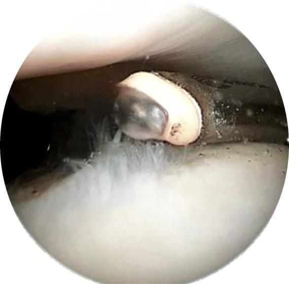
This clinical photograph is an intraoperative arthroscopic view demonstrating the use of a Coblation (controlled ablation) wand on articular cartilage within a human joint, likely the knee. The image shows the distal tip of the Coblation device, characterized by a light-beige ceramic insulator and a dark metallic electrode. A distinctive, semi-transparent dome-shaped plasma layer is visible at the working end of the wand, illustrating the radiofrequency-excited field used to dissolve tissue. The wand is in close proximity to a white, pearlescent cartilaginous surface. Visible pathology includes fine, filamentous white structures representing chondral fibrillation, typical of Grade 2 or 3 chondromalacia. The educational focus is the visualization of the plasma-mediated debridement process (chondroplasty), where the device is used to smooth frayed articular surfaces while minimizing thermal damage to underlying healthy chondrocytes and the extracellular matrix. This procedural image is highly relevant for orthopedic surgical training and sports medicine education regarding joint preservation techniques.
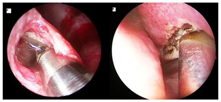
This composite diagnostic image features two endoscopic clinical photographs (A and B) demonstrating coblation turbinoplasty techniques for the treatment of inferior turbinate hypertrophy. Image A illustrates an intraturbinoplasty (submucosal) approach, showing a metallic coblation wand with a wire-like tip inserted into a submucosal channel of the inferior turbinate. The surrounding nasal mucosa is pink and intact, with minimal visible bleeding at the entry site. Image B illustrates an extraturbinoplasty (surface ablation) approach, where the coblation instrument is applied directly to the mucosal surface. The surgical site in B shows visible tissue disruption, charring, and thermal ablation effects, characterized by a brownish, irregular texture on the lateral aspect of the turbinate. Both images highlight the use of radiofrequency energy to reduce turbinate volume. This material is intended for otolaryngology surgical training, specifically demonstrating different technical applications of plasma-mediated ablation in endoscopic endonasal surgery.
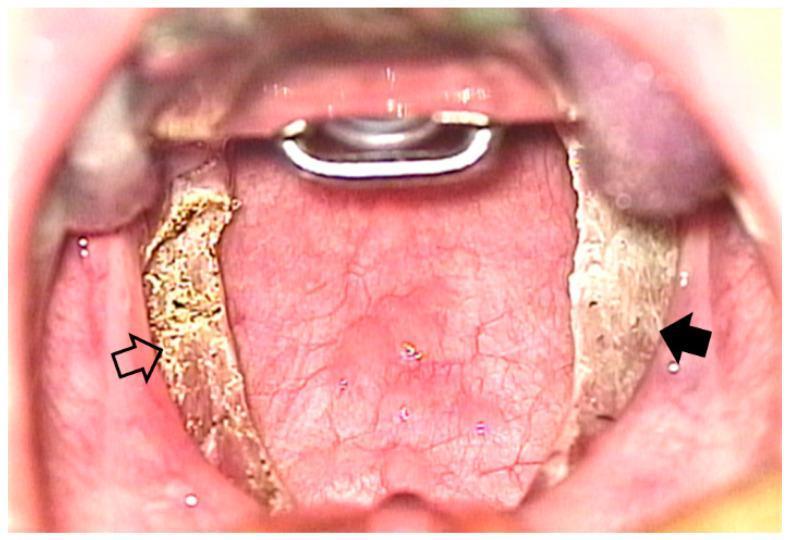
This intraoral clinical photograph provides a comparison chart view of the oropharynx immediately following bilateral extracapsular tonsillectomy. The image demonstrates two distinct surgical techniques applied to the left and right tonsillar fossae. The patient's right tonsillar fossa (indicated by a solid black arrow) shows the result of coblation tonsillectomy; the surgical bed appears relatively smooth, clean, and predominantly pinkish-red, indicating minimal thermal damage to the underlying tissue. In contrast, the patient's left tonsillar fossa (indicated by an open arrow) shows the result of monopolar electrocautery tonsillectomy. This area exhibits a yellowish-tan color with a more irregular, textured surface and visible surgical debris, consistent with higher thermal effect and tissue carbonization (slough). The comparison illustrates the different immediate postoperative tissue characteristics between plasma-mediated coblation and traditional electrocautery. Key landmarks visible include the posterior pharyngeal wall, the soft palate, and a surgical mouth gag in place for exposure. This visual serves as an educational tool for otolaryngologists to evaluate surgical site morphology and potential healing outcomes associated with different dissection modalities.
microdebrider powered nasal instrument ENT sinus surgery
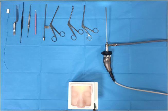
A clinical photograph showcasing a specialized surgical simulation set for Endoscopic Sinus Surgery (ESS). The visual contains three primary components arranged on a blue sterile field: a 3D-printed anatomical simulator, standard otorhinolaryngological (ENT) surgical instruments, and an endoscopic imaging system. The simulator at the bottom features a realistic, flesh-toned silicone facial skin model housed in a white frame, with a focus on the nasal anatomy, including the bridge and nostrils. The instrument array at the top includes metallic forceps, a Cottle dissector/elevator with a black handle, a pink-handled probe, and multiple long-reach, angled nasal grasping forceps (such as Blakesley or Takahashi style). To the right, a rigid endoscope is connected to a camera head and a fiber-optic light cable, which is essential for performing and viewing the simulated endonasal tasks. This educational setup is designed for training surgeons in tasks such as identifying landmarks, removing foreign bodies, and performing septal mucosal incisions in a non-destructive environment.
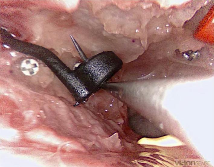
This endoscopic clinical photograph depicts an endonasal surgical procedure within a sinus cavity, likely the maxillary or sphenoid sinus. The visual field shows the moist, erythematous, and textured mucosal lining of the nasal passage. A specialized surgical instrument, appearing as a matte black fine wound retractor with a hooked tip, is actively manipulating a small, ring-shaped foreign object (EDGES™ Zig Aligna™). Several metallic, cross-headed surgical screws or navigation markers are embedded into the surrounding bony landmarks to assist with intraoperative navigation. The image demonstrates a precision task in endoscopic sinus surgery, focusing on the retrieval of small objects from deep paranasal structures. The educational focus highlights surgical dexterity, the use of specialized instrumentation in confined anatomical spaces, and the integration of surgical navigation systems in Otolaryngology (ENT).
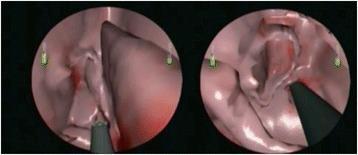
This Virtual Reality (VR) surgical simulation image displays two endoscope-view frames of the sinonasal cavity during Functional Endoscopic Sinus Surgery (FESS) tasks. The left frame illustrates an ethmoidectomy, showing the computer-rendered pinkish-white mucosal surfaces of the ethmoid air cells. A microdebrider, characterized by its gray shaft and dark suction-cutting tip, is seen interacting with the soft tissue within the narrow surgical corridor. The right frame illustrates a sphenoidotomy, showing a wider cavity corresponding to the sphenoid sinus. Both views utilize a 0-degree endoscope perspective and include small green vertical indicators on the periphery for orientation. Reddish shading on the virtual mucosa mimics clinical tissue manipulation or minor hemorrhage. This simulation material is designed for Otolaryngology (ENT) education to train medical students and residents on the anatomical landmarks and instrument handling required for safe sinus surgery, specifically focusing on the microdebrider's use near critical structures like the skull base and lamina papyracea.
harmonic scalpel vessel sealing LigaSure thyroid neck dissection ENT
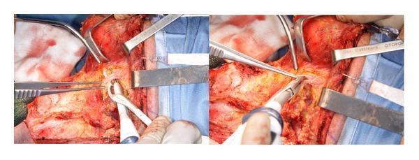
This clinical photograph displays an intraoperative view of a neck dissection, specifically focusing on levels II and III. The image shows a side-by-side comparison of surgical maneuvers using an Ultracision harmonic scalpel. In the surgical field, multiple metal retractors are positioned to provide exposure of the deep cervical structures, including the plane of the internal jugular vein and the sternocleidomastoid muscle. The white, curved-tip harmonic scalpel is seen actively engaging with the facial vessels for ultrasonic sealing and dissection. Forceps are used concurrently for tissue manipulation and tension. The surrounding tissue demonstrates detailed dissection of fibrofatty and muscular layers, with the blue surgical drape defining the sterile perimeter. This visual serves to illustrate advanced hemostatic techniques in otolaryngological surgery, highlighting the replacement of conventional hand-tie ligation with harmonic technology for vessel sealing and tissue coagulation.
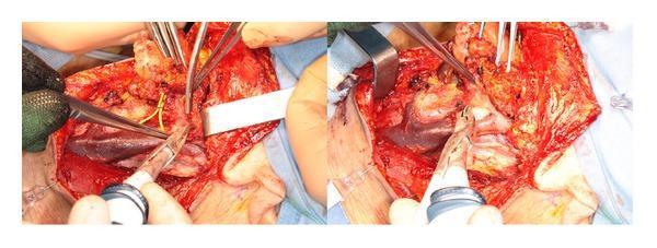
This clinical photograph shows an intraoperative view of a neck dissection (levels II-III) performed on a human patient. The surgical field focuses on the plane of the internal jugular vein (IJV), which is clearly visible as a large, dark-purple vascular structure. The procedure demonstrates the use of a Focus Ultracision harmonic scalpel (HS) for tissue dissection and hemostasis. Key anatomical features include the internal jugular vein and its associated tributary vessels, which are shown being sealed and divided using the ultrasonic device. The harmonic scalpel's curved paddle blade is positioned near the vascular structures, illustrating coaptive coagulation. Surgical instruments in the field include the harmonic scalpel, multiple fine surgical forceps used for tissue retraction and exposure, and a metal retractor maintaining the surgical window. Visible yellow nerve structures are being carefully isolated using clamps to avoid injury. The image serves as an educational resource for head and neck surgery, specifically illustrating the application of ultrasonic energy for vessel sealing and precise dissection along critical vascular planes during oncological or diagnostic neck procedures.
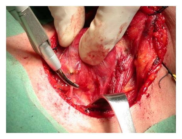
A close-up clinical photograph showing an intraoperative view of a total thyroidectomy. The surgical site displays exposed deep neck tissues, including the thyroid lobe being medially rotated by a surgeon's gloved hand. A Focus Ultracision Harmonic Scalpel, characterized by its metallic body and curved active blade tip, is positioned to seal and divide the inferior thyroid artery. The surgical field exhibits characteristic features of active dissection: red vascularized tissue, fibrous bands, and some peripheral bruising at the skin incision edges. A metal retractor is visible at the bottom of the frame, maintaining exposure of the operative field, while black silk sutures are noted at the superior aspect for tissue traction or marking. This image demonstrates the application of ultrasonic energy for simultaneous cutting and coagulation (hemostasis) in head and neck surgery, specifically targeting significant vascular structures like the thyroid arteries to minimize blood loss and operative time.
suture material absorbable vicryl chromic surgical ENT tonsil wound closure
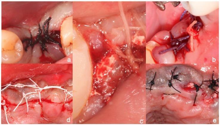
A composite of five clinical intraoral photographs (a–e) demonstrating different surgical suture materials used for wound closure in dental or maxillofacial surgery. Each panel illustrates specific material properties and knotting techniques: (a) Black braided silk suture showing a multifilament texture with multiple throws; (b) Violet-colored Vicryl (polyglactin 910), a coated synthetic absorbable suture; (c) Translucent gut suture, a natural collagenous material; (d) White PTFE (polytetrafluoroethylene) monofilament suture, characterized by its non-wicking, hydrophobic appearance and simple interrupted knots; (e) Dark-colored monofilament polyamide (nylon) suture. The images focus on tissue approximation, suture bite size, and the physical interaction between the thread and the oral mucosa. This visual guide serves as a pedagogical resource for identifying suture types by color, texture, and handling characteristics while highlighting clinical application in periodontal and surgical sites.
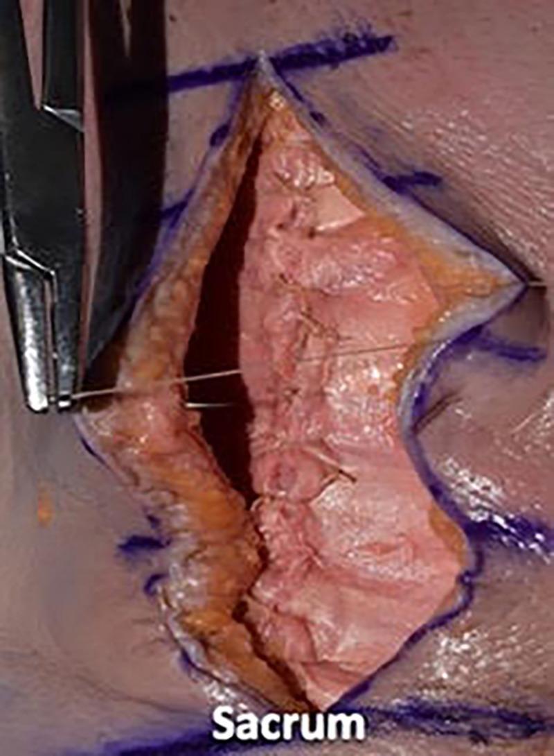
A clinical photograph demonstrating an intermediate stage of surgical wound closure in the lumbar region of a cadaveric specimen. The image shows a deep, longitudinal midline incision near the sacrum, with skin and subcutaneous fat layers retracted. The underlying deep fascial layer has been reapproximated using simple interrupted sutures, while the current step focuses on dermal closure. Specifically, 2-0 Vicryl sutures are being placed in an inverted, interrupted pattern to create buried knots, facilitating the reapproximation of the dermal edges without piercing the epidermal surface. Blue preoperative surgical markings are visible on the surrounding skin to guide the incision and alignment. A needle driver is shown on the left side of the frame, actively manipulating the suture material. This visual illustrates surgical techniques for layered wound closure, emphasizing deep-to-superficial tissue management and the use of absorbable sutures for dermal layers.
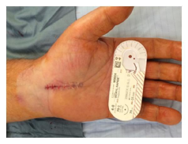
A clinical photograph of a human hand showing a post-operative carpal tunnel release incision. The linear incision, approximately 3 cm in length, is located on the proximal palmar surface, extending from the distal wrist crease toward the mid-palm. The wound is closed using interrupted horizontal mattress sutures with visible, absorbable multifilament material (Vicryl Rapide), as indicated by the suture packaging placed adjacent to the hand for scale and material identification. The surgical site displays mild perisutural erythema and localized bruising distal to the incision line, which are typical early inflammatory findings. A transparent adhesive film dressing partially covers the proximal aspect of the surgical site. The image demonstrates standard surgical technique for carpal tunnel decompression and immediate postoperative wound appearance using absorbable suture material for skin closure in hand surgery.
coblation tonsillectomy microdebrider ENT outcomes
harmonic scalpel vessel sealing head neck surgery
Surgical Devices & Suture Materials in ENT: A Detailed Modern Guide
1. COBLATION (Controlled Ablation)
What It Is
Key point: This is NOT a heat-based device. It is plasma-mediated tissue dissolution.
Temperature Comparison
| Method | Operative Temperature |
|---|---|
| Monopolar electrocautery | ~400°C |
| Harmonic scalpel | 70-80°C |
| Coblation | ~60°C |
| Cold steel | 0°C (no heat) |
How It Works (Mechanism)
- RF current flows between two electrodes on the wand tip
- Current passes through normal saline applied to the tissue surface
- This excites the sodium ions into a plasma field (thin layer of ionized particles)
- Plasma field breaks molecular bonds in tissue at LOW temperature
- Hemostasis occurs by forming a protein coagulum/plug in cut vessel ends
- Built-in suction removes water, blood, and debris
ENT Applications
- The Coblation wand is the only device that can perform both tonsillectomy and adenoidectomy with the same instrument
- Can do extracapsular (subcapsular) tonsillectomy - removing tissue outside the capsule entirely
- Can do intracapsular tonsillotomy - partial removal leaving the capsule, reducing pain and bleeding risk (especially preferred in children for sleep-disordered breathing)
- Significantly less postoperative pain vs electrocautery; comparable to cold steel
- A Cochrane review of 9 trials comparing coblation with other techniques found no definitive pain advantage over cold steel, but consistent advantage over electrocautery
- Submucosal (intraturbinoplasty): Wand inserted into the turbinate submucosa, ablating from inside, preserving mucosal surface
- Surface (extraturbinoplasty): Direct mucosal application, visible ablation
- Preferred because it preserves ciliary function better than electrocautery
- Uvulopalatoplasty (LAUP/CAPSO) for snoring/sleep apnea
- Tongue base reduction for OSA
- Nasal polypectomy
- Glomus tympanicum tumor excision (Salem et al., 2024 - demonstrated feasibility with no recurrence at 1 year)
- Nasal valve therapy (TCRF - Targeted Coblation of Nasal Valve) - 2025 RCT (Han et al.) confirmed 3-year durability for nasal airway obstruction
Wand Design
- Contains both suction and bipolar cautery in the same instrument
- Malleable curved shaft - easy maneuverability
- Integrated saline delivery (some models)
- Power is variable - lower settings for ablation, higher for coagulation
- Larger vessels not controlled by plasma can be managed by the wand's built-in electrocautery (use lowest effective setting to limit collateral damage)
Advantages
- Lowest thermal spread of all energy-based devices in ENT
- Less postoperative pain than electrocautery
- Minimal scarring
- Can do intracapsular tonsillotomy (especially useful in pediatric OSA)
- Single instrument for both tonsil and adenoid
- Office-based procedures possible (e.g., turbinate reduction, nasal valve)
Disadvantages / Limitations
- More expensive per-use than cold steel
- Slightly slower than electrocautery
- Cochrane review (9 RCTs) found no definitive superiority over cold steel regarding pain
- Learning curve for correct tissue plane identification
2. MICRODEBRIDER (Powered Shaver)
What It Is
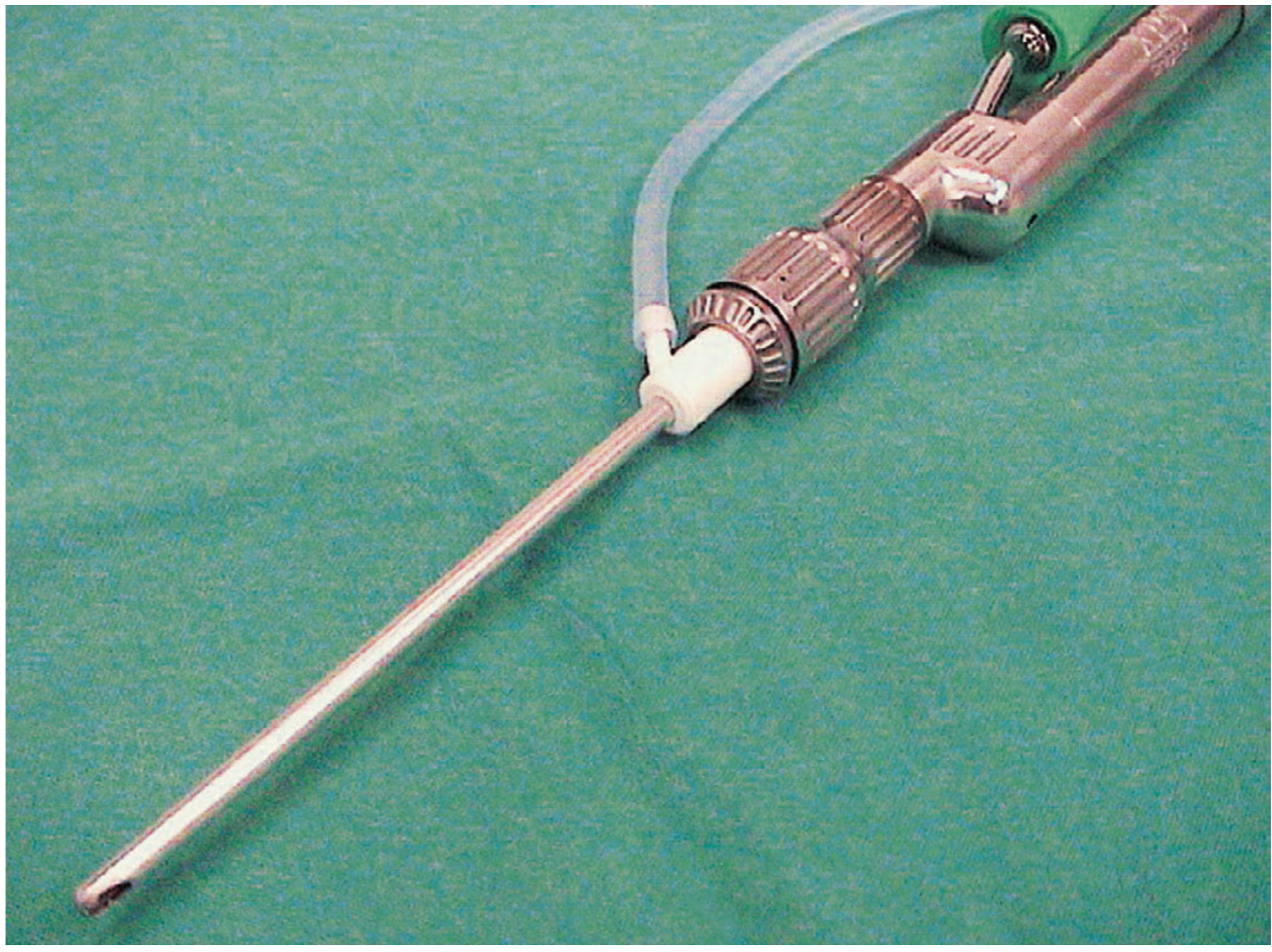
Mechanism
- A motor-driven inner rotating blade spins at adjustable speeds (typically 500-6000 RPM)
- The tissue is drawn into the aperture by suction
- The rotating blade cuts and aspirates simultaneously
- Tissue is collected in a suction trap for histopathology if needed
- No heat is generated - purely mechanical cutting
- Blade tips come in multiple angles (0°, 40°, 60°, 90°) and sizes for different anatomical areas
ENT Applications
- The primary workhorse of modern FESS
- Removes polyps, diseased mucosa, and soft tissue with precision
- Used for ethmoidectomy, maxillary antrostomy, sphenoidotomy
- Allows surgeon to maintain endoscopic visualization while removing tissue
- Avoids stripping of healthy mucosa compared to older forceps techniques
- Integrated with image guidance (navigation) systems for real-time positional feedback during complex sinus or skull base surgery
- Removes polyps while simultaneously clearing the surgical field with suction
- Histological sample collection possible
- Reduces need for multiple instrument exchanges
- Endoscopically controlled adenoidectomy under direct visualization
- Allows complete, precise removal compared to blind curettage
- Reduces adenoid regrowth rates
- A 2026 comparative cohort study (Prince Sultan Military Hospital) confirmed comparable outcomes between microdebrider and coblation for endoscopic adenoidectomy
- Turbinoplasty (submucosal tissue removal)
- Juvenile nasal angiofibroma debulking
- Inverted papilloma surgery
- Skull base and pituitary surgery (with image guidance)
- Laryngeal papillomatosis (laryngeal microdebrider blades)
- Benign laryngeal lesion removal
Microdebrider Components
| Component | Function |
|---|---|
| Handpiece | Motor-driven housing |
| Rotating inner cannula | Cutting element |
| Outer sheath | Guard, protects surrounding tissue |
| Blade tip | Interchangeable, various angles |
| Suction tubing | Continuous tissue aspiration |
| Speed control | Variable RPM (oscillate/rotate modes) |
| Irrigation port | Some models have saline irrigation |
Key Blade Types
- Straight blades: Anterior nasal cavity, inferior turbinate
- Angled blades (40-90°): Middle meatus, ethmoid sinus, adenoid
- Soft tissue blade: Polyps, mucosa
- Bone blade/rasp: Bony partitions (though less common - usually preserved)
- Laryngeal blades: For airway papillomatosis (smaller diameter)
Advantages
- No thermal damage (purely mechanical)
- Clear operating field via constant suction
- Precise - removes diseased tissue while preserving healthy mucosa
- Histopathological sampling possible
- Works alongside rigid endoscopy
- Multiple blade options for any ENT cavity
Disadvantages
- No hemostatic capability - bleeding must be managed separately
- Requires constant monitoring to avoid injury to critical structures (orbit, skull base, optic nerve, carotid)
- Cost of disposable blades
- Requires powered console unit
3. VESSEL SEALING DEVICES
A. Harmonic Scalpel (Ultrasonic Energy Device)
- Uses high-frequency ultrasonic vibrations (55,000 Hz / 55 kHz)
- A piezoelectric transducer converts electrical energy to mechanical vibration
- The vibrating blade denatures proteins in tissue (creates a protein coagulum that seals vessel ends)
- Also generates frictional heat (70-80°C at blade tip) which contributes to hemostasis
- Cuts and coagulates simultaneously - single-instrument hemostatic dissection
- Reliably seals vessels up to 3-5 mm diameter
- Larger vessels (>5 mm) may need supplemental ligation
- Tonsillectomy - one of the most popular techniques; cuts and seals in one pass along the peritonsillar plane
- Total/Hemi-thyroidectomy - seals superior/inferior thyroid vessels, reduces need for ties
- Parotidectomy - careful dissection and hemostasis around facial nerve
- Neck dissection (levels I-VI) - seals fascial and vascular pedicles, replaces hand-ties for smaller vessels
- Laryngectomy/Pharyngolaryngectomy - vessel management
- Oral cavity tumor excision
- Active blade: 70-80°C
- Lateral thermal spread: 0-3 mm (much less than monopolar electrocautery)
- Hemostasis via protein denaturation and coagulum, not tissue charring
- Hook blade: Fine dissection, superior tonsillar pole, thin tissue
- Flat/paddle blade: Better hemostasis, inferior tonsillar pole (major vessels), thyroid
- Curved blade: Angled access
B. LigaSure / Advanced Bipolar Vessel Sealers
- Uses advanced bipolar radiofrequency energy with pressure
- The device applies controlled RF energy while a tissue feedback system (impedance monitoring) automatically cuts off energy when sealing is complete
- Fuses collagen and elastin in vessel walls, creating a permanent, durable seal
- The sealed segment can then be cut (some devices have integrated blade)
- Can reliably seal vessels up to 7 mm diameter (greater than harmonic)
- Suitable for larger named vessels
- Thyroid surgery: Sealing superior thyroid pedicle, inferior thyroid artery - has replaced traditional ties in many centres
- Neck dissection: Sealing vascular pedicles during level II-IV dissection
- Tonsillectomy: Less common than harmonic but used in some centres
- Salivary gland surgery
- Parathyroid surgery
| Feature | Harmonic Scalpel | LigaSure / Bipolar Sealer |
|---|---|---|
| Energy type | Ultrasonic vibration | Advanced bipolar RF |
| Mechanism | Protein denaturation + heat | Collagen/elastin fusion |
| Max vessel size | 3-5 mm | 5-7 mm |
| Temperature | 70-80°C | Lower (adaptive feedback) |
| Lateral spread | 0-3 mm | Minimal (feedback-controlled) |
| Cutting | Simultaneous cut+seal | Seal then cut (or integrated blade) |
| Cost | Moderate | Higher |
| Best for | Fine dissection, tonsil, thyroid | Larger pedicle vessels, thyroid |
C. General Advantages of Vessel Sealers Over Traditional Ligation
- No suture material left in wound
- Faster operative time (fewer tie-cut steps)
- Reduced postoperative infection risk (less foreign material)
- Proven hemostasis for vessels ≤5-7mm
- Reduced blood loss in thyroid and neck dissection surgery
4. SUTURE MATERIALS IN ENT
Classification
SUTURES
├── ABSORBABLE
│   ├── Natural (Plain Gut, Chromic Gut)
│   └── Synthetic (Vicryl, PDS, Monocryl, Vicryl Rapide, Biosyn)
└── NON-ABSORBABLE
    ├── Natural (Silk, Linen, Cotton)
    └── Synthetic (Nylon/Prolene, Mersilene, PTFE/Gore-Tex, Stainless Steel)
ABSORBABLE SUTURES
Natural Absorbable
| Suture | Material | Absorption Mechanism | Duration | ENT Use |
|---|---|---|---|---|
| Plain Gut | Sheep/cattle submucosa (collagen) | Enzymatic (proteolysis) | 7-10 days | Rarely used now; oral mucosa, minor lacerations |
| Chromic Gut | Collagen + chromic salt treatment | Enzymatic (slower) | 10-21 days | Tonsil bed hemostasis (figure-of-eight), oral mucosa, skin (pediatric) |
- Chromic suture is the traditional choice for tonsil bed bleeding control using figure-of-eight sutures after cold steel tonsillectomy
- The chromic salt treatment reduces antigenic response and slows absorption
- Disadvantage: unpredictable absorption, tissue reaction, smell during degradation
Synthetic Absorbable (Preferred in Modern ENT)
| Suture | Material | Brand | Absorption | Strength Duration | Common ENT Use |
|---|---|---|---|---|---|
| Polyglactin 910 | Braided polyester (glycolide/lactide) | Vicryl | Hydrolysis | 3-4 weeks | Closure of pharyngeal mucosa, tonsil fossa, neck deep closure, parotid |
| Polyglycolic acid | Braided PGA | Dexon | Hydrolysis | 3-4 weeks | Similar to Vicryl |
| Poliglecaprone 25 | Monofilament | Monocryl | Hydrolysis | 1-3 weeks | Skin closure in neck (cosmetic), paediatric |
| Polyglactin 910 Rapide | Faster-absorbing Vicryl | Vicryl Rapide | Hydrolysis | 7-10 days | Oral mucosal closure, tonsil, adenoid |
| Polydioxanone (PDS) | Monofilament | PDS II | Hydrolysis | 4-6 weeks | Thyroid/laryngeal cartilage repair, deep neck layers, fascial closure |
| Polyglyconate | Monofilament | Biosyn/Maxon | Hydrolysis | 3-4 weeks | Fascial closure, neck dissection |
- Laryngotracheal reconstruction (cartilage approximation)
- Thyroid and parathyroid surgery deep layers
- Fascial closure in neck dissection
- Scenarios requiring prolonged tissue support (4-6 weeks tensile strength)
NON-ABSORBABLE SUTURES
| Suture | Material | Properties | ENT Use |
|---|---|---|---|
| Silk | Braided natural protein | Easy handling, excellent knot security, soft | Tying pedicles (older practice), vessel ligation (traditional thyroid/neck surgery), skin |
| Nylon (Ethilon) | Monofilament polyamide | Strong, inert, minimal tissue reaction, slight memory | Neck/facial skin closure, septoplasty intradermal |
| Polypropylene (Prolene) | Monofilament | Most inert, low tissue drag, excellent vascular use | Vessel anastomosis (microvascular free flap), skin closure, cartilage suture (otoplasty) |
| PTFE (Gore-Tex) | Expanded PTFE | Non-wicking, soft, no tissue reaction | Laryngeal framework surgery, vocal cord medialization, tympanoplasty |
| Mersilene | Braided polyester | Strong, permanent | Cartilage support, facial suspension sutures, fascia lata |
| Stainless steel wire | Metal | Maximum strength, permanent | Mandible/maxilla fixation (after orthognathic or trauma), tracheostomy tube fixation |
Suture Size Selection in ENT
| Size | Application |
|---|---|
| 6-0 / 7-0 (Prolene, Nylon) | Microsurgery - free flap vessel anastomosis, facial nerve repair |
| 5-0 (Prolene, Nylon) | Fine facial skin closure, delicate repairs |
| 4-0 (Vicryl Rapide, Monocryl) | Oropharyngeal mucosal closure, tonsil fossa |
| 3-0 (Vicryl, PDS) | Pharynx, thyroid deep closure, neck muscle |
| 2-0 / 1-0 (Vicryl, PDS, Silk) | Fascial closure, drain fixation, larger pedicle ligation |
| 2-0 Chromic / Vicryl | Tonsil bed figure-of-eight for bleeding control |
Modern Trends in ENT Suture Use
- Reduced suture use overall - vessel sealers (harmonic, LigaSure) have replaced many traditional ties
- Shift from natural to synthetic absorbables - Vicryl/PDS preferred over Chromic gut (more predictable absorption, less reaction)
- Barbed sutures (V-Loc, Quill) - increasingly used in ENT for pharyngeal/neck closure, especially after total laryngectomy, to provide evenly distributed tension without knot-tying
- Absorbable sutures for all intraoral/pharyngeal closure - no non-absorbable materials in saliva-contaminated fields
- Prolene monopoly in microvascular ENT - free flap reconstruction uses exclusively 8-0 to 9-0 prolene for vessel anastomosis
- PTFE for laryngeal framework surgery - thyroplasty, cricothyroid approximation
Quick Comparison Summary
| Device | Energy | Temperature | Primary ENT Use | Key Advantage |
|---|---|---|---|---|
| Coblation | Bipolar RF + saline (plasma) | 60°C | Tonsil, adenoid, turbinate, OSA surgery | Lowest thermal spread, intracapsular option |
| Microdebrider | Mechanical (no energy) | None | FESS, polyps, adenoid, larynx | Precise cutting + suction, no thermal damage |
| Harmonic Scalpel | Ultrasonic vibration | 70-80°C | Tonsil, thyroid, neck dissection | Cut + seal simultaneously, fast |
| LigaSure/Bipolar Sealer | Advanced bipolar RF | Feedback-controlled | Thyroid, neck pedicles | Largest vessel sealing (up to 7mm) |
- Pfenninger and Fowler's Procedures for Primary Care, 3rd Ed. (Chapter 83: Tonsillectomy and Adenoidectomy)
- Cummings Otolaryngology Head and Neck Surgery, (Chapter on Surgical Techniques)
- Bailey and Love's Short Practice of Surgery, 28th Ed. (FESS/Nasal Polyps)
- Schwartz's Principles of Surgery, 11th Ed. (Pharyngeal and Adenotonsillar Disease)
- Salem et al. (2024) - Coblation for glomus tympanicum tumors (retrospective, 28 patients)
- Han et al. (2025) - 3-year RCT of TCRF nasal valve treatment (Aetna/PubMed)
- Lao J et al. (2025) - Tonsillectomy vs Tonsillotomy meta-analysis [PMID: 39295458]