
hunter syndrome
Hunter syndrome MPS II mucopolysaccharidosis clinical features
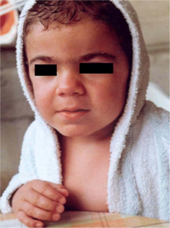
Clinical photograph of a 3-year-old child illustrating the early phenotypic manifestations of Mucopolysaccharidosis type II (MPS II), also known as Hunter syndrome. The image focuses on the patient's head and upper torso, demonstrating mild coarsening of facial features characteristic of lysosomal storage disorders. Key visual findings include a slightly flattened midface with a depressed nasal bridge, full and rounded cheeks, and thickened lips, particularly the upper lip. The hair appears somewhat coarse and dark. The eyes are obscured for patient privacy. This clinical visual serves as an educational tool for medical professionals to recognize subtle facial dysmorphism that precedes the more severe somatic and neurological symptoms of MPS II. The identification of such features at an early age is critical for timely diagnostic intervention and metabolic evaluation in pediatric populations.
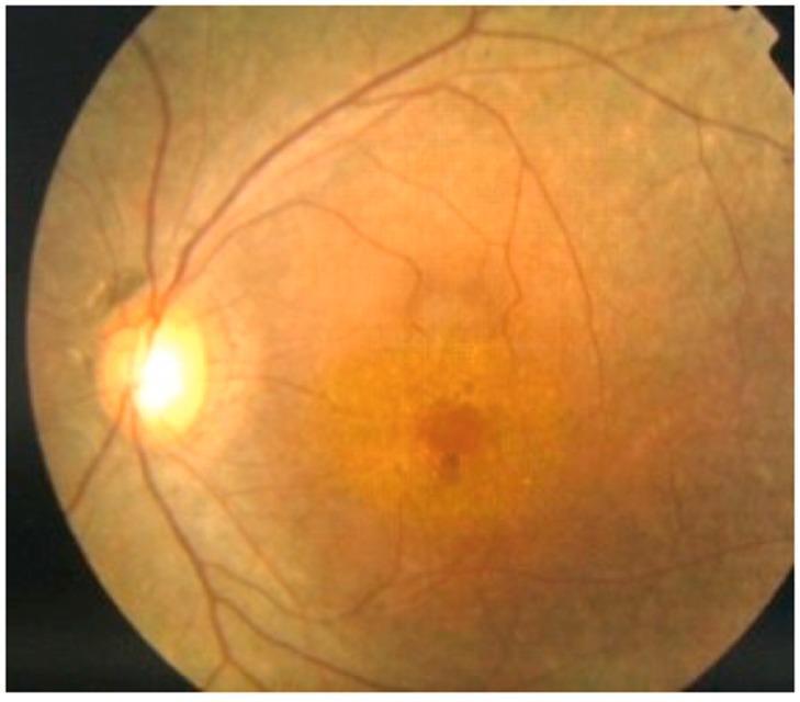
This clinical photograph displays a fundus view of the retina, identifying key diagnostic features associated with retinitis pigmentosa in a patient with Mucopolysaccharidosis Type II (MPS II or Hunter Syndrome). The image shows a well-defined optic disc with clear margins. The retinal vasculature exhibits significant attenuation, with thinning of both the arterioles and venules as they radiate toward the periphery. Centrally, the macula demonstrates a pigmented lesion and areas of atrophy, indicating macular involvement. The background retina reveals uneven pigmentation with a granular appearance, characteristic of tapetoretinal degeneration. These visual findings provide clinical evidence of retinopathy, which is a significant posterior chamber manifestation of MPS II, leading to nyctalopia and progressive visual field loss. The imaging is essential for ophthalmologists monitoring metabolic disorders where glycosaminoglycan (GAG) deposition can impact ocular structures.
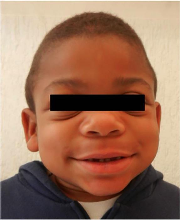
This clinical photograph of a pediatric patient demonstrates the classic 'coarse facial features' characteristic of mucopolysaccharidosis (MPS), specifically Hunter syndrome (MPS II). The visible dysmorphic features include a broad, flattened nasal bridge and a wide nasal tip. There is significant mid-face fullness with large, rounded cheeks and a prominent forehead. The lips are notably thickened (macrocheilia), and the patient exhibits a wide smile that reveals slightly irregular dental spacing. The overall facial contour is rounded with a lack of sharp definition, typical of the storage-related soft tissue and bony changes seen in lysosomal storage disorders. The ears appear slightly low-set. This image serves as a teaching tool for recognizing the phenotypic manifestations of metabolic diseases involving glycosaminoglycan accumulation.
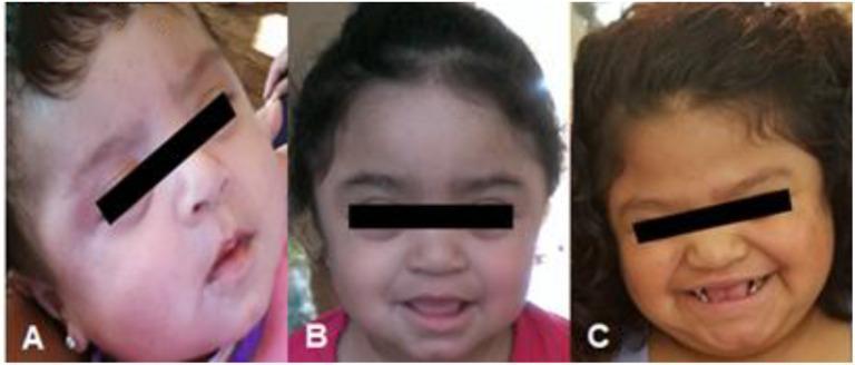
A series of three longitudinal clinical photographs (A, B, C) documenting the progressive facial phenotype of a female patient with Mucopolysaccharidosis type II (MPS II), or Hunter syndrome. Image A shows the patient as an infant with subtle facial features. Image B depicts the patient as a toddler, where a transition toward coarser facial features begins to emerge. Image C shows the patient at the time of presentation with the classic 'coarse' facies characteristic of severe MPS II. Visible features include a thickened, prominent forehead, a flattened nasal bridge with a broad nose, full lips, and macroglossia. Notable dental findings in image C include widely spaced, peg-shaped teeth and gingival hypertrophy. The hair appears progressively coarser and thicker throughout the timeline. This progression illustrates the metabolic accumulation of glycosaminoglycans (GAGs) in connective tissues, leading to the distinctive dysmorphic features used in the clinical diagnosis of lysosomal storage diseases.
"Hunter syndrome" OR "mucopolysaccharidosis II" treatment
Hunter Syndrome (Mucopolysaccharidosis Type II)
At a Glance
| Feature | Detail |
|---|---|
| Type | MPS II |
| Inheritance | X-linked recessive |
| Deficient enzyme | Iduronate-2-sulfatase (iduronate sulfate sulfatase) |
| Gene | IDS gene |
| Accumulated substrate | Dermatan sulfate + heparan sulfate |
| Affected | Almost exclusively males |
Pathophysiology
- Emery's Elements of Medical Genetics and Genomics
Clinical Features
Age of Presentation
Systemic Findings
- Coarse facial features - broad flat nose, full lips, thickened gingiva, prominent forehead, large tongue (macroglossia)
- Hepatosplenomegaly
- Joint stiffness with limited range of motion
- Short stature and high body mass index
- Large head circumference (macrocephaly)
- Hearing loss (both conductive and sensorineural)
- Recurrent upper respiratory infections
- Diarrhea
- Abnormal vertebral shape on spinal radiographs
Neurological
- Progressive intellectual deterioration (in severe form)
- Behavioral problems
- Death usually in adolescence in severe cases; milder forms can survive into adulthood
Dermatologic (Classic and Distinctive)
-
Extensive Dermal Melanocytosis (Mongolian spots) - large, atypical Mongolian spots in unusual locations (not just over sacrum/buttocks); often the first presentation to a dermatologist in infancy, when children are otherwise asymptomatic
-
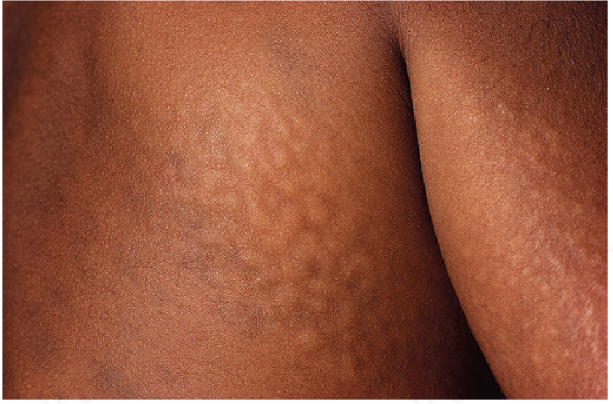
Pebbly/Cobblestone skin lesions - occur in ~10% of patients on the upper back, neck, chest, proximal arms, or thighs. They are firm, flesh-colored to white papules and nodules that coalesce into a cobblestone or reticular pattern (connective tissue nevus)
- Histology: increased dermal mucin + metachromatic granules in dermal fibroblasts, eccrine sweat glands, and epidermal keratinocytes
- Andrews' Diseases of the Skin
Ocular
- Retinitis pigmentosa pattern with retinal atrophy and vascular attenuation
- Progressive visual field loss and night blindness
Clinical Photographs

Diagnosis
- Urine GAG screening - elevated dermatan sulfate + heparan sulfate
- Enzyme assay - absent or markedly reduced iduronate-2-sulfatase activity in leukocytes or serum (confirmatory)
- Molecular genetics - direct IDS gene analysis
- Emery's Elements of Medical Genetics and Genomics; Andrews' Diseases of the Skin
Treatment
Enzyme Replacement Therapy (ERT)
- Idursulfase (Elaprase) - intravenous weekly infusions; the main disease-modifying treatment
- Reduces urinary GAG excretion, improves somatic features (organomegaly, joint mobility, respiratory function), and slows disease progression
- A 2024 systematic review (PMID 39201256) confirmed ERT with IV idursulfase reduces hepatosplenomegaly and improves functional outcomes but has limited CNS penetration (does not address neurological decline in severe phenotype)
Hematopoietic Stem Cell Transplantation (HSCT)
- Can be useful in appropriately evaluated patients, especially when done early
- Better neurological outcomes if performed before significant cognitive decline
Intrathecal / CNS-targeted therapy
- Emerging investigational approaches include intrathecal iduronate-2-sulfatase and antibody-enzyme fusion proteins targeting the transferrin receptor to improve CNS delivery (early efficacy data - Goodman & Gilman's; [Okuyama et al., 2019])
- A 2025 network meta-analysis (PMID 40317013) across MPS subtypes examined comparative efficacy of ERT vs. HSCT vs. combination strategies
Supportive
- Hearing aids, airway management (tracheostomy if needed), cardiac surveillance (valve disease is common), orthopedic interventions for joint disease, neurodevelopmental support
Comparison: Severe vs. Attenuated Phenotype
| Feature | Severe (MPS IIA) | Attenuated (MPS IIB) |
|---|---|---|
| CNS involvement | Severe progressive | Minimal or absent |
| Life expectancy | Usually die in adolescence | May survive into adulthood |
| Somatic features | Marked | Moderate |
Key Distinguishing Points vs. Hurler Syndrome (MPS I)
- Hunter is X-linked recessive (Hurler is autosomal recessive)
- Hunter accumulates dermatan + heparan sulfate (Hurler accumulates the same, but different enzyme: iduronidase)
- Hunter classically lacks corneal clouding (corneal clouding is a hallmark of Hurler)
- Hunter has the distinctive pebbly skin lesions and atypical Mongolian spots