
Atrial tachycardia
I'll read the medical library skill first, then research atrial tachycardia comprehensively.
Reading File
Now I'll run parallel searches across the library for atrial tachycardia content.
Finding Sources
Finding Sources
Searching PubMed
"atrial tachycardia"[MeSH Terms] AND management
Excellent. I have multiple high-quality sources. Now I'll read the key sections in parallel from Braunwald's, Goldman-Cecil, Fuster & Hurst, and Rosen's.
Reading File
Reading File
Reading File
Reading File
Now let me read the Braunwald's reentrant section and get the Guyton physiology entry, plus the management details from Fuster & Hurst, in parallel.
Reading File
Reading File
Loading Image
I now have comprehensive material from multiple authoritative sources. Let me compile the full answer.
Atrial Tachycardia (AT)
Definition
Atrial tachycardia is a group of supraventricular tachycardias (SVTs) arising from focal anatomic locations in the atria or great cardiac veins, with impulses propagating in a centrifugal pattern - distinct from sinus tachycardia, atrial flutter, and atrial fibrillation. The rate is typically >100 bpm and rarely exceeds 200 bpm. - Goldman-Cecil Medicine, p. 3266
Classification
| Type | Key Feature | Common Setting |
|---|---|---|
| Focal AT | Single site of origin; centrifugal spread | Any age; common near pulmonary veins, crista terminalis |
| Reentrant AT (Macroreentrant) | Circuit around fixed anatomic obstacle | Post-cardiac surgery, prior AF ablation scars |
| Multifocal AT (MAT) | ≥3 distinct P wave morphologies; irregular | Advanced pulmonary disease (COPD in ~60% of cases) |
| Paroxysmal AT | Sudden onset/termination | Young, otherwise healthy individuals |
Mechanisms
Three distinct electrophysiologic mechanisms underlie AT - Braunwald's Heart Disease, p. 492; Fuster & Hurst, p. 1141:
- Enhanced/abnormal automaticity - Spontaneous phase-4 depolarization in ectopic atrial myocytes outside the sinus node
- Triggered activity - Oscillations of membrane potential (early or delayed after-depolarizations), often drug/ischemia-related
- Reentry (micro or macro) - Micro-reentry in focal AT; macro-reentry around surgical scars or ablation lines in reentrant AT
Anatomic Origins
Common sites of focal AT cluster at: - Braunwald's Heart Disease, p. 479; Goldman-Cecil, p. 3266
- Pulmonary venous ostia (especially left superior pulmonary vein)
- Crista terminalis (right atrium)
- Atrial appendages (RA and LA) - associated with incessant tachycardia
- Tricuspid and mitral annuli
- Coronary sinus
- Atrial septa
- Aortomitral continuity
- Regions of surgical scar tissue
ECG Features
Focal AT:
- Regular rhythm at 100-200 bpm
- Discrete abnormal P waves (morphology differs from sinus P) with isoelectric intervals between them
- P wave axis and morphology reflect site of origin (e.g., negative P in inferior leads suggests inferior RA or septal origin)
- AV conduction ratio may be 1:1, 2:1, or higher depending on rate
- May show "warm-up" (gradual rate acceleration at onset) and "cool-down" at termination
- Long RP or short RP pattern depending on rate and AV conduction
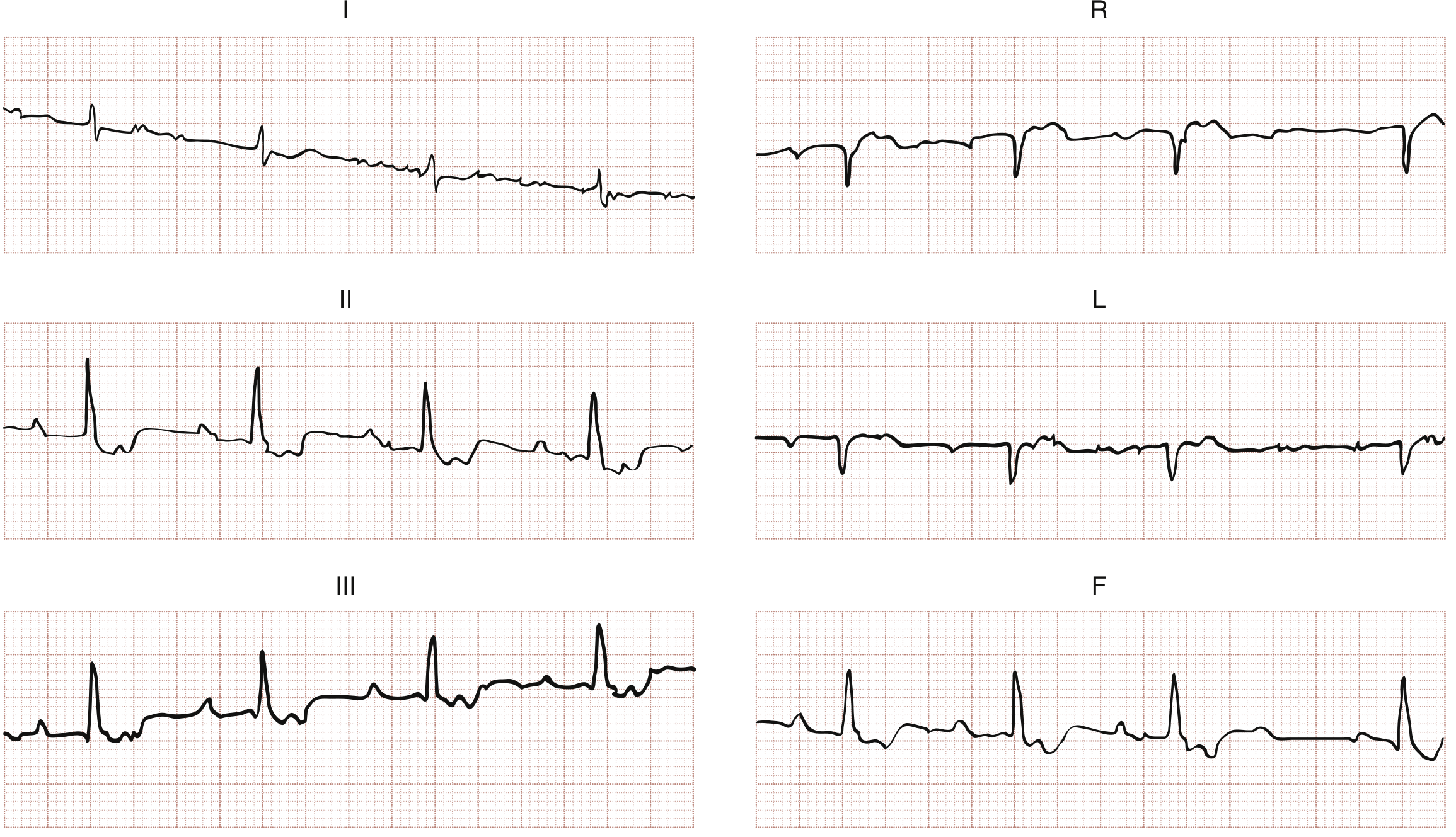
ECG showing atrial tachycardia with 2:1 AV conduction in a patient with digoxin toxicity - Rosen's Emergency Medicine
Multifocal AT:
- Irregular rhythm (often mistaken for AF)
- ≥3 distinct P wave morphologies
- Varying PR intervals and P-P intervals
- Distinguished from "wandering pacemaker" only by rate (MAT ≥100 bpm)
Clinical Presentation
Patients typically report: - Braunwald's Heart Disease, p. 492
- Palpitations
- Dizziness / presyncope (syncope unusual unless very rapid or structural heart disease present)
- Chest pain and dyspnea
- Fatigue
Special populations:
- AT is common in children and young adults with structural heart disease, often triggered by a premature atrial complex (PAC)
- In patients on digoxin, AT with 2:1 AV block is a hallmark sign of digoxin toxicity - Rosen's Emergency Medicine, p. 1052
- AT with rapid rates during sleep may be 40 bpm slower than waking rates
Tachycardia-Mediated Cardiomyopathy (TMC)
Incessant or near-incessant AT can cause a reversible dilated cardiomyopathy: - Braunwald's Heart Disease, p. 492
- Incidence ~10% in large focal AT series
- Occurs exclusively with incessant or very frequent paroxysmal tachycardia
- Affected patients: younger, more often male, slower mean tachycardia rate (~117 bpm)
- After successful ablation, LV function normalizes in virtually all patients
- Key locations causing incessant AT: RA/LA appendages and pulmonary venous ostia
Spontaneous Remission
- AT disappeared in 55% of patients under age 25 vs. only 14% of patients aged ≥26 after cessation of medical therapy - Braunwald's Heart Disease, p. 492
Management
Step 1 - Identify and Treat Precipitants
- Hypoxia/hypoxemia, electrolyte abnormalities (especially hypomagnesemia - give 2 g IV Mg over 5 min), drug toxicity (digoxin) - Rosen's Emergency Medicine, p. 1053
Step 2 - Acute Rate Control / Termination
- Vagal maneuvers and adenosine: unlikely to terminate AT (unlike AVNRT/AVRT), but useful to unmask atrial activity by transiently blocking AV conduction
- Beta-blockers: IV metoprolol for rate control
- Calcium channel blockers: IV diltiazem or verapamil for rate control
- DC cardioversion: can restore sinus rhythm acutely, but AT often recurs (especially MAT)
Step 3 - Chronic/Long-term Management
| Drug Class | Example | Note |
|---|---|---|
| Beta-blockers | Metoprolol 25 mg bid | First-line for rate control |
| Calcium channel blockers | Diltiazem 180 mg LA daily | First-line for rate control |
| Class IC antiarrhythmics | Flecainide, propafenone | Rhythm control; avoid with structural HD |
| Class III antiarrhythmics | Amiodarone, sotalol, dofetilide | Rhythm control; second-line |
- Goldman-Cecil Medicine, p. 3271
Catheter Ablation
- Increasingly offered as first-line therapy, especially for symptomatic or drug-refractory AT - Goldman-Cecil Medicine, p. 3271
- Uses activation mapping (earliest local activation precedes P wave onset by 15-40 ms) to localize the focus
- Success rate >90% with 3D electroanatomic mapping systems - Fuster & Hurst, p. 1141
- Phrenic nerve injury is a risk when ablating near the right superior pulmonary vein or right atrial appendage - must pace at high current to test for diaphragmatic contraction before ablation
- Multifocal AT is an important exception - very difficult to ablate; best managed by treating the underlying pulmonary/cardiac disorder
Anticoagulation
- Embolic events and stroke are rare with AT (unlike AF/flutter); anticoagulation is generally not indicated - Braunwald's Heart Disease
Special Case: Multifocal AT (MAT)
- Associated with advanced pulmonary disease in up to 60% (primarily COPD); also primary cardiac pathology
- Relatively unresponsive to antiarrhythmic drugs
- Cardioversion can restore sinus rhythm but MAT recurs quickly
- Treatment cornerstone: manage the underlying pulmonary disease
- If acute symptom control is essential: IV diltiazem, verapamil, or metoprolol
- Prognosis determined primarily by the underlying pulmonary disease - Goldman-Cecil Medicine, p. 3281
Key Distinctions from Other SVTs
| Feature | Focal AT | AVNRT | AVRT |
|---|---|---|---|
| P wave | Visible, abnormal morphology | Hidden in or just after QRS | Retrograde, shortly after QRS |
| Adenosine response | Transient AV block; AT continues | Terminates | Terminates |
| Warm-up/cool-down | Often present | Absent | Absent |
| Ablation target | Atrial focus | Slow pathway (AV node region) | Accessory pathway |
Sources: Braunwald's Heart Disease (15th ed.), Goldman-Cecil Medicine, Fuster & Hurst's The Heart (15th ed.), Rosen's Emergency Medicine, Guyton & Hall Textbook of Medical Physiology
Tell how the waves look in atrial tachycardia
The P wave morphology is the key ECG feature distinguishing AT from sinus tachycardia. Let me pull detailed waveform descriptions and ECG images from the textbooks.
Searching sources...
Finding Sources
Finding Sources
Good - I found several key sources and image URLs. Let me read the detailed waveform sections and fetch the ECG images in parallel.
Reading File
Reading File
Reading File
Loading Image
Now let me fetch the focal AT ECG from Braunwald's and the Harrison's diagnostic flowchart, plus the detailed P-wave localization text.
Loading Image
Loading Image
Reading File
I have all the material needed for a thorough, image-rich answer. Let me also grab the P-wave localization table from Braunwald's.
Reading File
Now I have everything needed for a detailed, image-rich answer on the ECG waves in atrial tachycardia.
ECG Waves in Atrial Tachycardia
The Core Principle
The entire ECG signature of AT revolves around one question: where is the P wave, what does it look like, and how does it relate to the QRS? The QRS and T waves are usually normal (the atria are sick, not the ventricles) - so the diagnostic action is entirely in the P wave.
1. The P Wave - The Defining Feature
Morphology
- The P wave is discrete and visible, with a shape different from the sinus P wave because the impulse spreads centrifugally from an ectopic atrial focus, not from the sinus node - Braunwald's Heart Disease, p. 492
- However, if the focus arises near the superior crista terminalis (high right atrium), the P wave may look almost identical to a sinus P wave - a key diagnostic trap
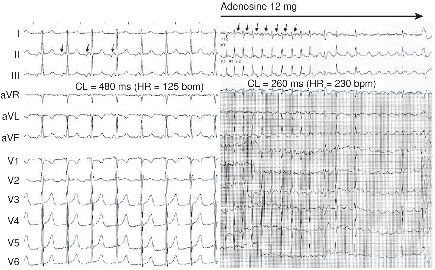
Left: Focal AT at 125 bpm. P waves are clearly visible before each QRS with an isoelectric interval. Note the biphasic negative-positive pattern in inferior leads - incompatible with sinus origin. This is a long RP tachycardia. Right: Rapid AT at 230 bpm after adenosine - AV conduction is blocked, but atrial tachycardia continues uninterrupted, proving the focus is in the atrium. - Braunwald's Heart Disease
P Wave Axis and Localization
The P wave axis tells you where the focus is - this is how electrophysiologists localise the tachycardia before ablation: - Braunwald's Heart Disease, p. 493
| Site of Origin | P wave in leads II, III, aVF | P wave in V1 | P wave in I/aVL |
|---|---|---|---|
| High right atrium (crista terminalis) | Upright (looks like sinus) | Biphasic or positive | Upright |
| Low right atrium / CS ostium | Inverted (negative) | Negative or flat | Variable |
| Left atrium (pulmonary veins) | Positive or biphasic | Positive/broad | Negative or flat |
| Left septal region | Biphasic negative-positive in inferior leads | Variable | Negative |
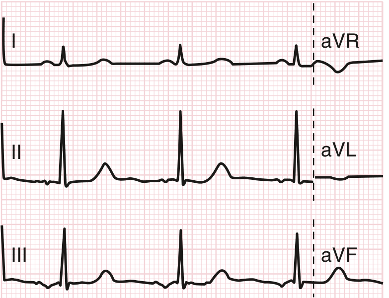
Ectopic atrial rhythm: Inverted P waves in leads II, III, and aVF indicate a non-sinus P wave originating in the low right atrium - Goldman-Cecil Medicine
2. The P-to-QRS Relationship (RP/PR Interval)
This is critical for distinguishing AT from AVNRT and AVRT: - Braunwald's Heart Disease, p. 493
Long RP Tachycardia (most common in AT)
- P wave comes well before the next QRS
- There is a clear isoelectric segment between the P wave and QRS
- The RP interval is longer than the PR interval
- Seen in slower ATs (e.g., 125-160 bpm)
Short RP Tachycardia (at rapid rates)
- At faster rates (e.g., 200-230 bpm), AV nodal conduction slows
- The P wave falls within or just after the preceding T wave
- The RP interval appears short, mimicking AVNRT
- Key differentiator: In AT, the RP-to-PR ratio can vary (called "unlinking") - this variability is virtually diagnostic of AT and does not occur in AVNRT/AVRT
The "Unlinking" Sign
- In AVNRT and AVRT, the VA (ventricle-to-atrium) time is fixed because the atria and ventricles are part of the same reentrant circuit
- In AT, the atria and ventricles are electrically independent - the tachycardia lives in the atrium and merely conducts through the AV node
- So the R-P interval can vary in AT even as the tachycardia rate stays constant - this is the key distinguishing feature
3. AV Conduction Ratio (P:QRS ratio)
Depending on the atrial rate, the AV node may not conduct every P wave: - Rosen's Emergency Medicine, p. 1052
| Atrial Rate | Typical AV Ratio | What you see on ECG |
|---|---|---|
| 100-160 bpm | 1:1 | Every P followed by a QRS |
| 160-200 bpm | Often 1:1, sometimes 2:1 | Every other P conducts |
| >200 bpm | 2:1 or higher | Multiple P waves per QRS |
- Digoxin toxicity classically produces AT with 2:1 AV block - a hallmark pairing on the ECG - Rosen's Emergency Medicine, p. 1052
4. The Isoelectric Interval
- In focal AT, there is a clear flat baseline between P waves (unlike atrial flutter where the baseline is never flat due to the sawtooth pattern)
- At very rapid rates, this isoelectric interval disappears and the tracing can resemble flutter
- The presence of an isoelectric interval strongly favours focal AT over macroreentrant AT or atrial flutter
5. Warm-Up and Cool-Down
A distinctive dynamic feature of AT: - Braunwald's Heart Disease, p. 492
- Warm-up: The rate gradually accelerates over 3-5 beats at the onset of the tachycardia (due to enhanced automaticity)
- Cool-down: The rate gradually slows over 3-5 beats at termination
- This is in contrast to AVNRT/AVRT which start and stop abruptly (on/off switch)
- Sudden onset is still possible in AT, particularly triggered activity forms
6. QRS Complex
- Usually narrow and normal - because ventricular conduction is unaffected
- Can become wide (aberrant) if there is rate-related bundle branch block or pre-existing BBB - this creates a wide-complex tachycardia that can mimic VT
- The QRS morphology itself gives no clue to the atrial origin
7. Multifocal AT (MAT) - A Special Pattern
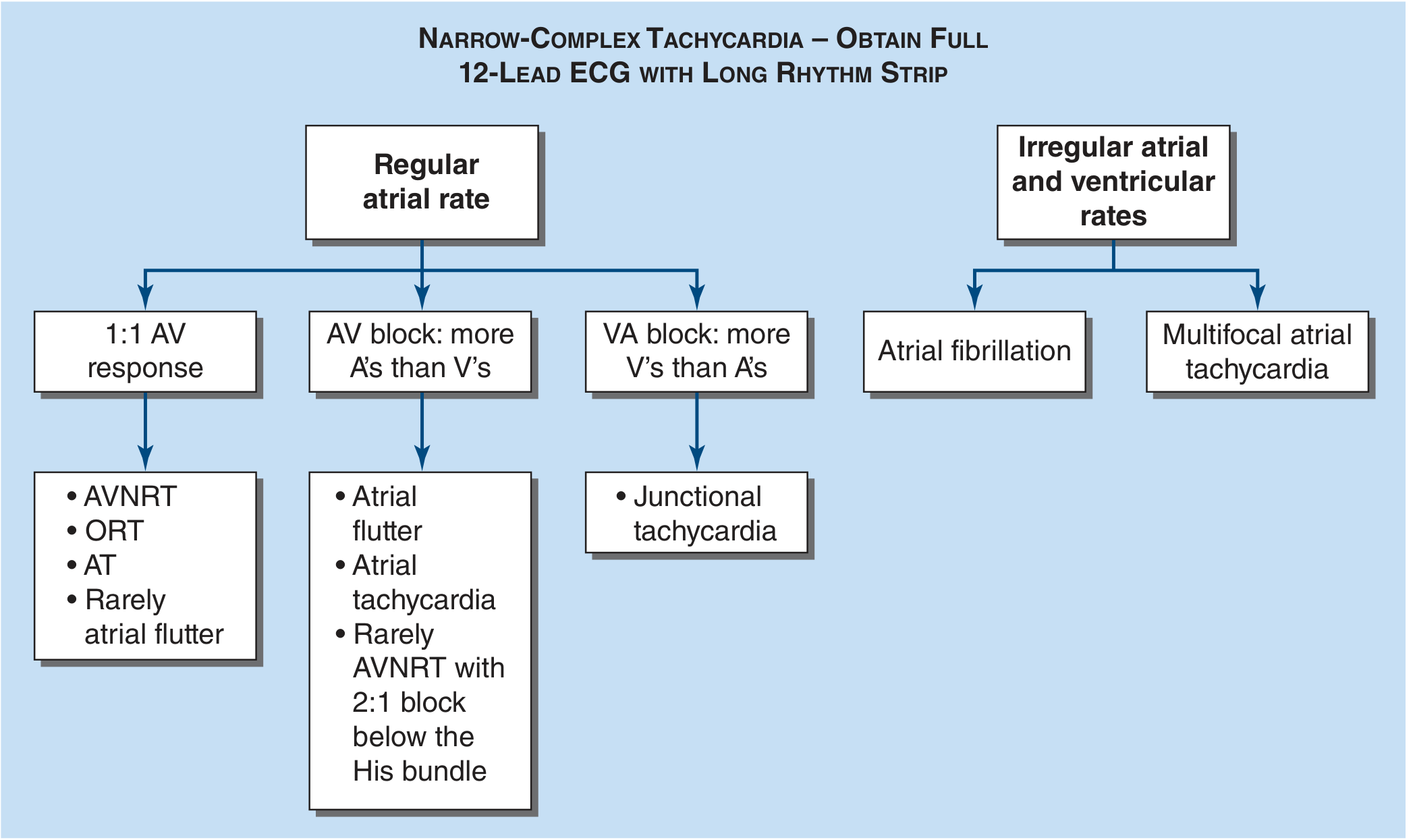
Diagnostic algorithm for narrow-complex tachycardia: MAT falls under "irregular atrial and ventricular rates" - Harrison's Principles of Internal Medicine
MAT has a completely different ECG appearance:
- Irregularly irregular rhythm - the hallmark feature (often confused with AF)
- Three or more distinct P wave morphologies in the same lead
- Varying PR intervals and P-P intervals between beats
- Rate >100 bpm (below 100 it is "wandering atrial pacemaker")
- Key differentiator from AF: in MAT, discrete P waves are visible with isoelectric intervals between them; in AF, the baseline is chaotic with no true P waves
Summary Table: AT vs Other SVTs on ECG
| Feature | Focal AT | AVNRT (typical) | AVRT | Atrial Flutter |
|---|---|---|---|---|
| P wave visible | Yes, discrete | Hidden in QRS | Just after QRS | Sawtooth, no isoelectric |
| P wave axis | Abnormal (non-sinus) | Retrograde | Retrograde | Fixed pattern |
| RP interval | Long or short, variable | Very short (<70 ms), fixed | Short-medium, fixed | N/A |
| Isoelectric baseline | Yes | N/A | N/A | No (sawtooth) |
| Warm-up/cool-down | Often | No | No | No |
| Adenosine effect | AV block; AT continues | Terminates | Terminates | Transiently unmasks flutter |
| Rate | 100-250 bpm | 150-250 bpm | 150-250 bpm | ~300 bpm atrial |
Sources: Braunwald's Heart Disease (15th ed.), Goldman-Cecil Medicine, Harrison's Principles (22nd ed.), Rosen's Emergency Medicine
This is a shared conversation. Sign in to Orris to start your own chat.