
tibia vara disease blound disease
Reading File
Blount disease (tibia vara) is a developmental condition causing progressive varus (bow-leg) deformity of the tibia due to abnormal growth of the medial proximal tibial physis. It primarily affects children, especially those who are obese or of African descent, and presents in two main forms: infantile (ages 1-3) and adolescent (ages 8-15).
Searching sources...
Finding Sources
Searching PubMed
"Blount disease" OR "tibia vara"
Good - I have sources from Campbell's Operative Orthopaedics 15th Ed (2026), Bailey & Love's Surgery, and Grainger & Allison's Radiology. Let me read all the key sections in parallel.
Reading File
Reading File
Reading File
Reading File
Now let me get the main Campbell's content on the clinical details and the Langenskiold classification.
Finding Sources
Finding Sources
Let me read the key clinical content section from Campbell's (the Langenskiold staging and clinical details section):
Reading File
Reading File
Reading File
Loading Image
Loading Image
Now let me get the Langenskiold staging detail and adolescent treatment section:
Reading File
I have all the information I need. Let me also fetch the Langenskiold staging image:
Loading Image
I now have all the material needed for a comprehensive answer. Here is a complete, well-organized overview of Blount disease (tibia vara):
Blount Disease (Tibia Vara / Osteochondrosis Deformans Tibiae)
Blount disease, first described by Walter Putnam Blount in 1937, is a progressive, pathological varus (bow-leg) deformity of the proximal tibia caused by disordered enchondral ossification of the posteromedial proximal tibial physis. The etiology remains unknown but is thought to involve abnormal mechanical loading of an already susceptible medial growth plate.
Classification: Two Distinct Forms
| Feature | Infantile | Adolescent |
|---|---|---|
| Onset | Before age 8 (typically 1-3 yrs) | Age 8 to skeletal maturity |
| Laterality | Bilateral 60%, Unilateral 40% | More often unilateral |
| Ethnicity | More common in Afro-Caribbean children | Affects all ethnic groups |
| Deformity course | Progressive without treatment | Worsens with time |
| Post-surgical recurrence | ~80-90% in children >4 yrs; <20% in children <4 yrs | Higher recurrence rate than infantile form |
| Inciting event | None | None |
(Campbell's Operative Orthopaedics 15th Ed 2026)
Pathophysiology
The disease involves abnormal mechanical stress on the medial tibial physis, leading to:
- Irregular and sloped medial physis
- Medial metaphyseal fragmentation (pathognomonic for progressive tibia vara)
- Progressive medial epiphyseal changes and eventual physeal bridge formation
- Concurrent femoral varus is a significant component in late-onset disease (average ~10 degrees of excess femoral varus, accounting for 34-76% of the total genu varum deformity)
Special variant - in severely obese children, an atraumatic "slip" of the proximal tibial epiphysis on the metaphysis can occur (dome-shaped metaphysis, inferomedial epiphyseal translation), which requires different treatment than conventional Blount disease.
Langenskiold Staging (Infantile Form)
Langenskiold described 6 progressive stages of epiphyseal change (Fig. 34.34 from Campbell's 2026):
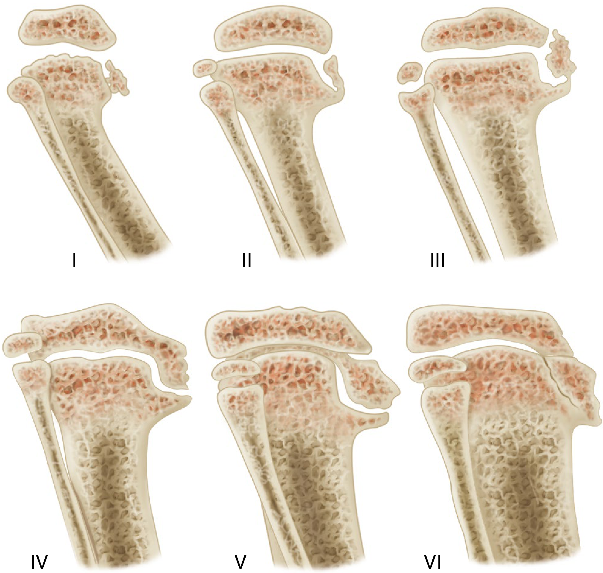
| Stage | Radiographic Features |
|---|---|
| I | Medial beaking of metaphysis, mild irregularity of physis |
| II | Medial metaphyseal beak more prominent, epiphysis wedged |
| III | Small medial ossification center fragment, physis slopes medially |
| IV | Medial epiphysis depressed below metaphysis, physis very irregular |
| V | Medial physis nearly fused, double epiphyseal contour |
| VI | Medial epiphysis fuses at 90-degree downward angle - complete physeal bar |
Stages I-II: may still correct spontaneously or with bracing; Stages III+: surgical intervention typically required.
Clinical Presentation
- Progressive, worsening genu varum (bow-legging) at or just below the knee
- Often associated with internal tibial torsion and significant in-toeing
- Palpable medial metaphyseal projection (though not itself diagnostic)
- Normal limb length early; leg length discrepancy develops in unilateral cases
- Children are typically obese and/or of African descent (infantile form)
Radiographic Findings
Standing full-leg radiographs are mandatory. Key features:
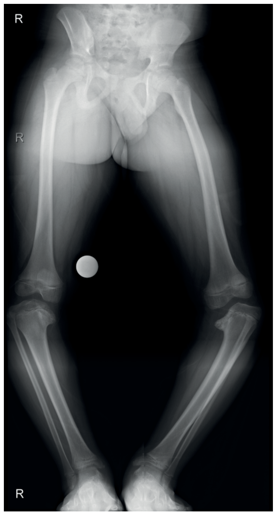
Bilateral Blount disease - both proximal medial tibial physes/epiphyses are abnormal (Bailey & Love's Surgery 28th Ed)
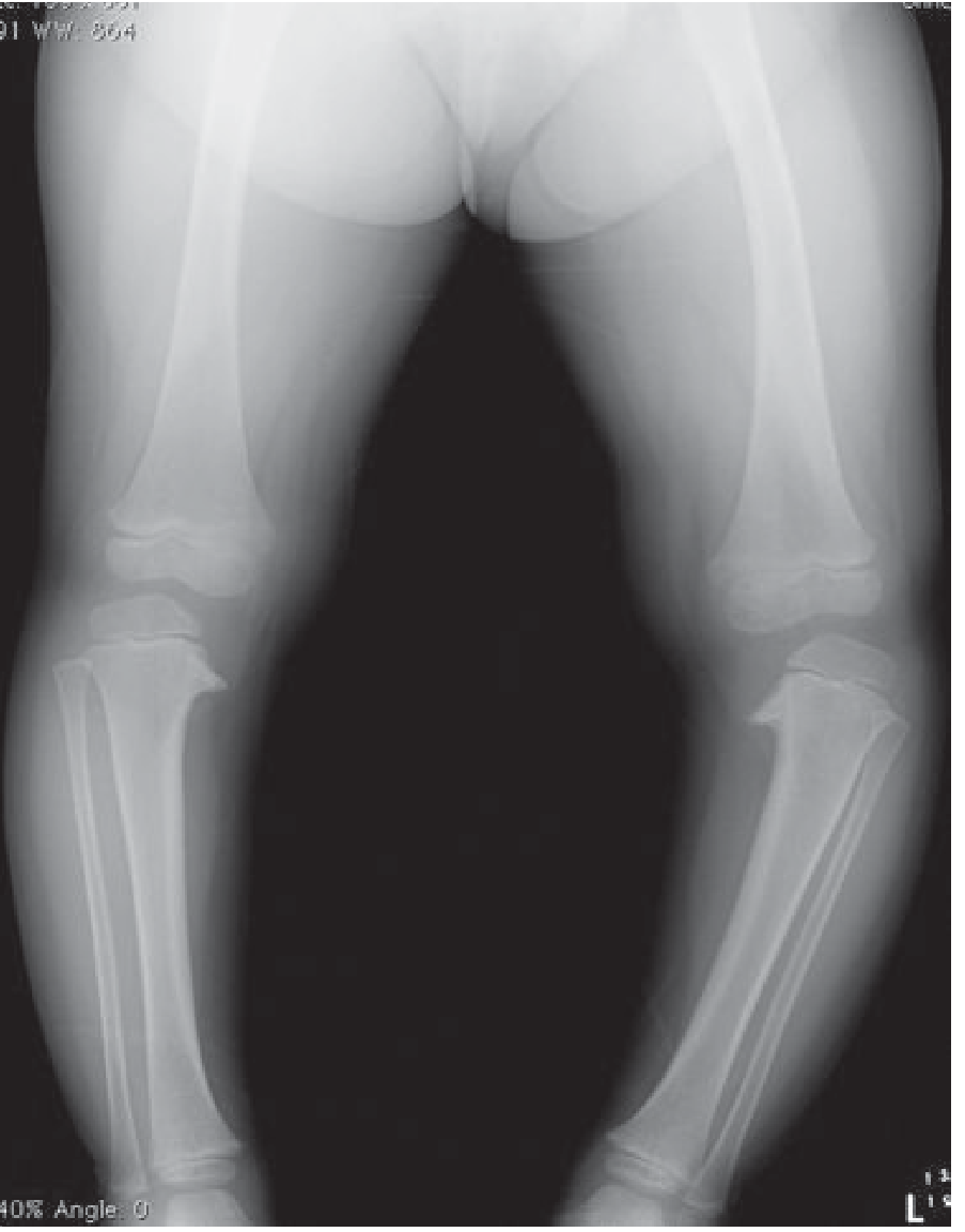
Blount disease: fragmentation of medial half of the left proximal tibial epiphysis with varus angulation (Grainger & Allison Radiology)
Key radiographic indicators:
- Metaphyseal-diaphyseal (Drennan) angle >11 degrees - most important early indicator
- Medial tibial epiphysis: short, thin, wedged
- Irregular, medially sloped physis
- Medial metaphyseal fragmentation (pathognomonic for progressive disease)
MRI findings (when needed):
- Increased thickness of medial chondroepiphysis
- Increased height and width of medial meniscus
- Abnormal medial femoral epiphysis
- Late resolution of tibial bowing
Differential Diagnosis of Tibia Vara
Other causes of tibial varus bowing that must be excluded:
- Physiological bowing (bilateral, self-resolving, 100% bilateral)
- Rickets
- Trauma / Infection
- Neurofibromatosis
- Ollier disease / Maffucci syndrome
- Fibrous dysplasia
- Focal fibrocartilaginous dysplasia (pathognomonic linear radiolucency, usually self-resolving)
(Grainger & Allison's Diagnostic Radiology)
Treatment
Infantile Blount Disease
| Age / Severity | Treatment |
|---|---|
| Ages 2-3 years (early disease) | Observation or KAFO bracing (knee-ankle-foot orthosis) |
| >3 years with progressive deformity | Valgus osteotomy of proximal tibia and fibula with mild overcorrection |
| Any age (growing support) | Guided growth with lateral tension band plates (eight-plate technique) |
Key points on osteotomy:
- Recurrence: ~80-90% in children >4 years, <20% in children <4 years
- Valgus osteotomy with mild overcorrection is recommended for children <4 years
- For more severe/older cases: combined tibial + fibular osteotomy; circular external fixation (Ilizarov) for complex multiplane deformities
Guided Growth (eight-plate/tension band):
- Minimally invasive; growing evidence base
- Implant failure requiring return to OR in ~12% of patients
- Mechanical failures more common in obese patients; four-screw or two eight-plate constructs if needed
- Recurrence after plate removal is related to slower medial physis growth rate
Adolescent Blount Disease
- Hemiepiphysiodesis (lateral physeal stapling or tension band plate) if sufficient growth remains
- Tibial osteotomy techniques include:
- Single-plane oblique osteotomy (Rab) - allows simultaneous varus + internal rotation correction
- Crescent-shaped chevron osteotomy (Greene) - maintains limb length
- Opening-wedge proximal tibial osteotomy
- Ilizarov circular external fixator with distraction osteogenesis for severe/complex deformities
- Femoral osteotomy may be needed in late-onset cases with significant femoral varus component
- Concomitant tibial lengthening (with external fixator) is often used for unilateral cases with shortening
For Langenskiold Stage V-VI (physeal bar present): physeal bar resection may be attempted if >50% of physis remains viable; otherwise, epiphysiodesis of the remaining physis at time of osteotomy to prevent recurrence.
Complications
- Deformity recurrence (main concern, especially in older children)
- Limb length discrepancy
- Lateral knee instability
- Premature osteoarthritis of the knee (if deformity uncorrected into adulthood)
- A 2025 systematic review (PMID 39987393) specifically examined total knee arthroplasty outcomes in adults with Blount disease or Blount-like deformity, confirming the long-term joint consequences of untreated disease.
Sources: Campbell's Operative Orthopaedics 15th Ed 2026 | Bailey and Love's Short Practice of Surgery 28th Ed | Grainger & Allison's Diagnostic Radiology
metaphysseal diaphyseal angle tibia
The metaphyseal-diaphyseal angle (MDA), also called the Drennan angle, is formed between a line perpendicular to the tibial longitudinal axis and a line through the two "beaks" of the proximal tibial metaphysis. An angle >11 degrees is the key early indicator distinguishing Blount disease from physiological bowing.
Searching sources...
Finding Sources
Searching PubMed
"metaphyseal diaphyseal angle" AND "tibia vara"
Good - there are two slightly different thresholds quoted (>11 degrees in Campbell's, >16 degrees in Miller's). Let me read both sections fully to clarify.
Reading File
Reading File
Reading File
Loading Image
Loading Image
Excellent - I have the diagrams from both textbooks. Now here is a complete answer:
Metaphyseal-Diaphyseal Angle (MDA) of the Tibia
Also Known As: Drennan Angle
The metaphyseal-diaphyseal angle (MDA) is a radiographic measurement on AP standing radiographs of the leg, used primarily to distinguish Blount disease (tibia vara) from physiological bowing in young children. It was described by Levine and Drennan (J Bone Joint Surg, 1982).
How to Measure It
Two lines are drawn on the AP radiograph of the proximal tibia:
- Line 1: Perpendicular to the longitudinal axis of the tibial diaphysis
- Line 2: Through the two "beaks" (medial and lateral prominences) of the proximal tibial metaphysis - this represents the transverse axis of the tibial metaphysis
The angle between these two lines = the metaphyseal-diaphyseal angle.
The diagrams below illustrate the technique compared to the tibiofemoral angle:
(From Miller's Review of Orthopaedics, 9th Ed):
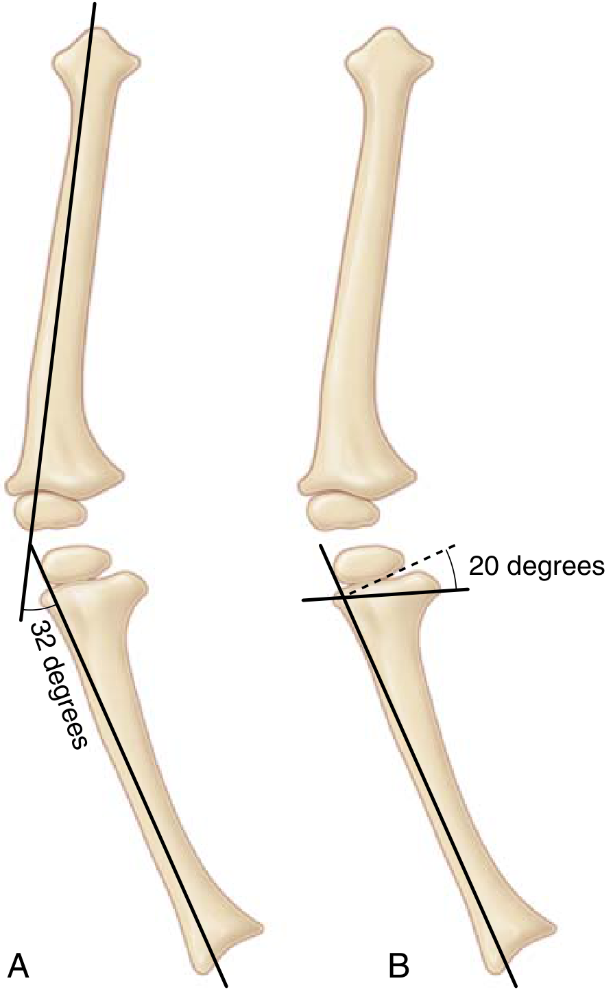
(From Campbell's Operative Orthopaedics, 15th Ed 2026):
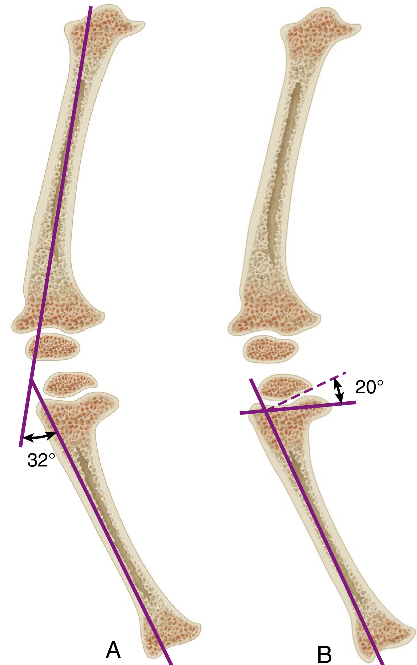
In both diagrams: (A) shows the tibiofemoral angle (32°) formed by the long axes of femur and tibia; (B) shows the MDA (20°) formed between the perpendicular to the tibial shaft and the metaphyseal beak line.
Interpretation / Cut-Off Values
| Source | Threshold | Interpretation |
|---|---|---|
| Campbell's Operative Orthopaedics 15th Ed 2026 | >11 degrees | Early indicator of Blount disease |
| Miller's Review of Orthopaedics 9th Ed | >16 degrees | Considered abnormal (Blount disease) |
Why the discrepancy? These reflect different thresholds used in the literature. The >11° threshold (Levine & Drennan, 1982) is the original, well-cited value for suspecting early Blount disease and prompting closer monitoring. The >16° threshold is used as the more definitive cut-off for calling the angle clearly pathological. An angle between 11-16° is a "gray zone" where the child needs serial radiographic follow-up rather than immediate intervention.
Physiological bowing generally shows <11 degrees and resolves spontaneously by age 3-4 years.
Clinical Significance
The MDA is most useful in children under 2 years where physiological bowing and early Blount disease look very similar on clinical examination.
Why the MDA beats the tibiofemoral angle in early disease:
- The tibiofemoral angle is influenced by both tibial AND femoral deformity, as well as patient positioning
- The MDA isolates the deformity specifically at the proximal tibial metaphysis - the site of Blount pathology
- It can detect early physeal involvement before Langenskiold stage changes are obvious
Key correlations:
- MDA >11°: warrants monitoring; one of four early indicators of Blount disease along with late MRI resolution of bowing, fibula-tibia length ratio, and severity of proximal vs. distal femoral angulation
- MDA >16°: strongly suggests pathological tibia vara
- Progressive increase in MDA on serial films: indicates active Blount disease requiring intervention
Limitations
- The angle has inter-observer variability, which is significant, especially when measured on digital images by less experienced observers
- It works best in the infantile form (ages <4 years); less useful in adolescent Blount disease where the metaphyseal beak may be less defined
- Must be measured on a true AP standing radiograph - rotation artifacts can falsely elevate or lower the angle
- Should always be interpreted alongside clinical findings (obesity, early walking, internal tibial torsion, progression of deformity)
Sources: Campbell's Operative Orthopaedics 15th Ed 2026 | Miller's Review of Orthopaedics 9th Ed (citing Levine & Drennan, J Bone Joint Surg 1982)
This is a shared conversation. Sign in to Orris to start your own chat.