
Stroma and parenchyma of the breast difference with pictorial
breast stroma parenchyma histology diagram
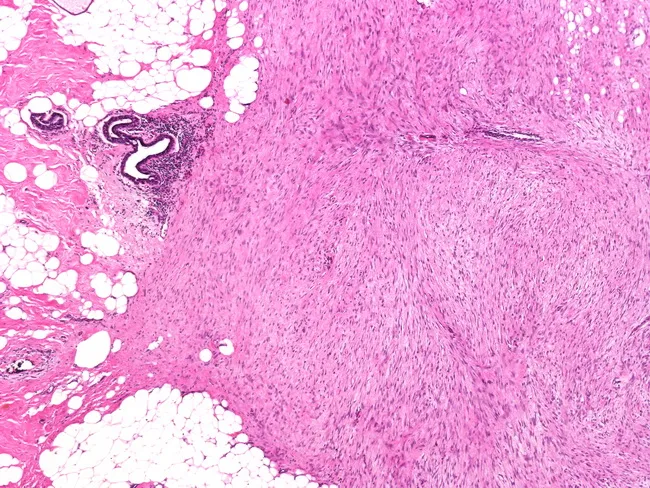
Breast tissue histology slide. Low-power observation reveals an ill-defined, infiltrative mass in mammary stroma with spindle cells arranged in sweeping fascicles and abundant dense collagen. The tumor abuts and extends into adjacent breast parenchyma and adipose tissue, producing stellate projections at the margins. Cellular morphology shows uniform, elongated spindle cells with slender elongated nuclei, minimal cytologic atypia, and low mitotic activity. The stroma is variably collagenized with scant vascularity. No overt epithelial malignancy is seen; ducts and lobules are entrapped by the desmoplastic, fibrous neoplasm. Immunohistochemistry (if performed) would favor a myofibroblastic phenotype with cytokeratin negativity and nuclear beta-catenin positivity supporting desmoid-type fibromatosis; smooth muscle actin (SMA) and vimentin positivity can be observed. The pattern can mimic carcinoma due to infiltrative growth and dense stroma, making correlation with clinical and radiologic findings essential. Clinically, fibromatosis of the breast presents as a firm, non-tender mass; radiographic appearance may mimic carcinoma; management requires wide local excision with negative margins to reduce recurrence. The lesion lacks metastatic potential but bears a notable risk of local recurrence. This image is educational for surgical pathology, spindle cell breast lesions, and radiologic-pathologic correlation in breast disease.
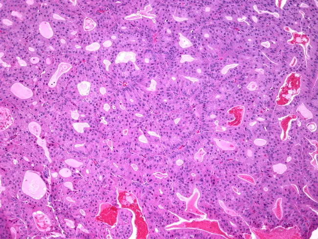
Breast tissue histology prepared as a formalin-fixed paraffin-embedded section stained with hematoxylin and eosin and examined by light microscopy. The specimen represents mammary parenchyma with ducts exhibiting papillary architecture consistent with papillary carcinoma; within several ducts there is apocrine metaplasia characterized by tall columnar cells with abundant eosinophilic granular cytoplasm and prominent apical snouts. The papillary fronds display fibrovascular cores, and the epithelial lining shows mild cytologic atypia without conspicuous mitotic activity in the field. The stroma is fibrous with scattered periductal lymphocytes; luminal spaces contain variable secretions. The differential diagnosis includes benign intraductal papilloma versus papillary carcinoma with apocrine differentiation; recognition of apocrine features is important to avoid misinterpretation as benign changes and to guide further workup. Clinically, papillary lesions of the breast require correlation with imaging and receptor studies; apocrine differentiation may be seen in older patients and can influence immunohistochemical profiling, including androgen receptor expression. This image is pertinent for education on breast tumor histopathology and serves as a reference for pathology residents, fellows, and radiology–pathology correlation cases. Potential applications include diagnostic training, differential diagnosis exercises, and quality assurance reviews in breast pathology. Correlation with gross findings and imaging enhances diagnostic confidence.
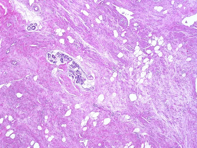
This case depicts a breast tissue histology slide prepared with Hematoxylin and Eosin stain, viewed at low to moderate magnification, illustrating a benign hamartomatous lesion (adenolipoma variant) within the mammary parenchyma. The lesion is well circumscribed, with a pseudocapsule created by compressed adjacent breast tissue. The mass consists of a heterogeneous admixture of mature adipose tissue intermingled with normal-appearing breast parenchyma, including ducts and lobules embedded in fibrous stroma. No cytologic atypia or mitotic activity is evident. The adipose component varies in proportion, reflecting the adenolipoma spectrum, while the epithelial elements form orderly acini and ducts surrounded by myoepithelial cells. The capsule-like margin and lack of invasion support a benign diagnosis. Clinically, hamartomas present as painless, movable breast nodules in premenopausal women but can occur across age groups. Radiologic imaging typically shows well-defined, round or oval masses with compressed breast tissue at the periphery. Pathological differential includes lipoma, fibroadenoma with lipomatous stroma, or other lipomatous or fibroepithelial lesions. Accurate histologic characterization is essential to distinguish from malignant neoplasms and to guide conservative surgical management when indicated. This description supports reproducible search terms for pathology education, breast disease registries, and radiology-pathology correlation studies to enhance case characterization and teaching.
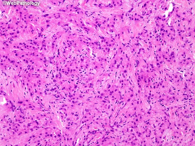
Histology by light microscopy of a breast tissue biopsy stained with Hematoxylin and Eosin (H&E). The specimen demonstrates invasive cells embedded in a dense desmoplastic stroma within the breast parenchyma. The primary neoplasm is described as invasive apocrine carcinoma, histiocytoid variant, characterized by plump epithelioid cells with abundant eosinophilic granular cytoplasm and prominent nucleoli, arranged in irregular cords and confluent nests. A differential diagnostic consideration is granular cell tumor, which can display similar eosinophilic granular cytoplasm but differs in cytologic detail and immunophenotype. In granular cell tumor, nuclei are small and hyperchromatic with minimal atypia, and tumor cells are strongly positive for CD68 and S-100, while negative for cytokeratin AE1/3. In contrast, apocrine carcinoma shows marked cytologic atypia, nuclear pleomorphism, and prominent nucleoli; immunophenotype typically demonstrates positivity for cytokeratin AE1/3 and is negative for CD68, with S-100 occasionally weakly positive. The biological significance lies in accurate classification, which guides prognosis and treatment. Clinically relevant implications include differential diagnosis between breast carcinoma with apocrine differentiation and granular cell lesion, interpretation in the context of receptor status, and implications for surgical management. An IHC panel including AE1/3, CD68, and S-100 is recommended to confirm lineage and differentiation, avoiding misdiagnosis and guiding therapy.
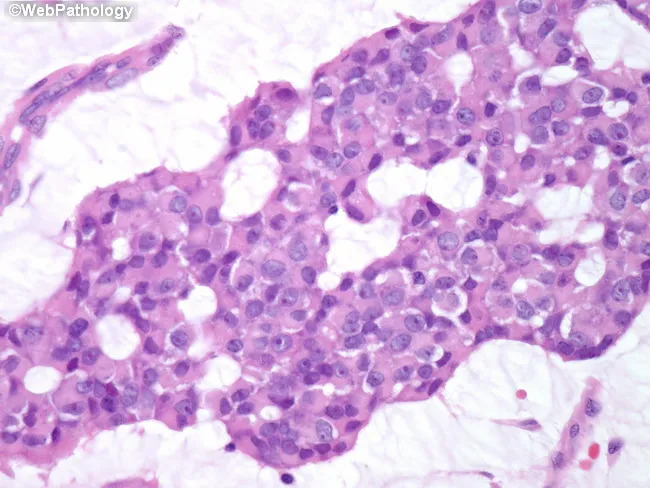
High-magnification bright-field histology image of breast parenchyma showing invasive mucinous (colloid) carcinoma with a mixture of solid and cribriform growth patterns. The tumor displays abundant extracellular mucin pools that create lakes in which clusters and occasional cords of neoplastic cells are dispersed. The neoplastic cells are relatively uniform, with small to medium-sized nuclei, round to oval contours, few conspicuous nucleoli, and minimal pleomorphism, consistent with well-differentiated histology. Mitotic activity is low and necrosis is not evident. Cribriform nests intersect the mucinous stroma, while solid foci demonstrate cohesive tumor islands embedded in mucinous matrix. In the surrounding breast parenchyma, fibrous stroma and adipose tissue may be present with mild stromal reaction but no extensive desmoplasia. The overall pattern aligns with mucin-producing adenocarcinoma of the breast, colloid type, which generally carries a favorable prognosis relative to conventional ductal carcinoma. Clinically relevant features include ER/PR positivity in many cases and a relatively indolent course; radiologic correlation may show a well-circumscribed, mucin-rich mass mimicking benign lesions. This image is valuable for pathology education, differential diagnosis with other mucinous and ductal carcinomas, and training in recognizing low-grade mucinous patterns at high magnification. Overall, this image captures characteristic mucin pools and uniform cellularity.
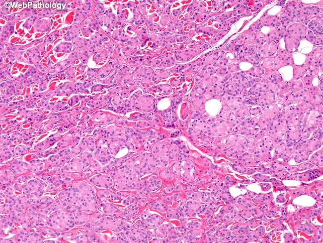
This image depicts a hematoxylin and eosin (H&E) stained histology slide of invasive apocrine carcinoma within breast parenchyma. Imaged with bright-field light microscopy at moderate magnification, the tissue shows nests and irregular cords of polygonal tumor cells infiltrating a dense, collagenous stroma. Cells characteristically possess abundant eosinophilic cytoplasm and apocrine-type features; nuclei are variably high in grade overall, though this exemplar demonstrates low-to-intermediate nuclear grade with occasional prominent nucleoli. The tumor nests are surrounded by desmoplastic stroma, and a chronic lymphocytic infiltrate is often present in the adjacent stroma. GCDFP-15 immunoreactivity is a supportive marker for apocrine differentiation, though it is not required to establish the morphologic diagnosis. The typical receptor profile includes estrogen receptor (ER) and progesterone receptor (PR) negativity with androgen receptor (AR) positivity; HER2 expression is negative in about half of pure apocrine carcinomas. Clinically, recognizing apocrine morphology and AR positivity can influence therapeutic considerations, including potential anti-androgen strategies, even when ER/PR are absent. Immunohistochemical workup is essential to distinguish from ductal carcinomas with apocrine features or metaplastic variants. This image exemplifies the morphological hallmarks and immunophenotype relevant to diagnosis, prognosis, and targeted treatment planning in breast oncology.
normal breast anatomy lobule terminal duct unit TDLU histology
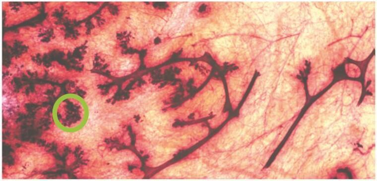
This whole mount preparation showcases the anatomical architecture of the adolescent human breast from a 15-year-old female. The image highlights a branching ductal network embedded within a matrix of translucent adipose tissue. The mammary ducts appear as dark, linear structures that radiate and subdivide throughout the tissue. Along the lateral edges and at the distal ends of these ducts are lobular structures characterized by increased staining density and granular morphology. A green circle highlights a Terminal Duct Lobular Unit (TDLU), which represents the primary functional unit of the breast where secretory activity occurs. The TDLU is visible as a compact, rounded cluster of buds emerging from a terminal duct. This histological overview illustrates the pubertal development of the mammary gland, specifically the expansion of the epithelial ductal system and the formation of complex terminal units in response to hormonal signaling. The clear visualization of the branching patterns and the high-contrast staining of the epithelial elements against the pale surrounding stroma provide an educational example of normal breast microanatomy during early reproductive development.
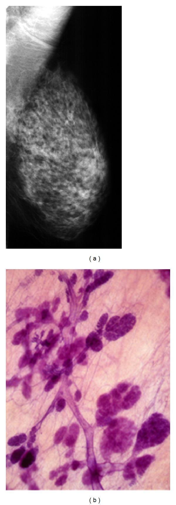
This composite educational material demonstrates a radiological-pathological correlation of normal breast anatomy. Image (a) is a mammogram of the left breast in a mediolateral oblique (MLO) projection. It illustrates nodular and oval-shaped opacities representing terminal duct lobular units (TDLUs), alongside linear densities corresponding to larger milk ducts. These structures are embedded within radiolucent (dark) adipose tissue, providing high natural contrast. Image (b) presents a large-section, subgross (3D) histology specimen stained with hematoxylin and eosin (H&E). The microscopy shows dark purple stained ductal networks and lobular clusters against a lighter pink connective tissue background. The comparison highlights how the 3D histological arrangement of lobules and ducts directly correlates with the radiographic densities seen on a mammogram. This material is designed for medical students and radiology residents to understand the structural basis of mammographic findings and the importance of large-section histology in visualizing contiguous breast anatomy.
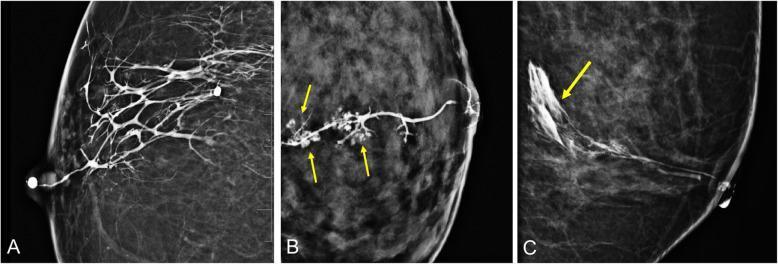
This composite of diagnostic images displays breast ductograms (galactograms) highlighting the ductal anatomy and contrast distribution. Image A shows a normal ductogram in a craniocaudal (CC) view, where radiopaque contrast material fills the primary lactiferous duct at the nipple and branches extensively throughout the breast parenchyma, demonstrating system patency. Image B is a close-up highlighting a 'lobular blush' (indicated by yellow arrows), which represents contrast material successfully filling the terminal ductal lobular units (TDLU), appearing as small, clustered, stippled areas of enhancement. Image C is a 90-degree mediolateral ductogram demonstrating iatrogenic extravasation (yellow arrow) where contrast material has leaked from the ductal system into the surrounding interstitial tissue, often caused by excessive injection pressure. These images are used in breast imaging to evaluate nipple discharge, identify intraductal lesions such as papillomas, and map ductal anatomy prior to surgical intervention. The modality is X-ray mammography following ductal cannulation and contrast injection.
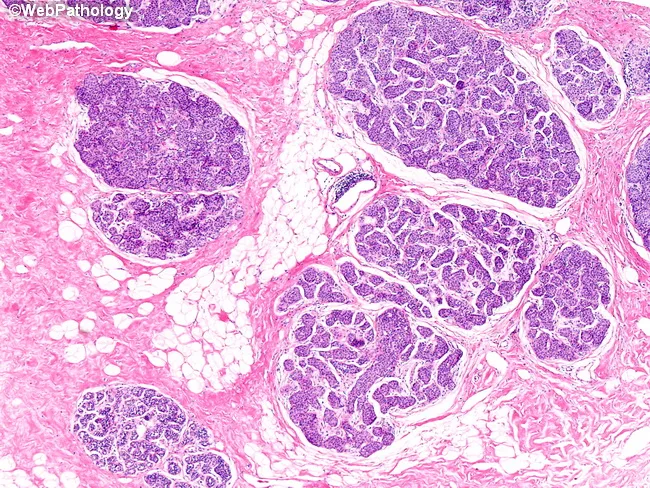
Imaging modality and technique: Light microscopy of hematoxylin and eosin stained formalin-fixed paraffin-embedded breast tissue. The field shows terminal duct-lobular units with lobules distended by uniform, medium-sized cells. Cells are noncohesive, with round to oval nuclei and minimal cytoplasm; chromatin is evenly distributed and nucleoli are inconspicuous. The architecture is intralobular, with proliferation confined to lobular acini and frequent preservation of the basement membrane, indicating lobular carcinoma in situ (LCIS). Involved lobules are expanded and packed with these cells, while an uninvolved lobule remains in the specimen. The lobular lesions are typically multicentric, as evidenced by multiple foci of LCIS in the same tissue fragment. Associated stroma is generally not desmoplastic, and there is no invasion into surrounding breast parenchyma. The appearance is consistent with classic LCIS: a diffuse, noninvasive proliferation that can be multicentric and bilateral in many cases. This histology correlates with the clinical concept that LCIS confers an elevated risk of subsequent breast carcinoma rather than an immediate invasive lesion. Recognition of this pattern is essential to distinguish LCIS from ductal carcinoma in situ and invasive lobular carcinoma, guiding surveillance strategies and chemoprevention decisions. Clinical implications include close radiologic surveillance and consideration of chemoprevention in high-risk patients.
breast fibrous stroma adipose tissue connective tissue anatomy
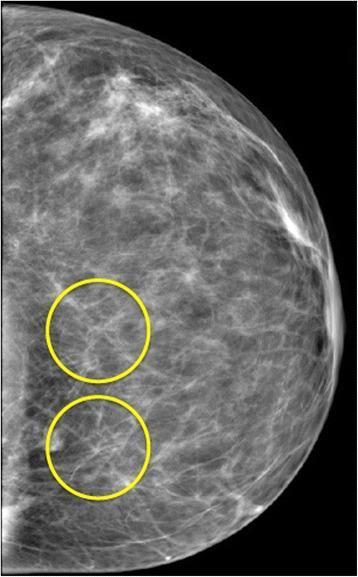
This diagnostic image is a mammogram, specifically a craniocaudal or mediolateral oblique view of the breast, rendered in a standard grayscale format. The image demonstrates the complex internal anatomy of the breast, characterized by heterogeneous fibroglandular tissue. Visible are fine, radiopaque thread-like filaments representing Cooper's ligaments and stromal connective tissue, which form a web-like or reticular pattern throughout the adipose background. Two yellow circles are superimposed on the lower-inner/central region of the tissue. These circles highlight areas of significant structural overlap where multiple thin filaments intersect. These points of intersection create localized focal densities that appear brighter (more radiopaque) than the surrounding stroma. Educationally, this image is used to illustrate the concept of anatomical superposition or 'summation artifacts' in 2D projection imaging, where overlapping normal structures can mimic pathological findings. The visual is relevant for radiologists and medical students learning to differentiate between true structural lesions, such as masses or architectural distortion, and the accidental superposition of benign fibrous tissues in a 3D volume.
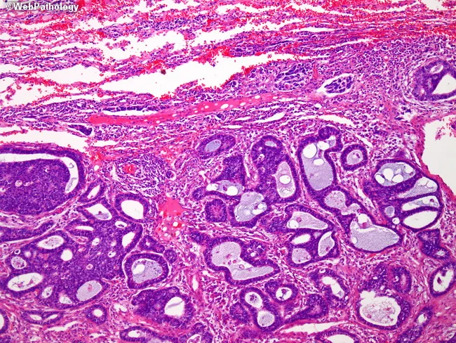
Imaging modality: Light microscopy of hematoxylin and eosin stained histology section from breast tissue. Section shows lobular and ductal glandular elements scattered among fibrous stroma and adipose tissue. At low magnification abundant heterogenous glandular structures display round to elongated lumens, lined by cuboidal to low columnar epithelium. The ducts vary in size with some dilated, cyst-like spaces; the surrounding stroma is fibrous with connective tissue strands and occasional inflammatory infiltrates not obvious. Adipose tissue occupies adjacent parenchyma, giving a mixed composition typical of breast. Cellular morphology reveals typical epithelial lining with basally oriented nuclei, inconspicuous nucleoli at this field; no convincing mitotic activity. No obvious invasion into surrounding stroma; myoepithelial layer presence cannot be assessed at this magnification. The image highlights normal breast architecture with glandular ducts, lobules, and supportive stroma and adipose tissue; features could reflect benign fibrocystic changes or ductal proliferations rather than malignancy in absence of nuclear atypia or stromal invasion. Clinically relevant for educational purposes: illustrating histologic breast tissue, fibrosis, ductal structures, and potential differential diagnoses including fibrocystic disease, fibroadenoma, DCIS, IDC. Useful for training pathology residents and for comparison with radiologic imaging findings and clinical breast exam in suspicion of benign vs malignant lesions.
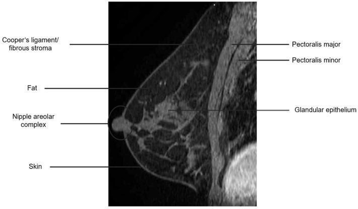
This diagnostic image is a sagittal T1-weighted fat-suppressed post-contrast MRI of the human breast, demonstrating normal anatomical structures. The fat suppression technique renders adipose tissue (fat) dark (low signal intensity), while the glandular epithelium and vascularized structures show enhancement (high signal intensity) following contrast administration. Key anatomical landmarks are labeled: the pectoralis major and pectoralis minor muscles are located posteriorly (deep) to the breast parenchyma; the pectoralis major is positioned anterior to the pectoralis minor. Within the breast tissue, the glandular epithelium (fibroglandular tissue) is visible as high-signal regions, supported by the fibrous stroma or Cooper’s ligaments, which appear as thin connective bands extending toward the dermis. The skin and nipple-areolar complex are also labeled, defining the anterior boundary of the breast. This image serves as an educational reference for breast MRI anatomy and the visual impact of fat-suppression and contrast enhancement on different tissue types during oncological imaging.
Breast Parenchyma vs. Stroma - A Detailed Comparison with Pictorial
Overview
1. PARENCHYMA - The Functional Glandular Tissue
Components:
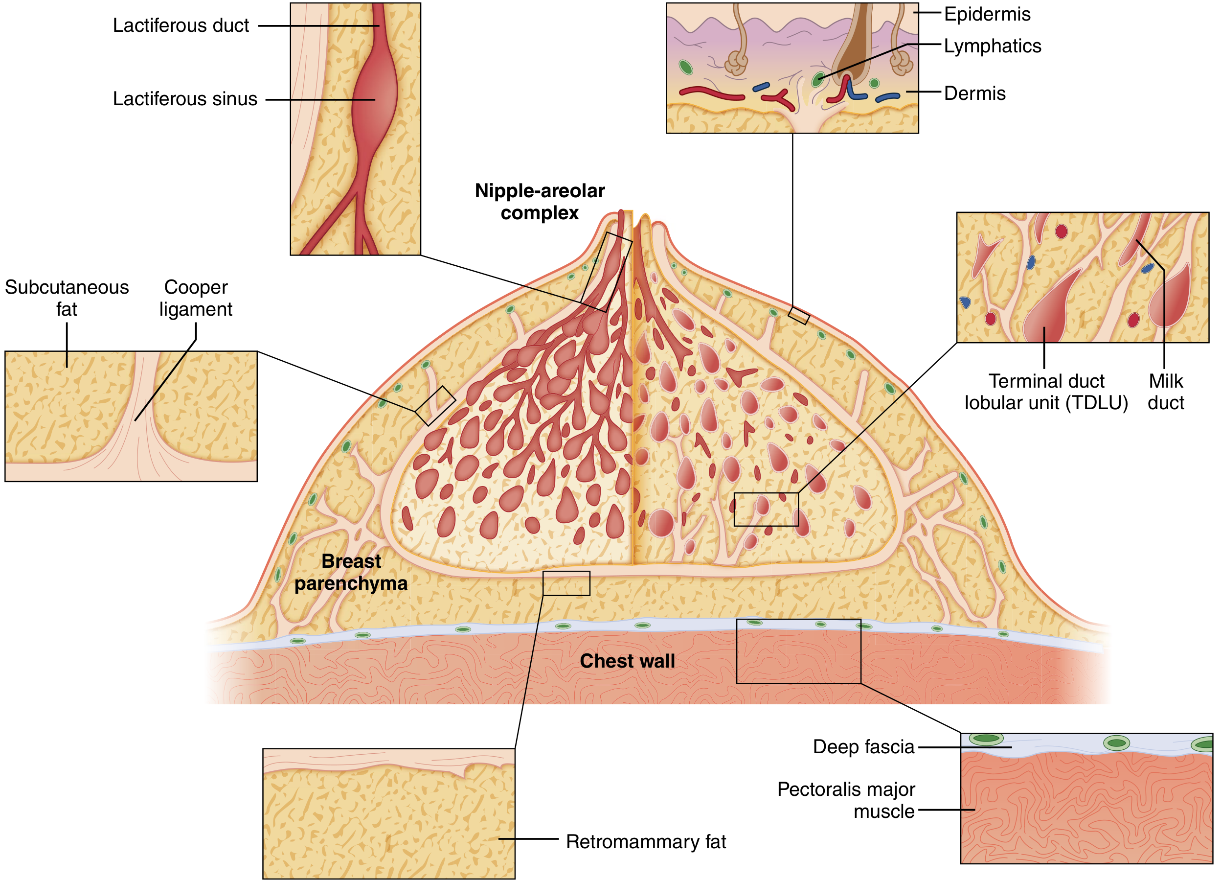
- Lactiferous ducts - 15-20 major ducts opening at the nipple
- Lactiferous sinus - dilated ampullary portion just beneath the nipple-areolar complex (NAC)
- Segmental and subsegmental ducts - progressively branching radially from the nipple
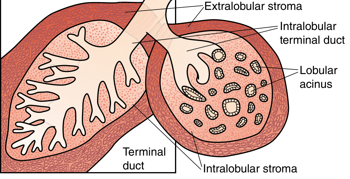
- Terminal duct lobular unit (TDLU) - the structural and functional unit of the breast; consists of:
- Terminal duct - smallest duct entering the lobule
- Lobular acini - blind-ended grape-like secretory sacs that produce milk
- Two epithelial cell layers in every duct and acinus:
- Inner luminal cells - columnar/cuboidal, secretory
- Outer myoepithelial cells - contractile, propel milk toward nipple; also produce basement membrane
Boundaries:
2. STROMA - The Supportive Connective Tissue Framework
A. Intralobular Stroma (specialized)
- Loose connective tissue within each lobule
- Surrounds the acini and terminal ductules inside the lobule
- Contains fibroblastic stromal cells that are hormonally sensitive (estrogen/progesterone responsive)
- Contains modest lymphocytes and plasma cells
- Gives rise to fibroadenoma and phyllodes tumor (biphasic tumors with epithelial and stromal components)
B. Interlobular (Extralobular) Stroma
-
Dense fibrous connective tissue between lobules
-
Contains larger breast ducts, blood vessels, lymphatics, and fat
-
Also called "extralobular stroma"
-
Not hormonally sensitive in the same way
-
Gives rise to mesenchymal tumors (lipoma, angiosarcoma, myofibroblastoma) - same as connective tissue elsewhere in the body
-
Contains Cooper's ligaments (suspensory ligaments) - fibrous bands running from the deep fascia to the dermis, giving the breast shape
-
Robbins, Cotran & Kumar, p. 967; Histology: A Text and Atlas, p. 2303
Pictorial: Gross Anatomy Diagram
Pictorial: Terminal Duct Lobular Unit - Intralobular vs. Extralobular Stroma
Pictorial: Adolescent Breast - Ductal Tree & TDLU in Whole Mount
Pictorial: Histology H&E - Parenchyma and Stroma
Pictorial: Mammogram vs. Histology Correlation
Summary Table
| Feature | Parenchyma | Stroma |
|---|---|---|
| Definition | Functional glandular tissue | Supportive connective tissue framework |
| Components | Ducts, lobules, acini, TDLU | Fibrous CT, adipose, vessels, lymphatics, Cooper's ligaments |
| Cell types | Luminal epithelial + myoepithelial cells | Fibroblasts, adipocytes, endothelial cells, lymphocytes |
| Subdivisions | Ducts (major → terminal) + Lobules | Intralobular stroma + Interlobular stroma |
| Hormone response | Estrogen/progesterone cause ductal branching, lobule formation | Intralobular stroma is hormonally sensitive; interlobular less so |
| On mammogram | White/dense (fibroglandular density) | Dark (fat) / interspersed fine white lines (fibrous CT) |
| On H&E | Dark purple glandular structures | Pink fibrous bands and clear adipocytes |
| Key boundary | Basement membrane separates parenchyma from stroma | - |
| Neoplasms arising | Carcinomas (from epithelial cells) | Fibroadenoma/phyllodes (intralobular); lipoma/angiosarcoma (interlobular) |
| Changes with age | Involutes post-menopause | Fat replaces glandular tissue post-menopause |
| Changes in pregnancy | Massive lobular expansion, nearly fills breast | Stroma reduced to "scant stroma" between lobules |
Key Clinical Correlations
- Peau d'orange (orange skin): Cancer or edema infiltrates Cooper's ligaments in the interlobular stroma, pulling on the dermis and causing skin dimpling.
- DCIS vs. Invasive cancer: DCIS stays within the parenchymal epithelium, bounded by the basement membrane. Once cells breach that membrane into stroma = invasive carcinoma.
- Breast density on mammogram: Dense breasts have more parenchyma and fibrous stroma relative to fat. Higher density = increased cancer risk and reduced mammographic sensitivity.
- Fibroadenoma: Arises from intralobular stroma (specialized stromal cells + proliferating lobular epithelium = biphasic tumor).
- Cooper's ligament tethering: Part of the interlobular stroma; surgically important as it can be divided to mobilize breast tissue.
Sources: Robbins, Cotran & Kumar - Pathologic Basis of Disease; Sabiston Textbook of Surgery (18th ed.); Histology: A Text and Atlas with Correlated Cell and Molecular Biology