
Give me 3 sets of 10 questions each for spotters containing ecg abg xray ct pocus and scoring system pertaining to emergency medicine with its answers
ECG inferior wall STEMI ST elevation leads II III aVF
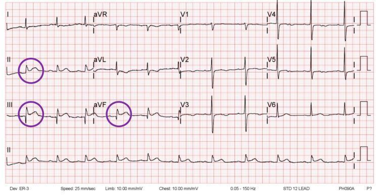
This diagnostic image is a standard 12-lead electrocardiogram (ECG) tracing on a red grid background, illustrating an acute inferior wall ST-elevation myocardial infarction (STEMI). The tracing displays the six limb leads (I, II, III, aVR, aVL, aVF) and the six precordial leads (V1-V6), with a continuous rhythm strip of lead II at the bottom. Purple circles highlight significant ST-segment elevation at the J-point in the inferior leads: II, III, and aVF. The ST elevation is approximately 1 mm or greater with a convex or upward-sloping morphology. Reciprocal ST-segment depression is visible in the high lateral leads (I and aVL), which is a classic finding in inferior STEMI. The precordial leads (V1-V6) show a normal R-wave progression without acute ST-segment changes. This ECG is a key educational tool for cardiovascular medicine, specifically for diagnosing acute coronary syndromes and identifying regional wall ischemia or infarction patterns.
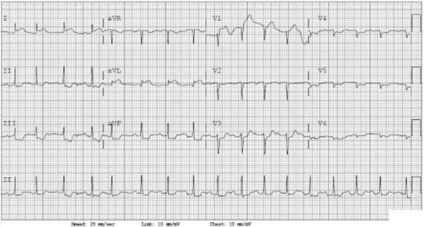
A diagnostic 12-lead electrocardiogram (ECG) displayed on a standard grid (25 mm/sec speed; limb 10 mm/mV, chest 20 mm/mV) demonstrating an inferolateral ST-segment elevation myocardial infarction (STEMI). The layout includes limb leads (I, II, III, aVR, aVL, aVF) and precordial leads (V1-V6), with a long Lead II rhythm strip at the bottom. Significant pathology is observed in the inferior leads (II, III, and aVF), which show marked ST-segment elevation. Additionally, lateral leads (V4-V6) exhibit ST-segment changes consistent with an extending infarct. Reciprocal ST-segment depression is present in the high lateral leads (I, aVL) and precordial leads V1-V3, further confirming the diagnostic pattern of an acute inferior wall STEMI. The ECG is essential for emergency department triage and rapid diagnosis of acute coronary syndrome (ACS) in clinical settings.
ECG hyperkalemia peaked T waves widened QRS
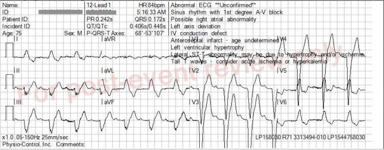
This diagnostic image is a 12-lead electrocardiogram (ECG) demonstrating multiple conduction and metabolic abnormalities. The tracing shows a sinus rhythm with a prolonged PR interval (0.242s), indicating first-degree atrioventricular (AV) block. The QRS complex is significantly widened (0.172s) with a morphology consistent with a left bundle branch block (LBBB) pattern, characterized by deep S waves in V1-V3 and broad, notched R waves in lateral leads such as V6 and aVL. Notably, the T waves are disproportionately tall, peaked, and 'tented,' particularly visible in the precordial leads V2-V5. These peaked T waves, when combined with QRS widening, are classic electrocardiographic manifestations of hyperkalemia. The image also displays left axis deviation and secondary ST-T wave discordance typical of LBBB. This ECG serves as a critical educational example of how metabolic disturbances like hyperkalemia can overlay and exacerbate underlying conduction system disease in a clinical emergency setting.
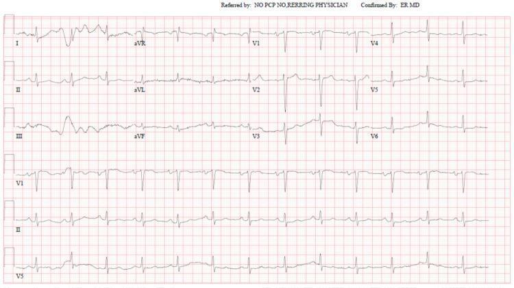
This diagnostic image is a standard 12-lead electrocardiogram (ECG) displayed on a red grid background. The tracing includes the limb leads (I, II, III, aVR, aVL, aVF) and precordial leads (V1-V6), along with rhythm strips for V1, II, and V5 at the bottom. The rhythm demonstrates a normal sinus rhythm with clearly visible, upright P waves in leads I, II, and V4-V6. The QRS complexes are narrow, and there is a normal R-wave progression from V1 through V6. The ST segments are isoelectric, and the T waves are largely upright, showing no acute signs of ischemia, infarction, or the classic manifestations of hyperkalemia (such as peaked T waves or widened QRS complexes). This ECG serves as a clinical example of a 'normal' tracing in a patient with pseudo-hyperkalemia, illustrating the absence of cardiac electrophysiological changes despite laboratory reports of severely elevated serum potassium levels. It is an important educational tool for medical professionals to differentiate between true electrolyte-induced cardiac emergencies and laboratory artifacts in the context of hematologic malignancies like chronic lymphocytic leukemia.
chest x-ray tension pneumothorax mediastinal shift
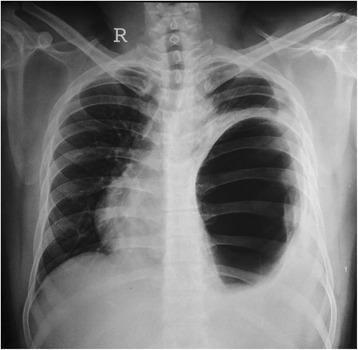
Antero-posterior (AP) chest X-ray demonstrating a large, well-defined area of marked hyper-translucency occupying the majority of the left hemithorax. This lucent region lacks normal bronchovascular markings, and its appearance mimics a massive pneumothorax or a large pulmonary bulla. However, the presence of a distinct curved superior border and the clinical context of traumatic diaphragmatic injury suggest a herniated, air-filled stomach (gastric bubble) within the thoracic cavity. There is significant mass effect evidenced by a prominent mediastinal shift to the right, displacement of the cardiac silhouette, and compression of the contralateral right lung. The left hemidiaphragm is obscured by the gas-distended structure. This imaging finding is characteristic of a large traumatic diaphragmatic hernia causing tension-like effects on mediastinal structures, requiring differentiation from tension pneumothorax to avoid inappropriate chest tube insertion.
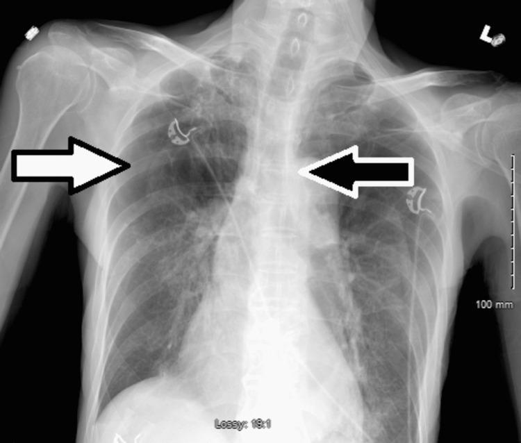
This diagnostic image is an anteroposterior (AP) chest X-ray demonstrating a large right-sided tension pneumothorax. The right hemithorax shows an extensive area of increased lucency (hyperlucency) with a complete absence of peripheral bronchovascular lung markings (indicated by the white arrow). Key diagnostic features include significant mass effect, characterized by a visible mediastinal shift to the left (indicated by the black arrow) and flattening of the right heart border and mediastinal contours. The trachea is also deviated toward the left. These findings are clinically significant as they represent a medical emergency where intrapleural pressure exceeds atmospheric pressure, compromising venous return and cardiac output. The image provides a clear example of tension physiology in the thoracic cavity following chest tube removal. The patient's background of COPD and COVID-19 pneumonia contributes to the underlying lung frailty and secondary spontaneous pneumothorax risk.
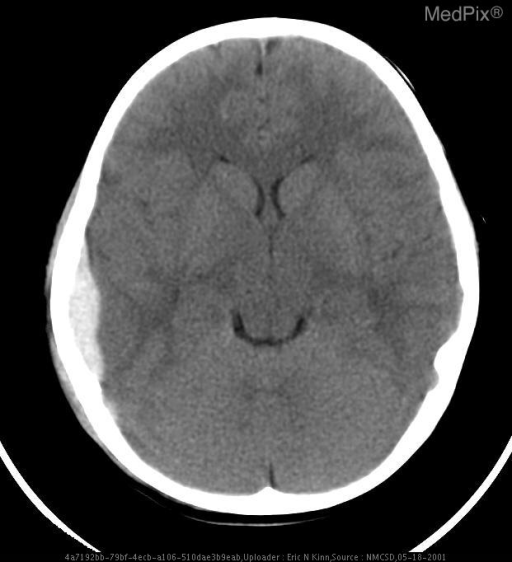
CT brain acute subdural hematoma crescent shaped
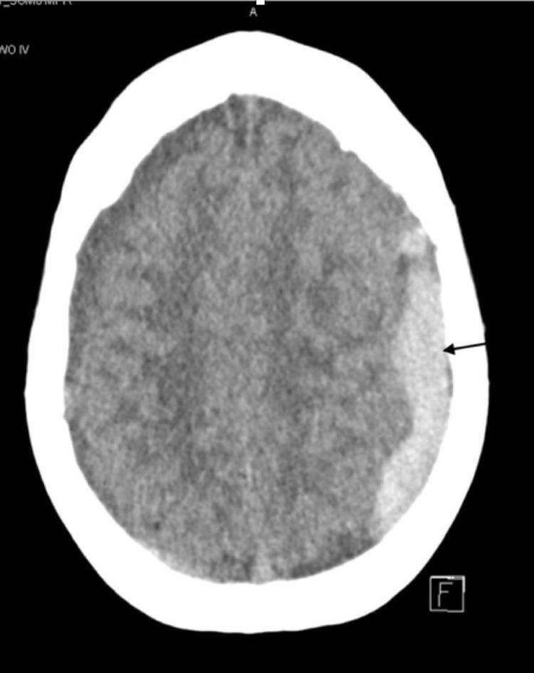
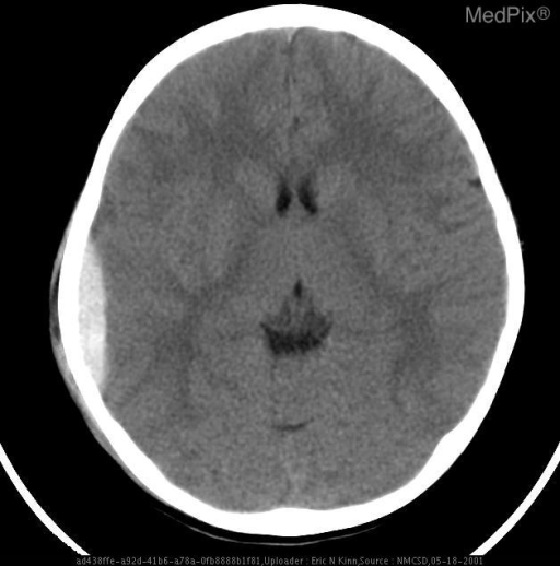
This diagnostic image is a non-contrast axial computed tomography (CT) scan of the brain. The primary finding is a large, hyperdense, crescent-shaped collection along the left cerebral convexity, characteristic of an acute subdural hematoma (SDH). The hematoma is well-demarcated and follows the contour of the inner table of the skull, extending across the frontoparietal region. There is a visible mass effect on the underlying brain parenchyma, evidenced by effacement of the adjacent sulci and a shift of midline structures toward the right side. The high attenuation (brightness) of the collection relative to the brain tissue is consistent with acute extravasation of blood. This scan illustrates a common neurosurgical emergency often associated with head trauma or anticoagulation therapy, demonstrating the visual hallmarks used to differentiate a subdural hematoma from the lenticular shape of an epidural hematoma.
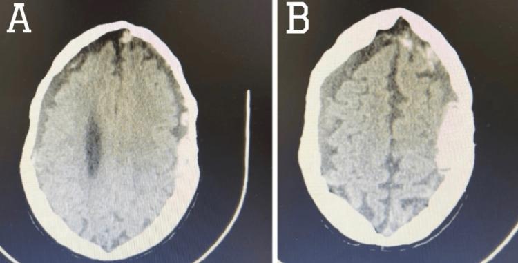
Two side-by-side axial sections of a non-contrast CT scan of the brain, demonstrating bilateral extra-axial fluid collections. Image (A) shows a hyperdense, crescent-shaped collection along the frontoparietal region, characteristic of an acute subdural hematoma (SDH). There is evidence of mild mass effect with compression of the underlying brain parenchyma and minimal midline shift. Image (B) demonstrates a contralateral collection that appears hypodense relative to acute blood, consistent with a chronic subdural hematoma. Additionally, focal areas of hyperdensity are visible within the sulci, indicating associated convexity subarachnoid hemorrhage (cSAH). These images serve as a clinical comparison of blood density at different stages of evolution (acute vs. chronic) within the subdural space. The diagnostic focus is on intracranial hemorrhage identification, mass effect evaluation, and the assessment of trauma-related or spontaneous vascular complications in a neuroradiological context.
FAST ultrasound Morison pouch free fluid hemoperitoneum
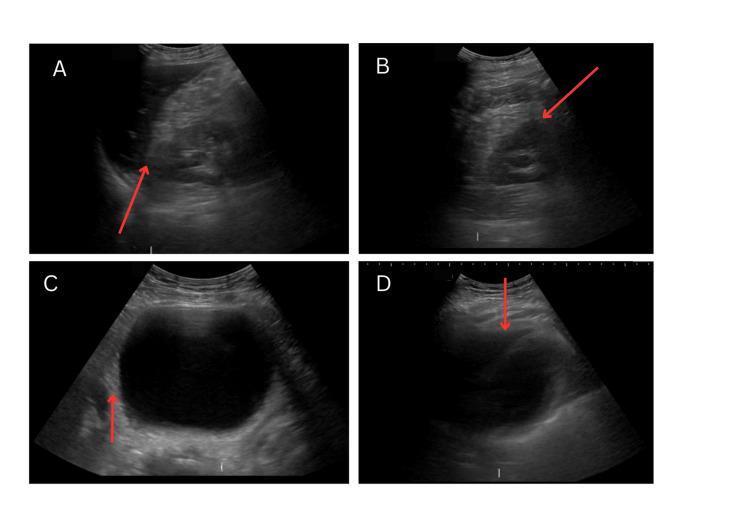
This diagnostic image series consists of four B-mode ultrasound frames representing a Focused Assessment with Sonography for Trauma (FAST) exam. Image A shows the right upper quadrant view, focusing on Morison’s pouch (the hepatorenal recess); red arrows indicate a clear interface between the liver and right kidney with no free anechoic fluid. Image B displays the left upper quadrant view, showing the splenorenal recess and the diaphragm interface, also devoid of fluid. Image C captures the pelvic view (suprapubic longitudinal or transverse), dominated by a large, well-circumscribed, anechoic urinary bladder; the red arrow points to the rectovesical space (in males) or pouch of Douglas (in females), which is negative for free fluid. Image D presents the subxiphoid or subcostal pericardial view, assessing the potential space between the visceral and parietal pericardium for effusion or tamponade, showing a negative result. Collectively, these images illustrate a normal FAST exam protocol used to rule out hemoperitoneum and hemopericardium in emergency clinical settings.
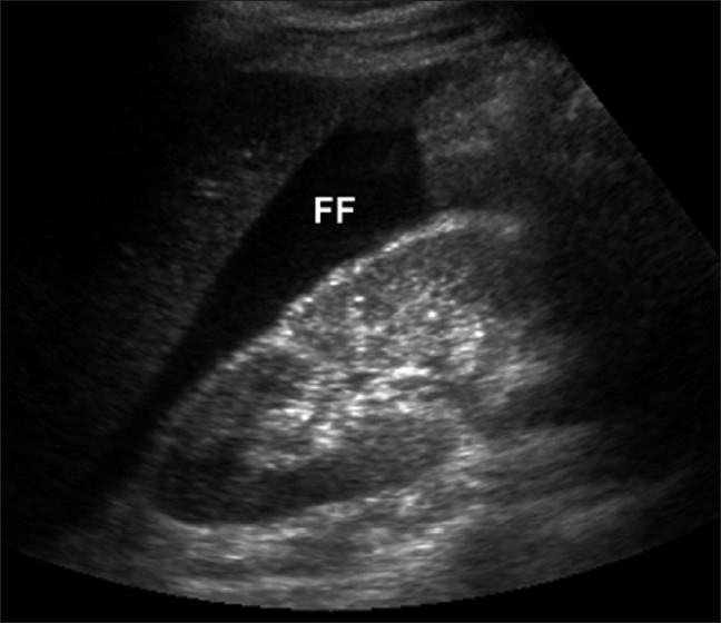
A diagnostic ultrasound image of the right upper quadrant (Morison's pouch) demonstrating a significant amount of free intraperitoneal fluid. The fluid is identified by a prominent anechoic (black) triangular region labeled 'FF', located in the potential space between the liver and the kidney. The kidney displays a typical reniform shape with a heterogeneous parenchymal echotexture and a hyper-echogenic renal sinus. The liver is visible as a more homogeneous, moderately echogenic structure adjacent to the fluid collection. This finding is clinically significant for hemoperitoneum, consistent with internal hemorrhage often seen in acute conditions such as a ruptured ectopic pregnancy. The image illustrates a key component of the Focused Assessment with Sonography for Trauma (FAST) exam, used to identify free fluid in the peritoneal cavity.
ECG torsades de pointes polymorphic ventricular tachycardia
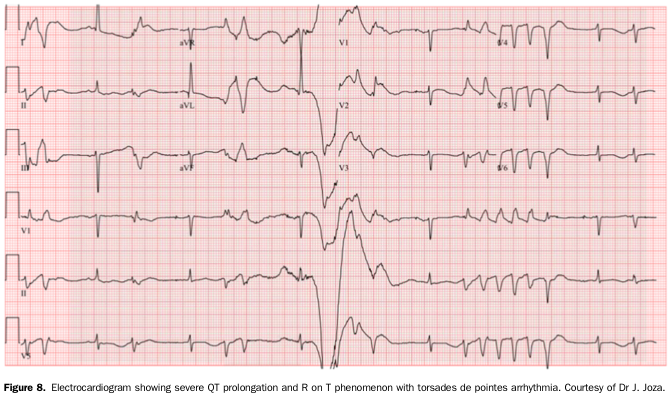
Summary : This figure presents a 12-lead electrocardiogram (ECG) demonstrating severe QT interval prolongation, the R on T phenomenon, and the onset of torsades de pointes arrhythmia.
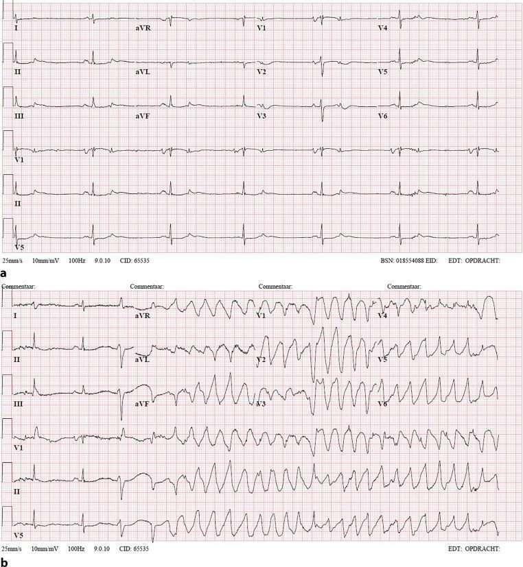
Two-part 12-lead electrocardiogram (ECG) comparison demonstrating drug-induced arrhythmia. (a) Initial ECG showing sinus bradycardia at approximately 40 bpm. Findings include narrow QRS complexes, prolonged QT interval, and T-wave abnormalities (inversions in inferior leads II, III, and aVF). This baseline demonstrates common findings in patients on QT-prolonging agents like sotalol. (b) ECG recorded during a syncopal event, displaying a transition from sinus rhythm into a polymorphic ventricular tachycardia (Torsades de Pointes). The tachycardia is characterized by wide QRS complexes (duration >120ms) with a shifting axis, rotating around the isoelectric line at a rapid rate of 150-200 bpm. The visual sequence illustrates the clinical progression from drug-induced QT prolongation to life-threatening ventricular arrhythmia. Educational focus is on identifying bradycardia, QT prolongation, and the morphology of polymorphic ventricular tachycardia in the context of syncope.
CT brain epidural hematoma lens shaped biconvex
echocardiogram pericardial effusion cardiac tamponade
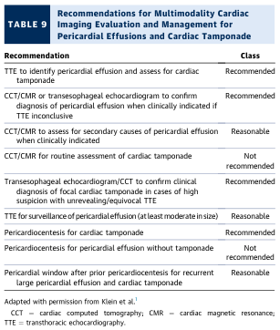
TABLE 9 Recommendations for Multimodality Cardiac Imaging Evaluation and Management for Pericardial Effusions and Cardiac Tamponade
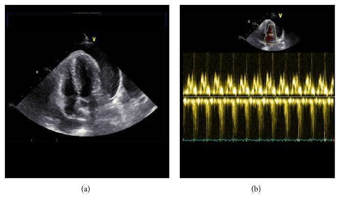
This medical visual consists of two ultrasound images depicting a cardiac evaluation for pericardial effusion. Panel (a) is a transthoracic echocardiogram in the apical four-chamber view. It reveals a significant, circumferential pericardial effusion, characterized by a large, anechoic (dark) space between the epicardium and the parietal pericardium. Despite the volume of fluid, the cardiac chambers, including the right ventricle and right atrium, appear to maintain their structural integrity without obvious diastolic collapse. Panel (b) shows a pulsed-wave Doppler echocardiogram of the mitral valve inflow. The spectral display shows regular, repetitive E and A waves oscillating around the baseline with stable velocities. This normal variation in mitral inflow indicates the absence of significant respiratory-related hemodynamic compromise (pulsus paradoxus) often seen in cardiac tamponade. Together, these images illustrate a clinically significant pericardial effusion without current evidence of right ventricular dysfunction or tamponade physiology.
chest x-ray pneumoperitoneum free air under diaphragm
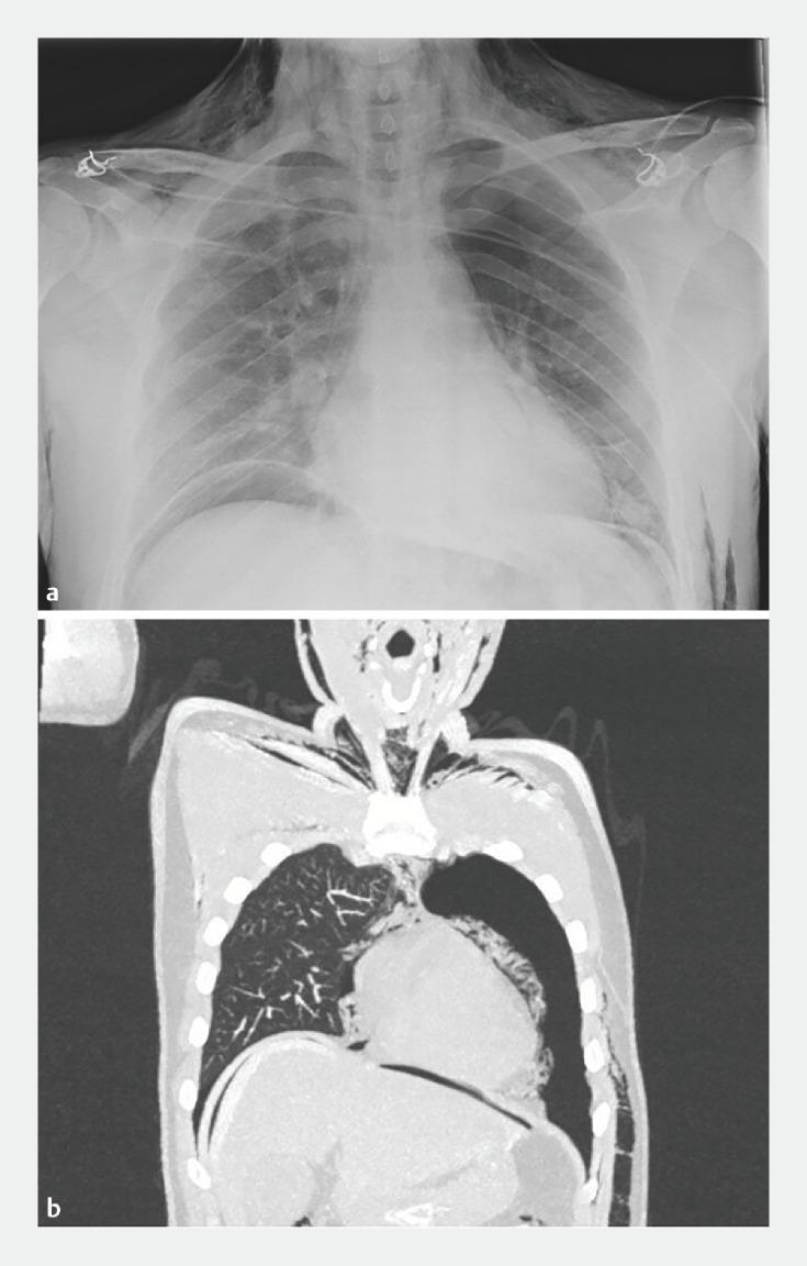
This composite figure displays diagnostic imaging of a patient exhibiting extensive extra-alveolar air following a Peroral Endoscopic Myotomy (POEM) procedure. Image A is a frontal chest X-ray demonstrating a large crescentic lucency beneath the diaphragm consistent with pneumoperitoneum, linear lucencies along the mediastinal borders indicating pneumomediastinum, and radiolucent striations in the soft tissues of the neck and chest wall signifying subcutaneous emphysema. Image B is a coronal CT scan reconstruction of the chest and upper abdomen, providing definitive visualization of a large left-sided pneumothorax with partial lung collapse. It further confirms pneumomediastinum with air tracking around the heart and great vessels, pneumoperitoneum with free air localized under the diaphragmatic domes, and extensive subcutaneous emphysema dissecting through the cervical and thoracic muscle planes. The images collectively illustrate common barotrauma or insufflation-related complications associated with endoscopic esophageal interventions.
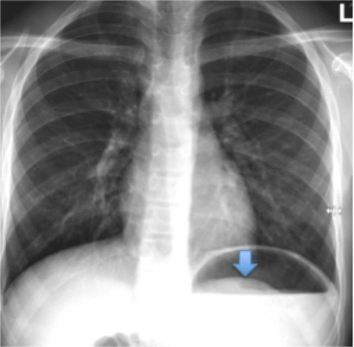
This diagnostic image is a posterior-anterior (PA) chest radiograph. The thoracic anatomy including the lungs, cardiac silhouette, and mediastinum appears within normal limits with no evidence of pulmonary opacities or lymphadenopathy. A blue arrow highlights the primary clinical finding in the left upper quadrant of the abdomen, just below the left diaphragm. Specifically, a large gastric air bubble is visible containing a curvilinear, soft-tissue density shadow projecting into the gas space. This appearance is characteristic of a gastric bezoar (trichobezoar), representing solid ingested material outlined by a rim of gastric air. The absence of free air under the diaphragm (pneumoperitoneum) suggests there is no gastric perforation. The image serves as an educational example of how abdominal pathology can be incidentally identified or subtly present on the lower portion of a standard chest X-ray.
chest x-ray pulmonary edema bilateral bat wing infiltrates
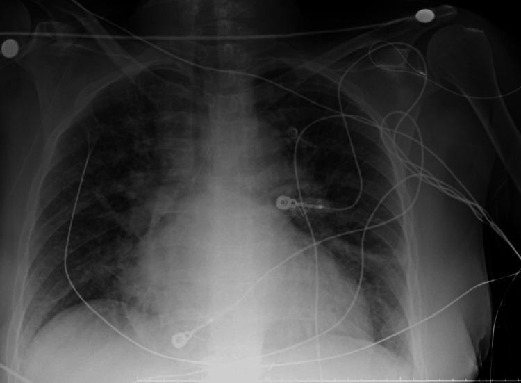
This anterior-posterior (AP) chest X-ray illustrates a case of acute pulmonary edema and cardiomegaly. The primary findings include diffuse, bilateral alveolar opacities and patchy infiltrates that exhibit a predominantly perihilar 'bat-wing' distribution. These densities obscure normal vascular markings and contribute to a mottled, ground-glass appearance throughout the mid-to-lower lung zones. The cardiac silhouette is significantly enlarged, with a widened mediastinum and indistinct heart borders consistent with congestive heart failure. Multiple supportive medical devices are present: a right-sided central venous catheter with its tip at the level of the superior vena cava, several circular radiopaque ECG electrode markers with associated lead wires traversing the anterior chest, and a vertical enteric tube passing through the lower mediastinum. The costophrenic angles are partially blunted by overlying infiltrates or potential pleural effusion. This diagnostic image serves as a classic representation of cardiogenic pulmonary edema for clinical educational purposes.
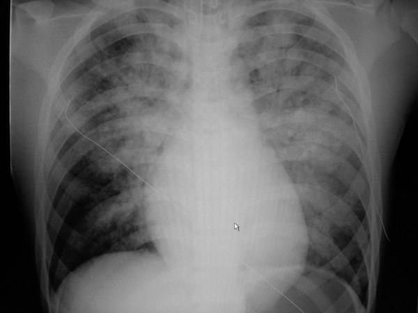
This diagnostic image is an anteroposterior (AP) chest X-ray demonstrating acute bilateral pulmonary edema. The radiograph reveals diffuse, hazy opacities and ill-defined infiltrates throughout both lung fields, exhibiting a characteristic perihilar 'bat-wing' distribution with prominent involvement of the upper and central lung zones. The lung parenchyma shows signs of alveolar consolidation and fluid accumulation. Cardiomegaly is evident, with an enlarged cardiac silhouette and indistinct borders. A thin, radiopaque monitoring line or catheter is visible crossing the right thoracic cavity toward the mediastinum. The mediastinal structures and bony thorax, including the clavicles and ribs, are otherwise unremarkable. This visual is clinically significant for students learning to identify radiological signs of heart failure and pulmonary congestion, as it highlights the hallmark features of cardiogenic pulmonary edema versus acute respiratory distress syndrome (ARDS).
Emergency Medicine Spotters — 3 Sets (10 Questions Each)
SET 1
SET 2
SET 3
Can you create a ppt of this sets
mkdir -p /tmp/workspace/em-spotters/assets && ls -la /tmp/workspace/
cd /tmp/workspace/em-spotters && python /tmp/skills/shared/scripts/fetch_images.py \ "https://cdn.orris.care/cdss_images/pmc_clinical_VQA_45e6159ca694f684efa9f491a17643e5e9da0870694f2324257ff36fbd36f575.jpg" \ "https://cdn.orris.care/cdss_images/pmc_clinical_VQA_05e30884f10a10ac914ad9908759c64ecc4970fc52b8bbef8f700d0d52f4e62d.jpg" \ "https://cdn.orris.care/cdss_images/pmc_clinical_VQA_6db0d76f6f9ac3659befbf54e718d995fd6e504ac2afc5f7a5a84d51f580bef3.jpg" \ "https://cdn.orris.care/cdss_images/pmc_clinical_VQA_347c13d9e2e94e9b1349ef7d699e37969922dfb3eb9c4554ad0bc2689b923163.jpg" \ "https://cdn.orris.care/cdss_images/pmc_clinical_VQA_0529938690e9a70b5a22c9362283464e00bbf4d0e5c7fbd074ee6cbd71755a33.jpg" \ "https://cdn.orris.care/cdss_images/GLGCA_4566687_1766513421210_0af75e19-e865-460b-b2a2-efd1d553a1aa_598c090f-7c71-44dc-90f9-9fa11c4bcd3d.png" \ "https://cdn.orris.care/cdss_images/medpix_vqa_6b9b24a5-28d8-4cb7-8361-d9e5d3101a44_1766301794560.png" \ "https://cdn.orris.care/cdss_images/pmc_clinical_VQA_531decad40c988e356ff83e58ee00a69dffeaac4da9c83f19935e0542690404b.jpg" \ "https://cdn.orris.care/cdss_images/pmc_clinical_VQA_35a9bdbbf849eab9162c6ba929958dc8bf2bbc561662647860786f686c27f18b.jpg" \ > images_result.json python -c " import json data = json.load(open('images_result.json')) for d in data: print(d['url'][-40:], 'OK' if d.get('base64') else 'FAIL', d.get('error')) "
cd /tmp/workspace/em-spotters && python -c " import json, base64 data = json.load(open('images_result.json')) names = ['stemi','tension_ptx','sdh','fast','pneumoperitoneum','torsades','edh','pericardial_effusion','pulm_edema'] for d, name in zip(data, names): b64 = d['base64'] header, encoded = b64.split(',', 1) ext = 'png' if 'png' in header else 'jpg' with open(f'assets/{name}.{ext}', 'wb') as f: f.write(base64.b64decode(encoded)) print(name, ext) " ls -la assets/
cd /tmp/workspace/em-spotters/assets && file *.jpg
cd /tmp/workspace/em-spotters/assets && mv edh.jpg edh.png && mv torsades.jpg torsades.png && ls
~/em-spotters/build_deck.py
#!/usr/bin/env python3
"""Build the Emergency Medicine Spotters PPTX deck (3 sets x 10 questions)."""
from pptx import Presentation
from pptx.util import Inches, Pt, Emu
from pptx.dml.color import RGBColor
from pptx.enum.text import PP_ALIGN, MSO_ANCHOR
from pptx.enum.shapes import MSO_SHAPE
import os

ASSETS = "/tmp/workspace/em-spotters/assets"

# ---------- Theme ----------
DARK_RED = RGBColor(0x8B, 0x1A, 0x1A)
NAVY = RGBColor(0x0B, 0x2545, )
NAVY = RGBColor(0x0B, 0x25, 0x45)
WHITE = RGBColor(0xFF, 0xFF, 0xFF)
LIGHT_BG = RGBColor(0xF4, 0xF6, 0xF8)
GREY_TXT = RGBColor(0x33, 0x33, 0x33)
ACCENT = RGBColor(0xC0, 0x39, 0x2B)
GREEN = RGBColor(0x1E, 0x7D, 0x32)

CAT_COLORS = {
    "ECG": RGBColor(0xC0, 0x39, 0x2B),
    "ABG": RGBColor(0x1B, 0x5E, 0x20),
    "CXR": RGBColor(0x1A, 0x5F, 0x9E),
    "CT": RGBColor(0x6A, 0x1B, 0x9A),
    "POCUS": RGBColor(0xE6, 0x7E, 0x22),
    "SCORE": RGBColor(0x2C, 0x3E, 0x50),
}

prs = Presentation()
prs.slide_width = Inches(13.333)
prs.slide_height = Inches(7.5)
BLANK = prs.slide_layouts[6]
SW, SH = prs.slide_width, prs.slide_height


def add_bg(slide, color=WHITE):
    rect = slide.shapes.add_shape(MSO_SHAPE.RECTANGLE, 0, 0, SW, SH)
    rect.fill.solid()
    rect.fill.fore_color.rgb = color
    rect.line.fill.background()
    rect.shadow.inherit = False
    slide.shapes._spTree.remove(rect._element)
    slide.shapes._spTree.insert(2, rect._element)
    return rect


def textbox(slide, x, y, w, h, text, size=18, bold=False, color=GREY_TXT,
            align=PP_ALIGN.LEFT, font="Calibri", anchor=MSO_ANCHOR.TOP, italic=False):
    tb = slide.shapes.add_textbox(Inches(x), Inches(y), Inches(w), Inches(h))
    tf = tb.text_frame
    tf.word_wrap = True
    tf.vertical_anchor = anchor
    lines = text.split("\n")
    for i, line in enumerate(lines):
        p = tf.paragraphs[0] if i == 0 else tf.add_paragraph()
        p.text = line
        p.alignment = align
        for r in p.runs:
            r.font.size = Pt(size)
            r.font.bold = bold
            r.font.italic = italic
            r.font.color.rgb = color
            r.font.name = font
    return tb


def add_footer(slide, text):
    textbox(slide, 0.4, 7.08, 8, 0.35, text, size=10, color=RGBColor(0x99, 0x99, 0x99))


def title_slide():
    slide = prs.slides.add_slide(BLANK)
    add_bg(slide, NAVY)
    # accent bar
    bar = slide.shapes.add_shape(MSO_SHAPE.RECTANGLE, 0, Inches(3.55), SW, Inches(0.08))
    bar.fill.solid(); bar.fill.fore_color.rgb = ACCENT; bar.line.fill.background()
    textbox(slide, 1, 2.5, 11.33, 1.0, "EMERGENCY MEDICINE SPOTTERS",
            size=44, bold=True, color=WHITE, align=PP_ALIGN.CENTER)
    textbox(slide, 1, 3.7, 11.33, 0.6, "3 Practice Sets  |  10 Questions Each",
            size=22, color=RGBColor(0xCC, 0xD6, 0xE0), align=PP_ALIGN.CENTER)
    textbox(slide, 1, 4.5, 11.33, 0.6, "ECG  \u2022  ABG  \u2022  X-Ray  \u2022  CT  \u2022  POCUS  \u2022  Scoring Systems",
            size=18, color=RGBColor(0xAA, 0xB8, 0xC8), align=PP_ALIGN.CENTER, italic=True)
    return slide


def divider_slide(set_num, n_q=10):
    slide = prs.slides.add_slide(BLANK)
    add_bg(slide, DARK_RED)
    textbox(slide, 1, 2.9, 11.33, 1.0, f"SET {set_num}", size=60, bold=True,
            color=WHITE, align=PP_ALIGN.CENTER)
    textbox(slide, 1, 4.0, 11.33, 0.6, f"{n_q} Spotter Questions", size=22,
            color=RGBColor(0xF0, 0xD0, 0xD0), align=PP_ALIGN.CENTER)
    return slide


def category_chip(slide, category, x=0.5, y=0.45):
    color = CAT_COLORS.get(category, ACCENT)
    w = Inches(1.7) if category != "SCORE" else Inches(2.0)
    chip = slide.shapes.add_shape(MSO_SHAPE.ROUNDED_RECTANGLE, Inches(x), Inches(y), w, Inches(0.45))
    chip.fill.solid(); chip.fill.fore_color.rgb = color
    chip.line.fill.background()
    chip.shadow.inherit = False
    tf = chip.text_frame
    tf.word_wrap = False
    tf.margin_left = 0; tf.margin_right = 0; tf.margin_top = 0; tf.margin_bottom = 0
    tf.vertical_anchor = MSO_ANCHOR.MIDDLE
    p = tf.paragraphs[0]
    label = category if category != "SCORE" else "SCORING SYSTEM"
    p.text = label
    p.alignment = PP_ALIGN.CENTER
    r = p.runs[0]
    r.font.size = Pt(14); r.font.bold = True; r.font.color.rgb = WHITE


def question_slide(set_num, q_num, category, stem, question, image_file=None):
    slide = prs.slides.add_slide(BLANK)
    add_bg(slide, LIGHT_BG)
    category_chip(slide, category)
    textbox(slide, SW.inches - 3.2, 0.45, 2.7, 0.45,
            f"SET {set_num} \u2022 Q{q_num}", size=16, bold=True, color=NAVY, align=PP_ALIGN.RIGHT)

    has_image = image_file and os.path.exists(os.path.join(ASSETS, image_file))
    if has_image:
        # Image on left, text on right
        img_path = os.path.join(ASSETS, image_file)
        pic = slide.shapes.add_picture(img_path, Inches(0.5), Inches(1.15), height=Inches(5.4))
        # constrain width if too wide
        if pic.width > Inches(7.2):
            ratio = Inches(7.2) / pic.width
            pic.width = Inches(7.2)
            pic.height = int(pic.height * ratio)
        # center image vertically in its zone
        pic.left = Inches(0.5)
        pic.top = Inches(1.15) + int((Inches(5.4) - pic.height) / 2) if pic.height < Inches(5.4) else Inches(1.15)

        text_x, text_w = 8.0, 4.9
    else:
        text_x, text_w = 0.6, 12.1

    textbox(slide, text_x, 1.15, text_w, 0.9, stem, size=16, bold=False,
            color=GREY_TXT, italic=True)
    textbox(slide, text_x, 2.15, text_w, 3.9 if has_image else 4.5, question,
            size=19 if has_image else 20, bold=True, color=NAVY)

    add_footer(slide, "Emergency Medicine Spotters \u2014 Question")
    return slide


def answer_slide(set_num, q_num, category, answer, image_file=None):
    slide = prs.slides.add_slide(BLANK)
    add_bg(slide, WHITE)
    category_chip(slide, category)
    textbox(slide, SW.inches - 4.0, 0.45, 3.5, 0.45,
            f"SET {set_num} \u2022 Q{q_num} \u2014 ANSWER", size=16, bold=True,
            color=GREEN, align=PP_ALIGN.RIGHT)

    bar = slide.shapes.add_shape(MSO_SHAPE.RECTANGLE, Inches(0.5), Inches(1.05), Inches(0.08), Inches(5.6))
    bar.fill.solid(); bar.fill.fore_color.rgb = GREEN; bar.line.fill.background()

    textbox(slide, 0.85, 1.1, 11.9, 5.6, answer, size=19, color=GREY_TXT, anchor=MSO_ANCHOR.TOP)
    add_footer(slide, "Emergency Medicine Spotters \u2014 Answer")
    return slide


# =====================================================================
# CONTENT — 3 sets x 10 questions
# Each item: (category, stem, question, answer, image_file_or_None)
# =====================================================================

SET1 = [
("ECG",
 "A 58-year-old man presents with crushing chest pain radiating to the jaw.",
 "Identify the abnormality and the culprit vessel.",
 "Acute inferior wall STEMI \u2014 ST elevation in leads II, III, aVF with reciprocal depression in I and aVL.\n\n"
 "Culprit vessel: right coronary artery (occasionally left circumflex).\n\n"
 "Next step: right-sided leads (V4R) to check for RV infarction before giving nitrates, then activate the cath lab.",
 "stemi.jpg"),
("ABG",
 "pH 7.12, PaCO2 18 mmHg, HCO3- 6 mEq/L, PaO2 98 mmHg, anion gap 28, glucose 480 mg/dL, ketones positive.",
 "Interpret this arterial blood gas.",
 "Severe high anion gap metabolic acidosis (diabetic ketoacidosis) with appropriate respiratory compensation "
 "(Winter's formula: expected PaCO2 = 1.5(6) + 8 \u00b1 2 = 17\u201321, matches).\n\n"
 "Management: IV fluids, insulin infusion, potassium replacement before insulin if K+ <3.3.",
 None),
("CXR",
 "A trauma patient becomes hypotensive with absent breath sounds on the right and distended neck veins.",
 "What does the film show and what is the immediate action?",
 "Right-sided tension pneumothorax \u2014 complete right-sided hyperlucency with loss of lung markings, mediastinal "
 "shift to the left, tracheal deviation left, flattened right heart border.\n\n"
 "This is a clinical diagnosis \u2014 do NOT wait for imaging in a decompensating patient; perform immediate needle "
 "decompression (2nd ICS midclavicular line, or 5th ICS anterior axillary line) followed by tube thoracostomy.",
 "tension_ptx.jpg"),
("CT",
 "An elderly patient on warfarin falls and presents with confusion.",
 "Describe the CT finding and its typical mechanism.",
 "Acute subdural hematoma \u2014 hyperdense, crescent-shaped (concave) collection crossing suture lines along the "
 "cerebral convexity, with sulcal effacement and midline shift.\n\n"
 "Mechanism: tearing of bridging cortical veins, common in elderly/anticoagulated patients even after minor trauma.\n\n"
 "Management: reverse anticoagulation (PCC + vitamin K), neurosurgical consult for evacuation if significant "
 "midline shift or clinical deterioration.",
 "sdh.jpg"),
("POCUS",
 "Right upper quadrant view during a FAST exam in a blunt trauma patient.",
 "What is seen and what is the significance?",
 "Anechoic (black) triangular stripe in Morison's pouch (hepatorenal recess) \u2014 positive free fluid, indicating "
 "hemoperitoneum until proven otherwise.\n\n"
 "In a hemodynamically unstable trauma patient, a positive FAST mandates immediate transfer to the OR for "
 "laparotomy rather than further imaging.",
 "fast.jpg"),
("SCORE",
 "A trauma patient opens eyes to pain, is confused when speaking, and localizes pain when stimulated.",
 "Calculate the score and state its significance.",
 "Glasgow Coma Scale = E2 + V4 + M5 = 11.\n\n"
 "GCS \u22648 is generally used as the threshold indicating need for airway protection/intubation "
 "(\"GCS 8, intubate\"). At GCS 11, intubation is not automatically required, but the patient needs close "
 "neurological observation.",
 None),
("ECG",
 "A hemodialysis patient who missed two sessions presents with weakness. ECG shows tall, tented T waves, "
 "widened QRS, and loss of P waves.",
 "Interpret and outline emergency treatment.",
 "Hyperkalemia-pattern ECG. Sequence of changes as K+ rises: peaked T waves \u2192 PR prolongation/flattened P "
 "waves \u2192 widened QRS \u2192 sine-wave pattern \u2192 VF/asystole.\n\n"
 "Treatment: IV calcium gluconate/chloride (membrane stabilization, immediate), insulin + dextrose and nebulized "
 "salbutamol (shift K+ intracellularly), and definitive removal via dialysis or kayexalate/patiromer.",
 None),
("ABG",
 "A COPD patient presents with worsening dyspnea. ABG: pH 7.30, PaCO2 68 mmHg, HCO3- 32 mEq/L, PaO2 55 mmHg "
 "on room air.",
 "Interpret this arterial blood gas.",
 "Chronic respiratory acidosis with acute-on-chronic decompensation (partially compensated) \u2014 elevated HCO3- "
 "shows chronic renal compensation, but pH is still acidotic indicating an acute exacerbation on top of chronic "
 "CO2 retention.\n\n"
 "Management: controlled oxygen (target SpO2 88\u201392%), NIV (BiPAP) if tolerated, bronchodilators, and steroids; "
 "avoid over-oxygenation which can worsen CO2 retention.",
 None),
("CXR",
 "An erect chest X-ray in a patient with sudden severe abdominal pain shows a crescentic lucency beneath "
 "the diaphragm.",
 "What is the diagnosis and next step?",
 "Pneumoperitoneum (free air under the diaphragm), classically from a perforated peptic ulcer or hollow viscus "
 "perforation.\n\n"
 "Next step: NPO, IV fluids, broad-spectrum antibiotics, urgent surgical consultation for laparotomy/laparoscopy.",
 "pneumoperitoneum.jpg"),
("SCORE",
 "A 65-year-old with pleuritic chest pain and dyspnea: HR 105/min, unilateral leg swelling, no alternative "
 "diagnosis, no hemoptysis, no prior DVT/PE, no recent surgery, no malignancy.",
 "Calculate the Wells score for PE and state the next step.",
 "Wells score = clinical signs of DVT (3) + HR >100 (1.5) + no alternative diagnosis as likely (3) = 7.5 \u2014 "
 "\"high probability\" (>6).\n\n"
 "Proceed directly to CT pulmonary angiography (or empiric anticoagulation if imaging delayed and no "
 "contraindication), rather than starting with D-dimer.",
 None),
]

SET2 = [
("ECG",
 "A patient on multiple QT-prolonging drugs develops recurrent syncope.",
 "Identify the rhythm and management.",
 "Torsades de pointes \u2014 polymorphic VT with a \"twisting\" QRS axis around the isoelectric line, preceded by "
 "QT prolongation and an R-on-T phenomenon.\n\n"
 "Management: IV magnesium sulfate (first line regardless of magnesium level), stop the offending drug, correct "
 "hypokalemia/hypomagnesemia, overdrive pacing or isoproterenol if recurrent; synchronized cardioversion/"
 "defibrillation if the patient becomes unstable or pulseless.",
 "torsades.png"),
("ABG",
 "A patient with persistent vomiting for 3 days: pH 7.52, PaCO2 46 mmHg, HCO3- 36 mEq/L, Cl- 82 mEq/L, "
 "K+ 2.9 mEq/L.",
 "Interpret this arterial blood gas.",
 "Metabolic alkalosis (chloride- and volume-responsive, e.g. from vomiting/NG suction) with mild appropriate "
 "respiratory compensation. Hypokalemia and hypochloremia are typical.\n\n"
 "Treatment: IV normal saline (0.9%) to correct volume/chloride deficit, potassium replacement, antiemetics.",
 None),
("CXR",
 "A febrile patient with productive cough \u2014 CXR shows a right lower lobe airspace opacity with air "
 "bronchograms, partially obscuring the right heart border.",
 "What is the diagnosis, and what severity score guides disposition?",
 "Lobar pneumonia (right lower/middle lobe consolidation with air bronchograms).\n\n"
 "Disposition is guided by CURB-65: Confusion, Urea >7 mmol/L, Respiratory rate \u226530, Blood pressure "
 "(SBP<90 or DBP\u226460), Age \u226565 \u2014 one point each.\n\n"
 "Score 0\u20131: outpatient treatment; 2: consider short inpatient stay; \u22653: hospitalize, consider ICU.",
 None),
("CT",
 "A young patient with a temporal bone fracture has a brief loss of consciousness, a lucid interval, then "
 "rapid deterioration.",
 "What does this CT show?",
 "Acute epidural hematoma \u2014 lens-shaped (biconvex) hyperdense collection in the right temporoparietal region, "
 "limited by cranial sutures, with overlying subgaleal hematoma and effacement of adjacent sulci/ventricle.\n\n"
 "Classic mechanism: middle meningeal artery injury from a temporal bone fracture, with the \"talk and "
 "deteriorate\" (lucid interval) presentation.\n\n"
 "This is a neurosurgical emergency requiring urgent evacuation.",
 "edh.png"),
("POCUS",
 "An apical four-chamber echocardiographic view in a hypotensive patient with muffled heart sounds and "
 "distended neck veins (Beck's triad).",
 "What is shown, and what additional finding would confirm tamponade physiology?",
 "Large, circumferential anechoic pericardial effusion.\n\n"
 "Tamponade physiology is confirmed by diastolic right ventricular collapse, right atrial systolic collapse, a "
 "plethoric IVC with minimal respiratory variation, and exaggerated respiratory variation in mitral/tricuspid "
 "inflow velocities.\n\n"
 "Management if decompensating: emergent pericardiocentesis.",
 "pericardial_effusion.jpg"),
("SCORE",
 "A 45-year-old smoker with sudden tearing chest pain radiating to the back, a blood pressure difference of "
 "25 mmHg between arms, and a widened mediastinum on CXR.",
 "What clinical decision tool is used and what does it guide?",
 "Aortic Dissection Detection Risk Score (ADD-RS): scores high-risk conditions (Marfan, connective tissue "
 "disease, family history, known aortic disease), high-risk pain features (abrupt onset, tearing/ripping, "
 "severe intensity), and high-risk exam findings (pulse deficit, BP differential, focal neuro deficit with "
 "pain, new murmur, hypotension/shock).\n\n"
 "A high score mandates urgent CT angiography of the aorta.",
 None),
("ABG",
 "A patient found unresponsive with pinpoint pupils: pH 7.18, PaCO2 70 mmHg, HCO3- 24 mEq/L, PaO2 60 mmHg.",
 "Interpret this arterial blood gas.",
 "Acute (uncompensated) respiratory acidosis \u2014 elevated PaCO2 with near-normal HCO3- (no time for renal "
 "compensation), consistent with opioid-induced respiratory depression.\n\n"
 "Management: naloxone, airway support, consider bag-mask ventilation or intubation if not responsive.",
 None),
("CXR",
 "A trauma patient's supine chest X-ray shows a widened mediastinum (>8 cm), loss of the aortic knob contour, "
 "and a left apical pleural cap.",
 "What is suspected, and what is the confirmatory test?",
 "Traumatic aortic injury (blunt aortic injury), classically at the aortic isthmus near the ligamentum "
 "arteriosum, from rapid deceleration.\n\n"
 "Confirmatory test: CT angiography of the chest. This is a surgical/endovascular emergency.",
 None),
("CT",
 "A 22-year-old with periumbilical pain migrating to the right lower quadrant, fever, and guarding. CT shows a "
 "dilated appendix (>6 mm) with wall thickening and periappendiceal fat stranding.",
 "What scoring system helps determine pretest probability before imaging?",
 "Alvarado score (migratory RIF pain, anorexia, nausea/vomiting, RIF tenderness, rebound tenderness, fever, "
 "leukocytosis, left shift \u2014 each scored).\n\n"
 "Score 7\u201310 = high probability (surgical consult); 5\u20136 = intermediate (imaging); 1\u20134 = low probability.\n\n"
 "CT findings here are diagnostic of acute appendicitis.",
 None),
("POCUS",
 "A hypotensive patient has a flat, collapsing IVC (<1 cm diameter, >50% collapsibility with respiration) on "
 "subxiphoid POCUS.",
 "What does this suggest and how does it change management?",
 "Suggests hypovolemia/volume responsiveness rather than cardiogenic shock or tamponade.\n\n"
 "Guides aggressive IV fluid resuscitation as first-line management, and helps differentiate causes of shock "
 "alongside cardiac and lung views in the RUSH (Rapid Ultrasound for Shock and Hypotension) exam protocol.",
 None),
]

SET3 = [
("ECG",
 "A patient with syncope has an ECG showing P waves marching independently of QRS complexes at a regular but "
 "slower rate, with no relationship between them (AV dissociation).",
 "Identify the rhythm and management.",
 "Complete (third-degree) AV block \u2014 atria and ventricles beat independently, ventricular rate usually "
 "30\u201340/min with a regular escape rhythm.\n\n"
 "Management: if unstable, atropine (often ineffective at this level), transcutaneous pacing, IV isoproterenol/"
 "dopamine as a bridge; definitive treatment is a permanent pacemaker.",
 None),
("ECG",
 "A young patient with palpitations has an ECG showing a short PR interval, a slurred upstroke at the start "
 "of the QRS (delta wave), and a wide QRS.",
 "Identify the diagnosis and a key drug class to avoid.",
 "Wolff-Parkinson-White (WPW) pattern \u2014 accessory pathway (bundle of Kent) causing pre-excitation.\n\n"
 "If this patient develops atrial fibrillation with a rapid, irregular, wide-complex rhythm, avoid AV-nodal "
 "blocking agents (adenosine, verapamil, diltiazem, beta-blockers, digoxin) since they can promote conduction "
 "down the accessory pathway and precipitate VF; use procainamide or synchronized cardioversion if unstable.",
 None),
("ABG",
 "A young woman on a fad diet with recurrent vomiting who also took aspirin for headaches: pH 7.44, "
 "PaCO2 22 mmHg, HCO3- 15 mEq/L, salicylate level elevated.",
 "Interpret this arterial blood gas.",
 "Mixed acid-base disorder classic for salicylate toxicity \u2014 primary respiratory alkalosis (direct "
 "stimulation of the medullary respiratory center) plus primary metabolic acidosis (high anion gap from "
 "uncoupling of oxidative phosphorylation/lactic and ketoacidosis), giving a near-normal pH despite two "
 "competing derangements.\n\n"
 "Management: alkalinize urine/serum with IV sodium bicarbonate to enhance salicylate excretion and prevent "
 "CNS penetration; hemodialysis for severe toxicity.",
 None),
("CXR",
 "A patient with acute dyspnea and orthopnea \u2014 CXR shows cardiomegaly, perihilar bilateral haziness in a "
 "\"bat-wing\" distribution, and blunted costophrenic angles.",
 "What is the diagnosis and immediate treatment?",
 "Acute cardiogenic pulmonary edema \u2014 cardiomegaly with bilateral perihilar \"bat-wing\" alveolar opacities "
 "and small effusions (blunted costophrenic angles).\n\n"
 "Treatment: sit patient upright, high-flow oxygen or NIV (CPAP/BiPAP), IV nitrates/diuretics (furosemide), "
 "treat the underlying cause (e.g. ACS, hypertensive emergency, arrhythmia).",
 "pulm_edema.jpg"),
("CT",
 "A patient with sudden right-sided weakness and slurred speech 2 hours after onset. Non-contrast CT head "
 "shows loss of grey-white differentiation in the left insular cortex and a hyperdense left MCA.",
 "What is the finding and time-critical treatment?",
 "Hyperdense MCA sign with early ischemic changes (insular ribbon sign) \u2014 indicates acute MCA territory "
 "ischemic stroke, likely from a thrombus/embolus in the MCA.\n\n"
 "Time-critical treatment: IV thrombolysis (alteplase/tenecteplase) if within window and no contraindications; "
 "evaluate for mechanical thrombectomy if large vessel occlusion confirmed on CT angiography, within eligible "
 "time windows.",
 None),
("POCUS",
 "A patient with severe epigastric pain radiating to the back and a history of alcohol use. Bedside "
 "ultrasound of the gallbladder is normal, but there is no clear pancreas view due to bowel gas.",
 "What score, calculated from clinical/lab data (not imaging), predicts severity?",
 "BISAP score (BUN >25 mg/dL, Impaired mental status, SIRS present, Age >60, Pleural effusion on imaging) \u2014 "
 "each worth 1 point.\n\n"
 "Score \u22653 predicts increased mortality/severe pancreatitis and need for closer monitoring, e.g. ICU-level "
 "care. (Ranson's criteria is the alternative severity score, using labs at admission and at 48 hours.)",
 None),
("ABG",
 "A septic patient in shock: pH 7.15, PaCO2 25 mmHg, HCO3- 9 mEq/L, lactate 8 mmol/L, anion gap 24.",
 "Interpret and outline initial management.",
 "High anion gap metabolic acidosis (lactic acidosis from sepsis/hypoperfusion) with appropriate respiratory "
 "compensation.\n\n"
 "Management: aggressive IV fluid resuscitation, early broad-spectrum antibiotics, source control, "
 "vasopressors (norepinephrine first-line) if fluid-refractory hypotension, per Surviving Sepsis Campaign "
 "bundles; treat the underlying cause rather than bicarbonate alone.",
 None),
("CXR",
 "A stab wound victim has a chest X-ray showing complete opacification of the left hemithorax with tracheal "
 "shift to the right. Bedside thoracic ultrasound shows no lung sliding and echogenic fluid.",
 "What is the diagnosis and initial management?",
 "Massive hemothorax \u2014 complete opacification with contralateral tracheal shift (versus ipsilateral shift "
 "seen with atelectasis/collapse).\n\n"
 "Management: large-bore tube thoracostomy; if initial output >1500 mL or ongoing bleeding >200 mL/hr, this "
 "meets criteria for emergent thoracotomy.",
 None),
("CT",
 "A postoperative patient with distension, absent bowel sounds, and vomiting. CT abdomen shows dilated small "
 "bowel loops with a \"transition point,\" collapsed distal loops, and no free air.",
 "What is the diagnosis?",
 "Mechanical small bowel obstruction, most likely from adhesions given the surgical history \u2014 dilated "
 "proximal loops (>2.5 cm) with a clear transition point to collapsed distal bowel, and air-fluid levels of "
 "differing heights on upright views.\n\n"
 "Management: NPO, NG decompression, IV fluids, correct electrolytes, surgical consultation, especially if "
 "signs of strangulation (fever, leukocytosis, peritonism, pneumatosis) are present.",
 None),
("SCORE",
 "A 55-year-old presents with chest pain: history is moderately suspicious, ECG shows mild ST depression (not "
 "diagnostic), age 55, one risk factor (hypertension), and initial troponin is 2x normal.",
 "Calculate the HEART score and state its implication.",
 "HEART score \u2014 History (1: moderately suspicious), ECG (1: nonspecific repolarization disturbance), "
 "Age (1: 45\u201365), Risk factors (1: one risk factor), Troponin (1: 1\u20133x normal). Total = 5.\n\n"
 "This places the patient in the intermediate-risk category (4\u20136), recommending admission for observation, "
 "serial troponins, and further risk stratification \u2014 rather than immediate discharge (low risk, 0\u20133) or "
 "immediate invasive management (high risk, 7\u201310).",
 None),
]

SETS = [SET1, SET2, SET3]

# ---------- Build ----------
title_slide()
for set_idx, questions in enumerate(SETS, start=1):
    divider_slide(set_idx, len(questions))
    for q_idx, (cat, stem, question, answer, img) in enumerate(questions, start=1):
        question_slide(set_idx, q_idx, cat, stem, question, image_file=img)
        answer_slide(set_idx, q_idx, cat, answer, image_file=img)

out_path = "/tmp/workspace/em-spotters/EM_Spotters_3Sets.pptx"
prs.save(out_path)
print("Saved:", out_path, "| slides:", len(prs.slides.__iter__.__self__._sldIdLst))
cd /tmp/workspace/em-spotters && python build_deck.py
~/em-spotters/build_deck.py
Successfully replaced 1 instance(s) of the string in '/tmp/workspace/em-spotters/build_deck.py'
cd /tmp/workspace/em-spotters && python build_deck.py
cd /tmp/workspace/em-spotters && python -c " from pptx import Presentation prs = Presentation('EM_Spotters_3Sets.pptx') print('Total slides:', len(prs.slides)) for i, slide in enumerate(prs.slides, 1): if i in (1,2,3,4,5): texts = [s.text_frame.text[:60] for s in slide.shapes if s.has_text_frame and s.text_frame.text.strip()] pics = [s.shape_type for s in slide.shapes if s.shape_type == 13] print(i, '| pics:', len(pics), '|', texts[:3]) " ls -la EM_Spotters_3Sets.pptx
EM Spotters 3Sets
Presentation · PPTX
- Slide 1: Title slide
- Slides 2, 22, 43: Set dividers (Set 1, Set 2, Set 3)
- Each question gets a Question slide (category tag, clinical stem, image where available — STEMI, hyperkalemia-pattern, tension pneumothorax, subdural hematoma, FAST free fluid, pneumoperitoneum, torsades de pointes, epidural hematoma, pericardial effusion, pulmonary edema) followed immediately by an Answer slide (green accent bar, full explanation/management)
- Color-coded category chips: ECG (red), ABG (green), CXR (blue), CT (purple), POCUS (orange), Scoring System (slate)