
Facilated diffusion
Facilitated diffusion is the passive transport of molecules across a cell membrane down their concentration gradient, aided by membrane proteins (no energy required).
Searching sources...
Reading File
Finding Sources
Finding Sources
Reading File
Reading File
Reading File
Loading Image
Loading Image
Loading Image
Here is a thorough breakdown of facilitated diffusion based on Guyton & Hall, Costanzo Physiology, and Harper's Biochemistry.
Facilitated Diffusion
Definition
Facilitated diffusion is the passive transport of molecules across the cell membrane down their electrochemical (concentration) gradient, with the help of specific carrier proteins or channel proteins embedded in the membrane. No metabolic energy (ATP) is consumed.
Key Characteristics
| Feature | Facilitated Diffusion | Simple Diffusion |
|---|---|---|
| Direction | Down concentration gradient | Down concentration gradient |
| Energy required | No | No |
| Membrane protein needed | Yes (carrier or channel) | No |
| Rate at high concentration | Saturates (reaches V | Continues to rise linearly |
| Specificity | Highly specific (stereospecific) | Non-specific |
| Competitive inhibition | Yes | No |
Mechanism (Carrier-Mediated)
The diagram below illustrates the postulated mechanism:
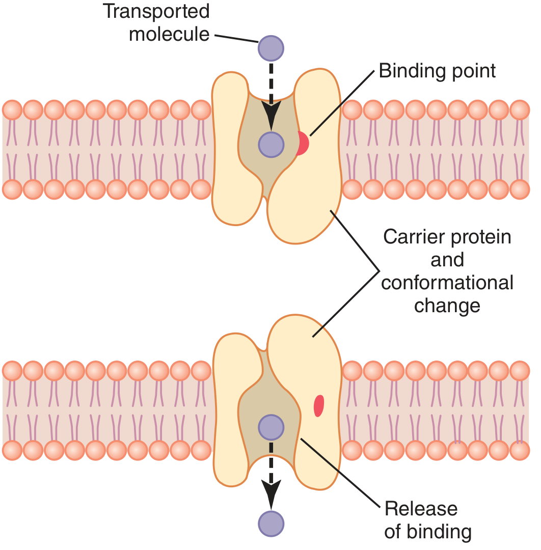
- The molecule enters a pore in the carrier protein and binds to a receptor site.
- A conformational change occurs in the carrier protein - the pore flips to open on the opposite side of the membrane.
- The weak binding is overcome by thermal motion, and the molecule is released on the other side.
- The carrier returns to its original conformation and the cycle repeats.
Because the rate is limited by how fast the carrier can cycle between its two states, there is a maximum transport rate (Vmax).
- Guyton and Hall Textbook of Medical Physiology, p. 68
Rate vs. Concentration: The Vmax Concept
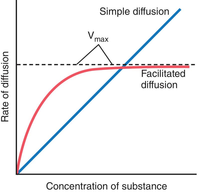
- At low concentrations: facilitated diffusion is faster than simple diffusion (the carrier "helps").
- At high concentrations: all carrier binding sites are occupied (saturated), and the rate plateaus at V
max. Simple diffusion has no such ceiling.
Properties of Carrier-Mediated Transport (shared with active transport)
From Harper's Biochemistry:
- Specific binding site for the solute
- Saturation - there is a finite V
max - Km - the transporter has an affinity constant for its substrate (like an enzyme)
- Competitive inhibition - structurally similar molecules compete for the same binding site
Transporters are analogous to enzymes but do not chemically modify their substrates.
- Harper's Illustrated Biochemistry, 32nd Ed, p. 2502
Types of Transporter Systems
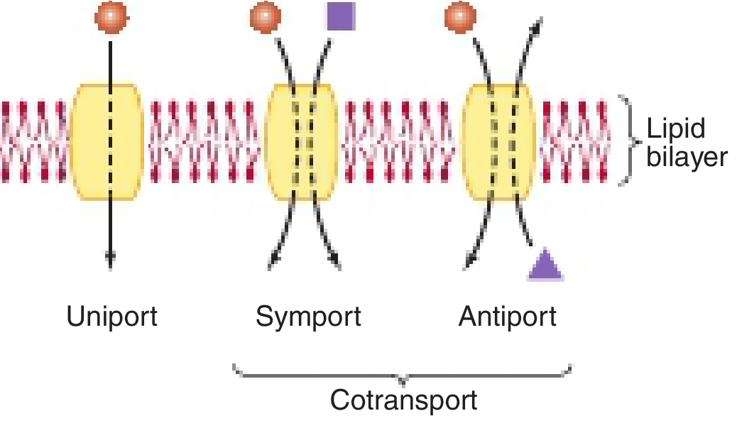
| Type | Movement | Example |
|---|---|---|
| Uniport | One molecule, bidirectional | GLUT transporters (glucose) |
| Symport | Two solutes, same direction | Na⁺-glucose cotransporter (SGLT) |
| Antiport | Two solutes, opposite directions | Na⁺/Ca²⁺ exchanger |
Symport and antiport together are called cotransport systems. Note that cotransport with Na⁺ (e.g., SGLT) is actually active transport (secondary), not facilitated diffusion, since it goes against the glucose gradient.
Key Examples
1. GLUT Transporters (Glucose)
- At least 14 members of the GLUT family have been identified.
- GLUT4 is found in skeletal muscle, adipose tissue, and heart.
- Insulin stimulates translocation of GLUT4 from intracellular vesicles to the plasma membrane, increasing glucose uptake 10-20 fold.
- D-galactose and phlorizin competitively inhibit glucose transport via GLUT4.
- The non-physiologic stereoisomer L-glucose is NOT transported (demonstrates stereospecificity).
2. Amino Acids
- Most amino acids cross cell membranes via carrier-mediated facilitated diffusion or Na⁺-dependent active transport.
3. Fructose
- Absorbed at the apical membrane of intestinal epithelium via facilitated diffusion (GLUT5).
4. Microbiology Note
- Facilitated diffusion is common in eukaryotic microorganisms (e.g., yeast) but rare in prokaryotes.
- Glycerol is one of the few molecules transported by facilitated diffusion in bacteria.
Transporters vs. Ion Channels
| Transporters | Ion Channels | |
|---|---|---|
| Mechanism | Bind solute + undergo conformational change | Form pores in membrane |
| Transport type | Passive (facilitated diffusion) AND active | Passive only |
| Speed | Slower | Faster (ions flow through open pore) |
- Harper's Illustrated Biochemistry, 32nd Ed
Summary
Facilitated diffusion is passive (no ATP), carrier-dependent, saturable, stereospecific, and subject to competitive inhibition. It allows hydrophilic molecules (like glucose and amino acids) that cannot cross the lipid bilayer freely to move rapidly down their concentration gradient. The hallmark difference from simple diffusion is the Vmax plateau seen at high substrate concentrations.
Secondary transport explain in easy language
Secondary transport uses the energy stored in one ion's gradient (usually Na⁺) to move another molecule - no ATP directly, but it piggybacks on work already done by the Na⁺-K⁺ pump.
Searching sources...
Finding Sources
Finding Sources
Reading File
Reading File
Reading File
Loading Image
Loading Image
Reading File
Loading Image
Finding Sources
Here is a simple, easy-to-understand explanation of Secondary Transport (Secondary Active Transport):
Secondary Transport - Simple Explanation
The Big Idea: "Riding on Na⁺'s Back"
Think of it like a toll road system:
- The Na⁺-K⁺ pump (primary active transport) uses ATP to constantly push Na⁺ out of the cell. This creates a high Na⁺ concentration outside and low concentration inside.
- This is like charging a battery - energy is now stored in that Na⁺ gradient.
- Secondary transport uses that stored energy to move another molecule. No ATP is needed directly!
That's why it's called secondary - it borrows energy indirectly from the Na⁺ gradient (which itself was created by ATP-driven primary transport).
The Two Types
1. Cotransport (Symport) - "Both go IN together"
Na⁺ rushes into the cell (going downhill), and it drags another molecule in with it - even if that molecule is going against its own gradient.
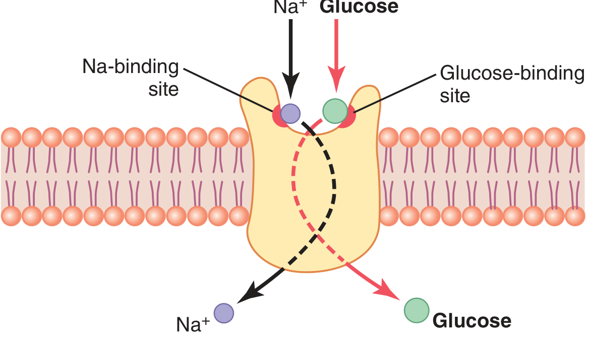
Best example - Na⁺-Glucose Cotransport (SGLT):
- The SGLT carrier has two binding sites - one for Na⁺, one for glucose.
- Both must bind for the carrier to rotate.
- Na⁺ flows in down its gradient → glucose gets carried in too, even against its own concentration gradient.
- This is how your intestine absorbs glucose from food and your kidney rescues glucose from urine.
Other cotransport examples:
- Na⁺ + amino acids (intestine and kidney)
- Na⁺ + K⁺ + 2Cl⁻ (NKCC transporter in kidney thick ascending limb)
2. Countertransport (Antiport/Exchange) - "One goes IN, other goes OUT"
Na⁺ still rushes in, but this time it pushes another molecule out in the opposite direction.
Best example - Na⁺/Ca²⁺ Exchanger:
- Na⁺ enters the cell → Ca²⁺ is pushed out of the cell.
- This keeps intracellular Ca²⁺ low (important for heart muscle relaxation).
Another example - Na⁺/H⁺ Exchanger:
- Na⁺ enters → H⁺ is pushed out.
- Important for regulating cell pH and kidney acid secretion.
The Full Picture: How it all connects
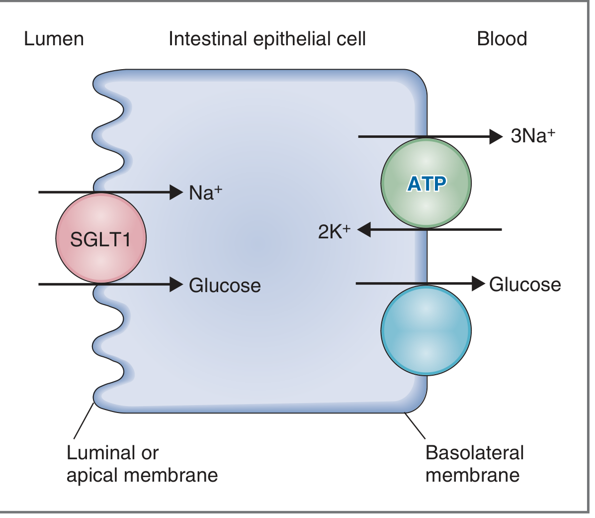
This diagram shows the complete picture in a gut cell:
- Luminal (apical) side: SGLT1 cotransports Na⁺ + glucose into the cell from the gut.
- Basolateral side: Na⁺-K⁺ ATPase uses ATP to pump Na⁺ out (keeps intracellular Na⁺ low, maintaining the gradient for SGLT1). Glucose exits by facilitated diffusion (GLUT) into the blood.
The Na⁺-K⁺ pump is constantly "recharging the battery" that powers SGLT.
What Happens if You Block the Na⁺-K⁺ Pump?
If you give ouabain (a poison that blocks Na⁺-K⁺ ATPase):
-
Intracellular Na⁺ rises → the gradient disappears → SGLT and all other secondary transporters stop working.
-
This shows that all secondary transport indirectly depends on ATP, just not directly.
-
Costanzo Physiology 7th Edition, p. 17-18
Quick Summary Table
| Feature | Secondary Transport |
|---|---|
| Direct ATP use? | No |
| Indirect ATP use? | Yes (via Na⁺ gradient) |
| Driving force | Na⁺ electrochemical gradient |
| Who creates gradient? | Na⁺-K⁺ ATPase |
| Cotransport (symport) | Both molecules go same direction |
| Countertransport (antiport) | Molecules go opposite directions |
| Key examples | SGLT (glucose), Na⁺/Ca²⁺, Na⁺/H⁺ |
One-line memory trick:
Primary transport = uses ATP directly. Secondary transport = uses the gradient ATP already built.
Homeostasis defination with negative feedback and positive feedback explain it an easy manner
Homeostasis is the body's ability to maintain a stable internal environment despite external changes. Negative feedback corrects deviations (most common), while positive feedback amplifies a response until a task is complete.
Searching sources...
Finding Sources
Finding Sources
Finding Sources
Finding Sources
Reading File
Reading File
Finding Sources
Finding Sources
Reading File
I have everything needed. Here is a simple, clear explanation:
Homeostasis - The Body's "Stay Normal" System
Definition
Homeostasis is the body's ability to maintain a stable, constant internal environment even when the outside world keeps changing.
Think of it like a thermostat in a room - no matter how hot or cold it gets outside, the thermostat keeps the room at a comfortable temperature.
The body does this for:
- Body temperature (37°C)
- Blood glucose levels
- Blood pressure
- Blood pH (7.35-7.45)
- Na⁺, K⁺, Ca²⁺ concentrations
- Oxygen and CO₂ levels
"Homeostasis is the control of a vital parameter." - Medical Physiology (Boron & Boulpaep)
The 4 Parts of Any Homeostatic System
Every control system in the body has these 4 parts:
| Part | Role | Example (Blood Glucose) |
|---|---|---|
| Sensor / Receptor | Detects the change | Beta cells sense high glucose |
| Set point | The "normal" target value | Normal glucose = 70-115 mg/dL |
| Control center | Compares signal to set point, decides response | Pancreas |
| Effector | Carries out the corrective action | Insulin released → cells take up glucose |
1. Negative Feedback - "The CORRECTOR" ✅
Simple Idea:
When something goes too high, the body brings it down. When something goes too low, the body brings it up. It opposes (goes negative to) the change. That's why it's called negative.
This is the most common type. The body uses it constantly, thousands of times per second.
Easy Analogy: Room Thermostat
- You set the thermostat to 25°C (set point).
- Room gets too hot → thermostat turns on AC → room cools down.
- Room gets too cold → thermostat turns on heater → room warms up.
- In both cases, the system corrects back to 25°C.
Medical Example 1: Blood Glucose
You eat food
    ↓
Blood glucose RISES above normal
    ↓
Pancreas SENSES the rise (sensor)
    ↓
Pancreas releases INSULIN (effector)
    ↓
Cells take up glucose → blood glucose FALLS back to normal
    ↓
Insulin release STOPS (feedback loop complete)
The high glucose triggered a response that opposed it (negative feedback).
Medical Example 2: Blood CO₂
- CO₂ rises in blood → chemoreceptors detect it → brain tells lungs to breathe faster → more CO₂ is expelled → CO₂ returns to normal.
- CO₂ falls too low → breathing slows down → CO₂ builds back up.
Always correcting. Always opposing the change.
Key Point: Gain
The gain tells you how effectively the system corrects. Higher gain = better correction. Body temperature control has a gain of about 33 (very effective). If the body temperature rises 1°C above normal, the corrective response brings it back so that only 1/33rd of that rise remains.
2. Positive Feedback - "The AMPLIFIER / FINISHER" 🔁
Simple Idea:
When something goes up, the body makes it go even higher. It pushes in the same direction - amplifying the change instead of correcting it. It does NOT return to a set point. Instead it builds and builds until a task is completed, then stops.
This is rare compared to negative feedback. The body only uses it for specific "all-or-nothing" events.
Easy Analogy: Microphone Feedback
When a microphone gets too close to a speaker, the sound goes into the mic → gets amplified → comes out louder → goes back into the mic → even louder → screeching noise! It keeps amplifying itself.
Medical Example 1: Childbirth (Labour)
Baby's head pushes on cervix
    ↓
Cervix stretches → signals sent to brain
    ↓
Brain releases OXYTOCIN
    ↓
Oxytocin causes stronger uterine contractions
    ↓
Stronger contractions push baby harder against cervix
    ↓
Even MORE oxytocin released...
    (keeps amplifying)
    ↓
Baby is BORN → stretch stimulus removed → cycle ends ✅
The positive feedback loop ends naturally once the baby is delivered.
Medical Example 2: Blood Clotting
Blood vessel is injured
    ↓
A small amount of thrombin (clotting factor) is formed
    ↓
Thrombin triggers MORE thrombin formation
    ↓
More thrombin = more clot formation
    ↓
Clot grows rapidly until the wound is sealed ✅
    ↓
Bleeding stops → positive feedback ends
Once clotting is part of an overall system, it serves the goal of restoring normal bleeding-free state - so it is actually nested inside a larger negative feedback process.
"In each case in which positive feedback is useful, the positive feedback is part of an overall negative feedback process." - Guyton and Hall Textbook of Medical Physiology
Positive Feedback Gone Wrong: Vicious Cycles ⚠️
Positive feedback can be dangerous if it is not controlled:
- Haemorrhagic shock: Losing blood → heart gets weaker → pumps less blood → organs get less oxygen → heart gets even weaker → more blood loss... → death.
- The body loses 2+ litres of blood without intervention → this vicious cycle cannot be stopped on its own.
Side-by-Side Comparison
| Feature | Negative Feedback | Positive Feedback |
|---|---|---|
| Direction | Opposes the change | Amplifies the change |
| Goal | Return to set point (stability) | Complete a task (all-or-nothing) |
| Frequency | Very common | Rare |
| End point | Steady state maintained | Event completed then stops |
| Examples | Body temp, blood glucose, BP, CO₂ | Childbirth, blood clotting, LH surge (ovulation) |
| If uncontrolled | Fine - body stays stable | Dangerous - can cause vicious cycles |
One-Line Memory Tricks
-
Negative feedback = "Fix it and calm it down" (like a THERMOSTAT 🌡️)
-
Positive feedback = "Keep going until the job is done" (like a DOMINO effect 🁢)
-
Guyton and Hall Textbook of Medical Physiology, pp. 25-28
-
Medical Physiology (Boron & Boulpaep), p. 16
How insulin help in in glucose take up
Insulin binds to its receptor on cell surfaces and triggers a signaling cascade that moves GLUT4 transporters to the cell membrane, allowing glucose to enter the cell.
Searching sources...
Finding Sources
Finding Sources
Reading File
Reading File
Reading File
Reading File
Loading Image
Finding Sources
Reading File
Loading Image
Loading Image
Here is a simple, step-by-step explanation of how insulin helps glucose enter cells:
How Insulin Helps Glucose Enter Cells
The Basic Problem First
Glucose is a large, hydrophilic (water-loving) molecule. It cannot cross the fatty cell membrane on its own. It needs a door - and that door is the GLUT4 transporter protein.
But here is the trick: GLUT4 doors are hidden inside the cell (stored in vesicles) most of the time. Insulin is the key that brings these doors to the cell surface.
Step 1: First, How is Insulin Released?
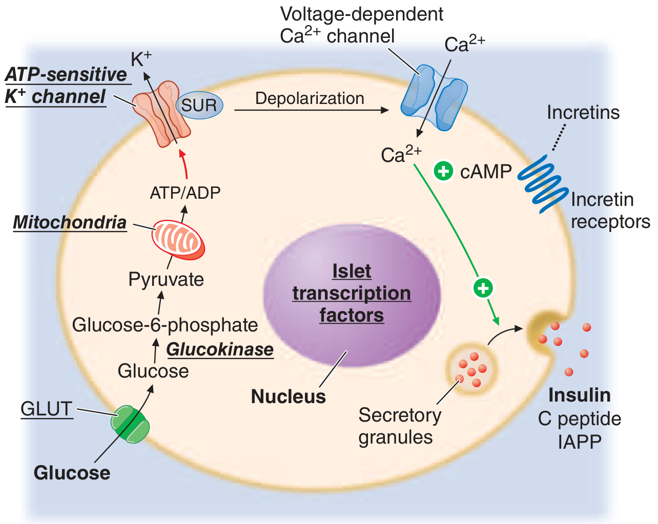
When you eat food and blood glucose rises:
- Glucose enters the pancreatic beta cell via GLUT transporters
- Glucose is metabolised → ATP is produced
- High ATP closes K⁺ channels → cell membrane depolarises
- Depolarisation opens Ca²⁺ channels → calcium floods in
- Calcium triggers insulin secretion from secretory granules
Incretins (GLP-1, GIP) from the gut also boost this process after a meal.
Step 2: Insulin Travels to Target Tissues
Insulin enters the blood and reaches its main targets:
- Skeletal muscle (biggest glucose consumer)
- Adipose tissue (fat cells)
- Liver
Step 3: Insulin Binds to Its Receptor - The Signal Cascade
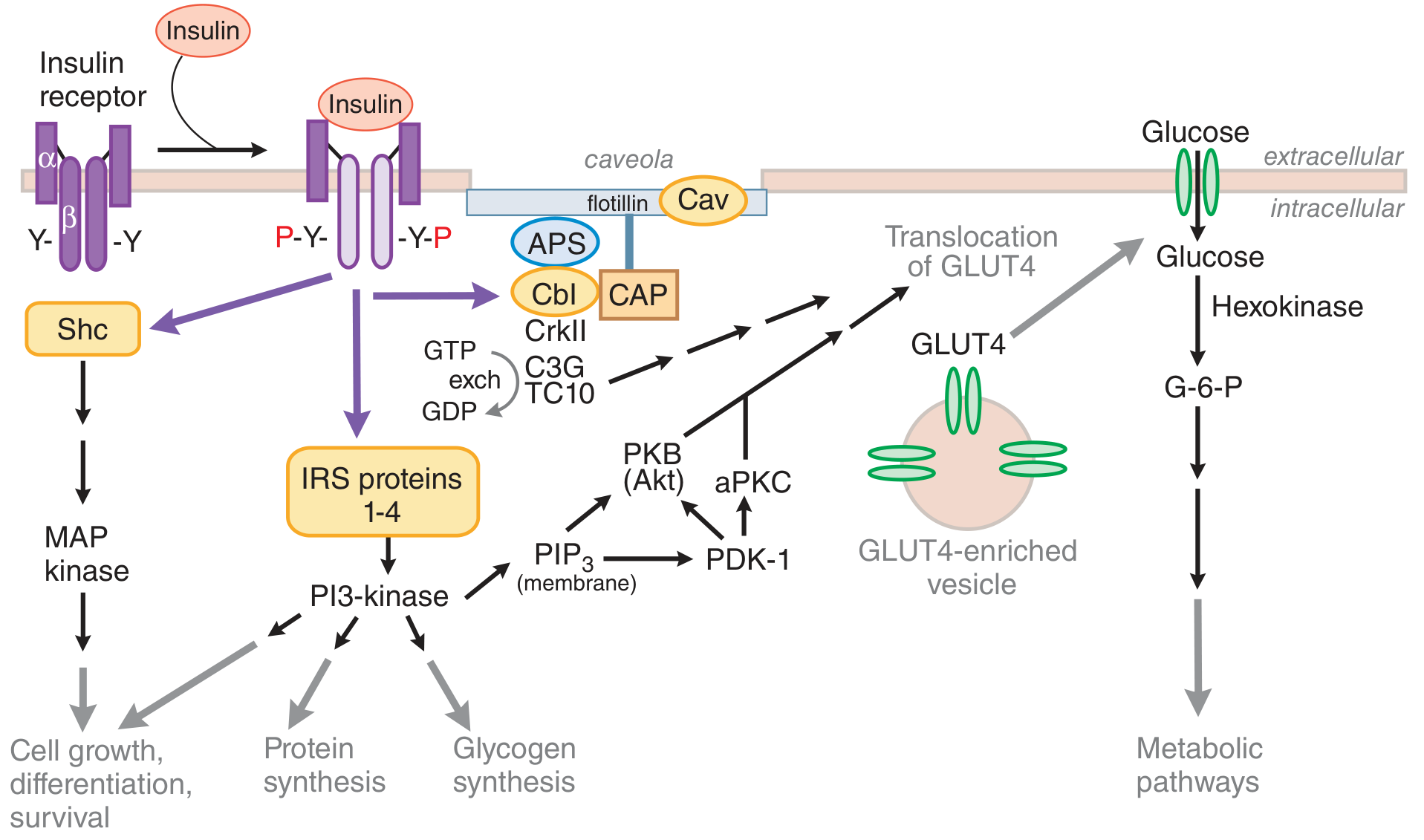
Think of this like a chain of dominoes falling:
Insulin binds to Insulin Receptor (α subunit)
        ↓
Receptor's β subunit activates its own Tyrosine Kinase
        ↓
Receptor AUTO-phosphorylates itself (turns itself ON)
        ↓
IRS proteins (Insulin Receptor Substrates) get phosphorylated
        ↓
IRS activates PI3-Kinase (PI3K)
        ↓
PI3K makes PIP3 (a lipid messenger)
        ↓
PIP3 activates PDK-1
        ↓
PDK-1 activates Akt2 (PKB)
        ↓
Akt2 signals GLUT4 vesicles to MOVE to the cell surface
        ↓
GLUT4 transporters appear on the plasma membrane 🚪
        ↓
Glucose flows IN via facilitated diffusion ✅
- Harrison's Principles of Internal Medicine 22E, p. 3242
- Goodman & Gilman's Pharmacological Basis of Therapeutics, p. 1046
Step 4: Once Inside - What Happens to Glucose?
Once glucose enters the cell through GLUT4:
| Action | Enzyme | Result |
|---|---|---|
| Phosphorylated to G6P | Hexokinase II (also stimulated by insulin) | Glucose trapped inside cell |
| Stored as glycogen | Glycogen synthase (activated by insulin) | Energy storage |
| Used for energy | Glycolysis pathway | ATP production |
| Converted to fat | Lipogenesis | Long-term storage |
The GLUT Family - Different Doors for Different Tissues
| Transporter | Location | Special Feature |
|---|---|---|
| GLUT1 | Brain, RBCs, placenta | Always present, insulin-independent |
| GLUT2 | Liver, pancreas, intestine | High capacity, low affinity (senses glucose levels) |
| GLUT3 | Neurons | Very high affinity - brain always gets glucose first |
| GLUT4 | Muscle, fat, heart | Insulin-dependent - the key one for blood glucose control |
| GLUT5 | Small intestine | Transports fructose, NOT glucose |
- Tietz Textbook of Laboratory Medicine, 7th Ed
Simple Analogy - The Parking Garage
Imagine GLUT4 transporters are parked cars inside a garage (intracellular vesicles). They are ready but not doing anything.
- No insulin → garage door is shut → cars (GLUT4) stay inside → glucose cannot enter the cell
- Insulin arrives → garage door opens → cars (GLUT4) drive out to the street (plasma membrane) → glucose can now enter freely
When insulin levels drop back to normal → GLUT4 is pulled back inside (endocytosis) → glucose entry slows down again.
What Goes Wrong in Diabetes?
| Type | Problem | Effect |
|---|---|---|
| Type 1 Diabetes | Pancreas destroys beta cells → NO insulin made | No signal to open GLUT4 doors → glucose stays in blood |
| Type 2 Diabetes | Cells become resistant to insulin | Insulin binds but the signaling cascade (PI3K/Akt) is weakened → GLUT4 does not move to surface properly |
In Type 2, the PI3K-Akt2 pathway is impaired, often due to excess fatty acids causing abnormal serine phosphorylation of IRS proteins, which blocks the normal signal.
One-Line Summary
Insulin binds its receptor → activates PI3K → Akt2 → GLUT4 moves from inside the cell to the cell surface → glucose enters by facilitated diffusion → blood sugar drops. ✅
This is a shared conversation. Sign in to Orris to start your own chat.