
Easy diagrams to be drawn in university exam of Pterygopalatine fossa
Reading File
The pterygopalatine fossa (PPF) is a high-yield anatomy topic. Key diagrams commonly asked in exams include its boundaries, communications, contents, and the nerve/vessel courses through it. Let me pull detailed content from the medical library now.
Searching sources...
Finding Sources
Searching Images
pterygopalatine fossa diagram anatomy communications
Reading File
Reading File
Searching Images
pterygopalatine ganglion maxillary nerve branches
Loading Image
Reading File
Loading Image
Reading File
Loading Image
Reading File
Reading File
Loading Image
Reading File
I now have all the content needed from Gray's Anatomy for Students to give a thorough, exam-focused answer. Let me compile the complete response.
Pterygopalatine Fossa - Easy Diagrams for University Exams
Here are the most commonly asked and easiest-to-draw diagrams for the pterygopalatine fossa (PPF), sourced from Gray's Anatomy for Students and KJ Lee's Essential Otolaryngology.
Diagram 1: Boundaries of the PPF (Box Diagram - Easiest to Draw)
Draw a simple rectangular box with labeled walls. This is the most commonly asked and takes under 2 minutes to draw.
ROOF
(sphenoid body + palatine bone - orbital process)
┌─────────────────────────────┐
│ │
POSTERIOR│ PTERYGOPALATINE FOSSA │ANTERIOR
(pterygoid│ │(posterior wall of
process +│ │ maxillary antrum)
greater │ Inverted teardrop │
wing of │ shape │
sphenoid)│ │
└─────────────────────────────┘
FLOOR
(apex - opens into palatine canal)
MEDIAL: palatine bone / nasal mucoperiosteum
LATERAL: open → pterygomaxillary fissure → infratemporal fossa
Walls (bones forming them):
| Wall | Bone |
|---|---|
| Anterior | Posterior surface of maxilla |
| Medial | Lateral surface of palatine bone |
| Posterior + Roof | Sphenoid bone (pterygoid process + greater wing) |
| Lateral | Open gap = pterygomaxillary fissure |
Here is a textbook diagram showing the skeletal boundaries:
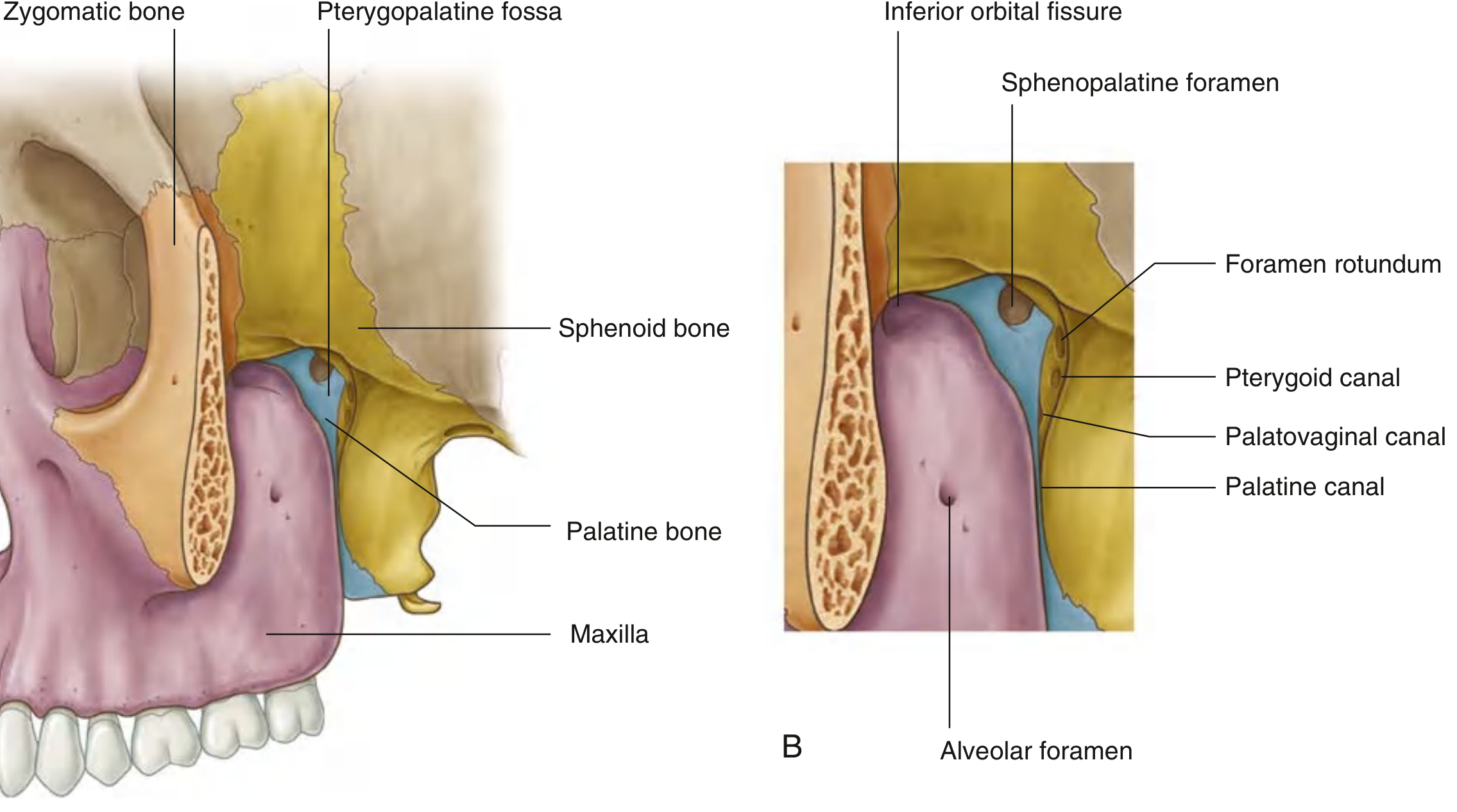
Diagram 2: Communications / Gateways of the PPF (Spider/Wheel Diagram - Most Exam-Favourite)
This is the single most asked diagram. Draw the PPF as a central box with 7 arrows pointing outward, each labeled with the gateway and where it leads.
Middle Cranial Fossa
↑ Foramen Rotundum (V2 enters)
↑ Pterygoid Canal (nerve of pterygoid canal)
Nasal Cavity ┌──────────────┐ Infratemporal Fossa
← Sphenopalatine │ │ ← Pterygomaxillary Fissure
Foramen │ PPF │ (maxillary artery enters;
│ (centre) │ post. sup. alveolar N. exits)
Orbit └──────────────┘
↑ Inferior │
Orbital Fissure ↓
Palatine Canal
(greater & lesser palatine N. + A.)
│
Roof of Mouth (Hard Palate)
Palatovaginal Canal → Nasopharynx
Here is the textbook "gateways" diagram:
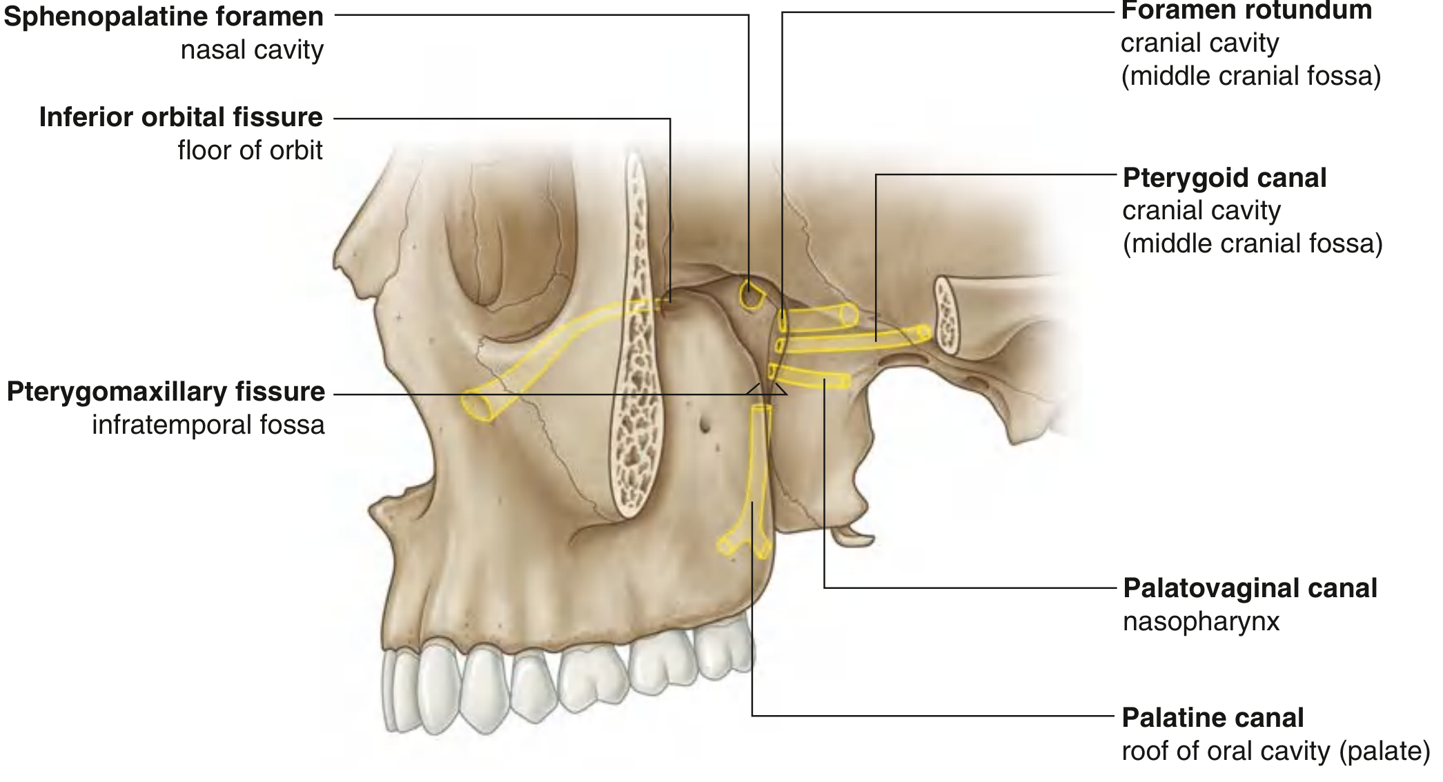
Summary table to write alongside the diagram:
| Gateway | Communicates With | Structure Passing Through |
|---|---|---|
| Foramen rotundum | Middle cranial fossa | Maxillary nerve V2 (enters) |
| Pterygoid canal | Middle cranial fossa | Nerve of pterygoid canal |
| Pterygomaxillary fissure | Infratemporal fossa | Maxillary artery (enters), post. sup. alveolar N. (exits) |
| Inferior orbital fissure | Floor of orbit | Infra-orbital N. + A., zygomatic N. |
| Sphenopalatine foramen | Lateral wall of nasal cavity | Sphenopalatine vessels, nasal branches |
| Palatine canal | Roof of oral cavity (hard palate) | Greater + lesser palatine N. + A. |
| Palatovaginal canal | Nasopharynx | Pharyngeal branch of maxillary N. |
Diagram 3: Contents of the PPF
Three main contents - the mnemonic is MAG (Maxillary artery, Autonomic ganglion/pterygopalatine ganglion, nerve - maxillary nerve V2):
┌─────────────────────────────────────┐
│ CONTENTS OF PPF │
│ │
│ 1. MAXILLARY NERVE (V2) │
│ - enters via foramen rotundum │
│ - purely sensory │
│ - exits as infra-orbital N. │
│ via inferior orbital fissure │
│ │
│ 2. PTERYGOPALATINE GANGLION │
│ (= sphenopalatine ganglion) │
│ - largest parasympathetic │
│ ganglion in head │
│ - hangs off V2 by 2 roots │
│ │
│ 3. MAXILLARY ARTERY (3rd part) │
│ - enters via pterygomaxillary │
│ fissure │
│ - terminal part of ext. carotid │
└─────────────────────────────────────┘
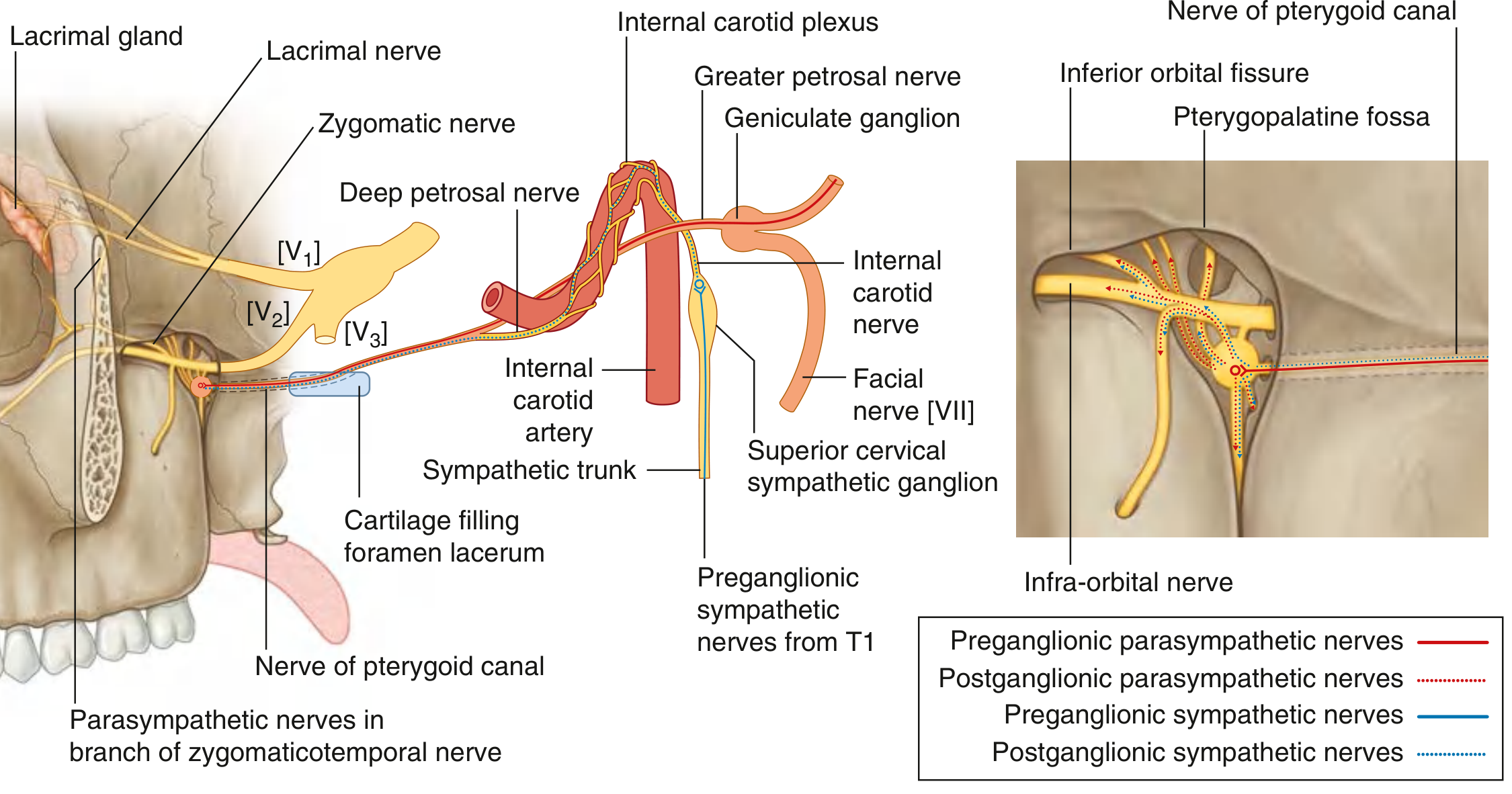
Diagram 4: The Pterygopalatine Ganglion - Roots & Branches (High-Yield Nerve Diagram)
This is the most intellectually demanding diagram but very commonly asked. Draw the ganglion as an oval with inputs coming in (roots) and outputs going out (branches).
ROOTS (3 functional inputs):
- Sensory root - 2 ganglionic branches from V2 (pass through without synapsing)
- Parasympathetic root - greater petrosal nerve (from geniculate ganglion of VII) + deep petrosal nerve (sympathetic, from internal carotid plexus) → unite = Nerve of Pterygoid Canal → SYNAPSE occurs here (preganglionic → postganglionic parasympathetic)
- Sympathetic root - deep petrosal nerve fibers (postganglionic from superior cervical ganglion, T1 origin) - pass through without synapsing
BRANCHES (outputs):
| Branch | Exits Via | Supplies |
|---|---|---|
| Orbital branches | Inferior orbital fissure | Orbital periosteum, ethmoidal + sphenoidal sinuses |
| Greater palatine N. | Greater palatine foramen | Mucosa of hard palate, gingiva |
| Lesser palatine N. | Lesser palatine foramen | Soft palate, palatine tonsil |
| Nasal branches (medial/lateral) | Sphenopalatine foramen | Nasal mucosa, septum |
| Pharyngeal branch | Palatovaginal canal | Nasopharyngeal mucosa |
| Zygomatic N. (back into V2) | Inferior orbital fissure | Skin over temple + cheek; carries parasympathetics to lacrimal gland via zygomaticotemporal N. |
Here is the textbook diagram showing the full nerve pathway through the pterygoid canal and pterygopalatine ganglion:
And the sphenoid bone showing the foramen rotundum and pterygoid canal:
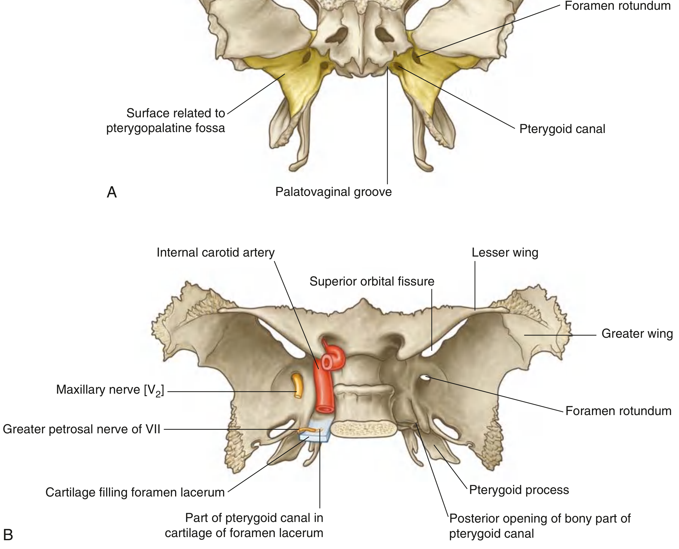
Diagram 5: Maxillary Artery in the PPF (3rd Part)
Draw the maxillary artery entering from the pterygomaxillary fissure and giving 5-6 named branches:
External carotid artery
↓
Maxillary artery
↓ (enters via pterygomaxillary fissure)
┌─── PPF (3rd part of maxillary artery) ───┐
│ │
├──→ Posterior superior alveolar A. │
│ (exits via pterygomaxillary fissure) │
├──→ Infra-orbital A. │
│ (exits via inferior orbital fissure) │
├──→ Greater palatine A. │
│ (exits via palatine canal) │
├──→ Sphenopalatine A. │
│ (exits via sphenopalatine foramen) │
├──→ Artery of pterygoid canal │
└──→ Pharyngeal A. │
(exits via palatovaginal canal) │
Quick Exam Tips
- Shape of PPF: Inverted teardrop / pyramid - narrow inferiorly, wide superiorly
- The only truly "open" wall is the lateral one (pterygomaxillary fissure) - all others are bony walls with specific foramina
- V2 is purely sensory - it does NOT synapse in the pterygopalatine ganglion; only preganglionic parasympathetics synapse there
- Pterygopalatine ganglion = sphenopalatine ganglion (same structure, two names)
- Largest parasympathetic ganglion in the head (the other three are ciliary, otic, submandibular)
- Lacrimation pathway: VII → geniculate ganglion → greater petrosal N. → nerve of pterygoid canal → PPF ganglion → zygomatic N. (V2) → zygomaticotemporal N. → lacrimal N. (V1) → lacrimal gland - damage anywhere = dry eye
All diagrams sourced from Gray's Anatomy for Students, Chapter 8 (Head and Neck).
Endoscopic approaches to Pterygopalatine fossa
Endoscopic approaches to the PPF include the transnasal-transpterygoid route (most common), the transmaxillary route via the posterior maxillary wall, and the transorbital approach. The goal is to access the fossa for tumor resection (juvenile nasopharyngeal angiofibroma, schwannomas), pterygopalatine ganglion block, or vascular control. Let me pull detailed content from the textbooks and recent literature.
Searching sources...
Finding Sources
Searching PubMed
endoscopic approach pterygopalatine fossa
Searching the Web
endoscopic approaches pterygopalatine fossa surgical techniques 2024
Finding Sources
Finding Sources
Reading Web Page
https://pmc.ncbi.nlm.nih.gov/articles/PMC5629706
Reading File
Finding Sources
Finding Sources
Finding Sources
Reading File
Reading File
Searching PubMed
endoscopic endonasal transpterygoid pterygopalatine infratemporal fossa
Reading Article
PMID: 40599854
Reading Article
PMID: 37350591
Loading Image
Now I have comprehensive material from both the textbooks and the latest literature. Here is a thorough answer:
Endoscopic Approaches to the Pterygopalatine Fossa
The PPF sits at a crossroads of six anatomical regions, making it accessible through several endoscopic corridors. The key principle across all approaches is that the PPF is most naturally reached through the nasal cavity because its medial wall - the palatine bone - directly abuts the lateral nasal wall at the sphenopalatine foramen. The table below summarises all major approaches, followed by step-by-step technique for each.
Overview of Approaches
| Approach | Entry Point | Best For | Lateral Reach |
|---|---|---|---|
| Endoscopic endonasal (EEA) standard | Sphenopalatine foramen | Medial PPF, sphenopalatine artery, V2 schwannomas | Moderate |
| Endoscopic transmaxillary (posterior antrostomy) | Posterior wall of maxillary sinus | Maxillary artery ligation, JNA, PPF masses | Good |
| Endoscopic transpterygoid (EETPA) | Pterygoid process after PPF | Petrous apex, Meckel's cave, lateral clivus, cavernous sinus | Excellent |
| Prelacrimal recess approach | Anteromedial maxillary sinus | Lateral PPF, infratemporal fossa | Good |
| Denker's approach (modified medial maxillectomy) | Anterior maxillary wall takedown | Large ITF/PPF tumors, JNA Stage III-IV | Best lateral reach |
| Transoral / transmaxillary (hybrid) | Oral vestibule + maxilla | ITF extension, when endonasal corridor is blocked | Lateral ITF |
1. Standard Endoscopic Endonasal Approach (EEA) to PPF
This is the workhorse approach for accessing the medial PPF and ligating the sphenopalatine artery.
Indications
- Posterior epistaxis (sphenopalatine artery ligation)
- Medial PPF tumors (V2 schwannoma, nerve sheath tumors)
- Pterygopalatine ganglion block / neurolysis
- Biopsy of PPF masses
Surgical Steps
- Positioning & prep: Patient supine, head elevated 15-20°. Decongestion with adrenaline-soaked pledgets. 0° and 30° endoscopes.
- Uncinectomy + maxillary antrostomy: Wide middle meatal antrostomy to expose the posterior wall of the maxillary sinus. The posterior fontanelle is the key landmark.
- Identify crista ethmoidalis: This bony ridge on the posterior lateral nasal wall marks the sphenopalatine foramen - the gateway to the PPF.
- Sphenopalatine foramen dissection: Elevate mucosa posterior to the crista ethmoidalis. The sphenopalatine artery (and its branches) exits here. For epistaxis, this is where ligation/clips are placed.
- Enter PPF: The periosteum of the PPF is incised. The first structure encountered is fat, then the vascular plexus (distal branches of maxillary artery - Vidian, descending palatal, palatovaginal branches), and deepest are the neural structures (V2, Vidian nerve, pterygopalatine ganglion).
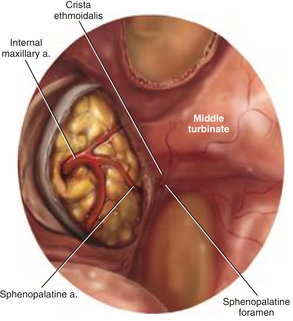
Layered anatomy encountered (medial to lateral, deep to superficial):
Nasal mucosa → crista ethmoidalis → sphenopalatine foramen
→ PPF periosteum → PPF fat → maxillary artery branches
→ pterygopalatine ganglion → V2 (maxillary nerve)
→ Vidian nerve (deepest, most medial, at pterygoid canal)
2. Endoscopic Transmaxillary Approach (Maxillary Artery Ligation)
Used when proximal vascular control is needed, or when a tumor occupies the medial corridor and blocks the standard approach.
Steps (from Cummings Otolaryngology)
- Wide maxillary antrostomy exposing the full posterior wall of the maxillary sinus.
- Strip mucosa from the posterior wall.
- Drill/Kerrison the posterior maxillary bone - preserve the periosteum underneath.
- Electrocauterize periosteum to expose the PPF fat.
- The maxillary artery is visible as a pulsatile structure traveling lateral to medial in the fat.
- Use a blunt probe to isolate the main trunk, dissect circumferentially.
- Place two clips proximally, one distally; apply bipolar between clips, then transect.
- Pack with dissolvable hemostatic material.
Risks specific to this approach: Vidian nerve injury (dry eye/decreased lacrimation), greater palatine nerve injury (palatal paraesthesia), infra-orbital nerve injury (facial numbness - the most common complication, seen in ~10% in large series).
3. Endoscopic Endonasal Transpterygoid Approach (EETPA)
This is the most extended endoscopic approach. After accessing the PPF, the pterygoid process is progressively drilled to reach deeper skull base targets. Finger et al. (2023) describe 5 variants of this approach.
Indications
- Petrous apex lesions (cholesterol granuloma, chondrosarcoma)
- Lateral clivus / inferior cavernous sinus
- Meckel's cave (trigeminal schwannoma)
- Jugular foramen tumors
- Deep infratemporal fossa tumors
Key Anatomic Landmarks (the "three foramina" of EETPA)
The EETPA is built around three bony foramina on the posterior wall of the PPF:
┌─────────────────────────────────────────────┐
│ POSTERIOR WALL OF PPF │
│ │
│ FORAMEN ROTUNDUM (superolateral) │
│ → V2 enters from middle cranial fossa │
│ │
│ VIDIAN (PTERYGOID) CANAL (medial) │
│ → nerve of pterygoid canal │
│ → KEY landmark: identifies floor of │
│ sphenoid sinus + ICA position │
│ │
│ PALATOVAGINAL CANAL (inferomedial) │
│ → pharyngeal branch to nasopharynx │
└─────────────────────────────────────────────┘
Procedural Steps (Finger et al., Operative Neurosurgery 2023)
- Maxillary antrostomy - standard wide exposure
- Posterior wall of maxillary sinus opened - exposes PPF
- Sphenopalatine artery cauterized at sphenopalatine foramen (prevents bleeding; destroys ipsilateral nasal septal flap reconstructive option - plan reconstruction BEFORE this step)
- PPF contents mobilized - fat, vessels, and pterygopalatine ganglion retracted to expose the pterygoid process
- Identify Vidian canal - this is the crucial landmark. The Vidian nerve runs medially at the floor of the sphenoid sinus and guides safe drilling toward the petrous ICA
- Drill pterygoid process stepwise between the Vidian canal (medially) and foramen rotundum (superolaterally) to expose deeper targets
- Variants depending on target:
- Extended PPF: remove pterygoid plate for lateral sphenoid recess
- Petrous apex: follow Vidian canal posteriorly
- ITF: drill lateral to foramen rotundum
Critical safety rule: The Vidian nerve points directly to the petrous ICA. Drilling medial to the Vidian canal risks catastrophic ICA injury.
4. Prelacrimal Recess Approach vs. Denker's Approach
Both expand the endoscopic corridor into the lateral PPF and infratemporal fossa. Li et al. (2022, Am J Rhinology) compared these two directly.
Prelacrimal Recess Approach
- A mucosal incision is made anterior to the inferior turbinate, preserving the nasolacrimal duct
- The anteromedial maxillary wall is dissected subperiosteally, gaining access to the anterior face of the PPF and lateral ITF without removing the anterior maxillary wall
- Advantage: Preserves nasolacrimal system; avoids facial incisions; good for moderate lateral extension
- Disadvantage: Limited field of view compared with Denker's; steep instrument angle
Denker's Approach (Modified Medial Maxillectomy / Extended Medial Maxillectomy)
- The anterior wall of the maxillary sinus is removed endoscopically to create a wide window
- Provides the broadest lateral exposure of any purely endonasal approach
- Used for large JNAs (Stage III-IV), large PPF/ITF schwannomas, or malignancies
- Advantage: Maximum lateral exposure, allows two-handed dissection, excellent for JNA
- Disadvantage: Loss of anterior maxillary wall support, epiphora risk, greater mucosal disruption
5. Stepwise Endonasal Approach: The 5-Step Protocol (Plzák et al., PMC 5629706)
The most systematic description for tumor resection in the PPF comes from Plzák's series:
| Step | Action | Purpose |
|---|---|---|
| Step 1 | Debulk nasal tumor (piecemeal if JNA blocking corridor) | Open the working corridor |
| Step 2 (Ethmoid step) | Anteroposterior ethmoidectomy + sphenoidotomy + wide antrostomy + partial middle turbinate resection | Create wide bimanual working space |
| Step 3 | Posterior maxillary wall removal - preserve PPF periosteum | Expose PPF without entering it prematurely |
| Step 4 | Incise PPF periosteum → identify fat → then vessels → then nerves | Respect the layer order; neural structures lie deepest |
| Step 5 | Drill Vidian canal root | Prevent JNA recurrence (JNA nidus is at the Vidian canal root) |
Outcomes & Evidence (2025 Systematic Review)
The largest recent series - Zoli et al. (2025, Frontiers in Oncology) - analyzed 100 patients over 20 years of EEA for PPF/ITF lesions:
- Most common histotypes: JNA (36%), malignancies (26%), chordomas (14%)
- Gross total resection: 88% of PPF/ITF tumors
- Most common complication: V2 hypoesthesia (facial numbness) - 10 cases (10%), with 3 being transient
- Factors predicting incomplete resection: tumor extension into the temporo-masseteric zone or tubo-pharyngeal space - these areas are beyond the reach of pure EEA and may require combined open approaches
Indications for Open vs. Endoscopic Approaches
| Feature | Favour Endoscopic | Favour Open / Combined |
|---|---|---|
| Tumor location | Medial PPF, central ITF | Temporo-masseteric zone, infratemporal fat pad |
| Vascular involvement | Pre-embolized JNA | Encasing ICA |
| Extension | Confined to PPF / nasal cavity | Cheek, masticator space, parapharyngeal space |
| Reconstruction | Small defects | Large dural defects, orbit |
| Surgeon experience | High-volume skull base center | Any center |
Common Pitfalls & Complications
- V2 injury (most common) - facial and palatal numbness; avoid by identifying V2 at foramen rotundum before dissecting laterally
- Vidian nerve sacrifice - dry eye (decreased lacrimation); the nerve lies medially at the pterygoid canal floor
- ICA injury - rare but catastrophic; always identify Vidian canal as ICA marker before deep drilling
- Ipsilateral nasal septal flap unavailable - the EETPA requires sphenopalatine artery cauterization, which devascularizes the Hadad-Bassagaisteguy flap; plan skull base reconstruction with contralateral flap or free flap
- JNA recurrence - failure to drill the Vidian canal root; tumor nidus sits at the junction of the pterygoid canal and the sphenopalatine foramen
Sources: Cummings Otolaryngology Head and Neck Surgery (Chapter 47); Scott-Brown's Otorhinolaryngology Head & Neck Surgery Vol. 2; Plzák J et al., PMC5629706; Zoli M et al., PMID 40599854 (Frontiers in Oncology, 2025); Finger G et al., PMID 37350591 (Operative Neurosurgery, 2023)
This is a shared conversation. Sign in to Orris to start your own chat.