
Mechanism of action of penicillin
Penicillin inhibits bacterial cell wall synthesis by blocking the transpeptidase enzymes (penicillin-binding proteins, or PBPs) responsible for cross-linking peptidoglycan strands. Without proper cross-linking, the cell wall weakens and the bacterium lyses due to osmotic pressure. It is bactericidal and works only on actively dividing cells.
Searching sources...
Reading File
Finding Sources
Finding Sources
Finding Sources
Finding Sources
Good - I have several relevant sources. Let me read the most pharmacology-focused ones: Lippincott (mechanism of action section) and Katzung (with the figure), plus check for resistance mechanisms.
Reading File
Reading File
Excellent - I have rich content from both Katzung and Lippincott. Let me also fetch the key diagram images and read a bit more for resistance mechanisms.
Loading Image
Loading Image
Reading File
I now have comprehensive content from two authoritative textbooks with diagrams. Let me compose the full answer.
Mechanism of Action of Penicillin
1. Target: The Bacterial Cell Wall
The bacterial cell wall is a rigid, cross-linked polymer called peptidoglycan (also called murein). It consists of long chains of alternating amino sugars - N-acetylglucosamine (NAG) and N-acetylmuramic acid (NAM) - with short pentapeptide side chains attached to the NAM residues. These side chains terminate in D-alanyl-D-alanine (D-Ala-D-Ala). For the wall to be structurally rigid and functional, these peptide side chains must be cross-linked to one another - a process called transpeptidation.
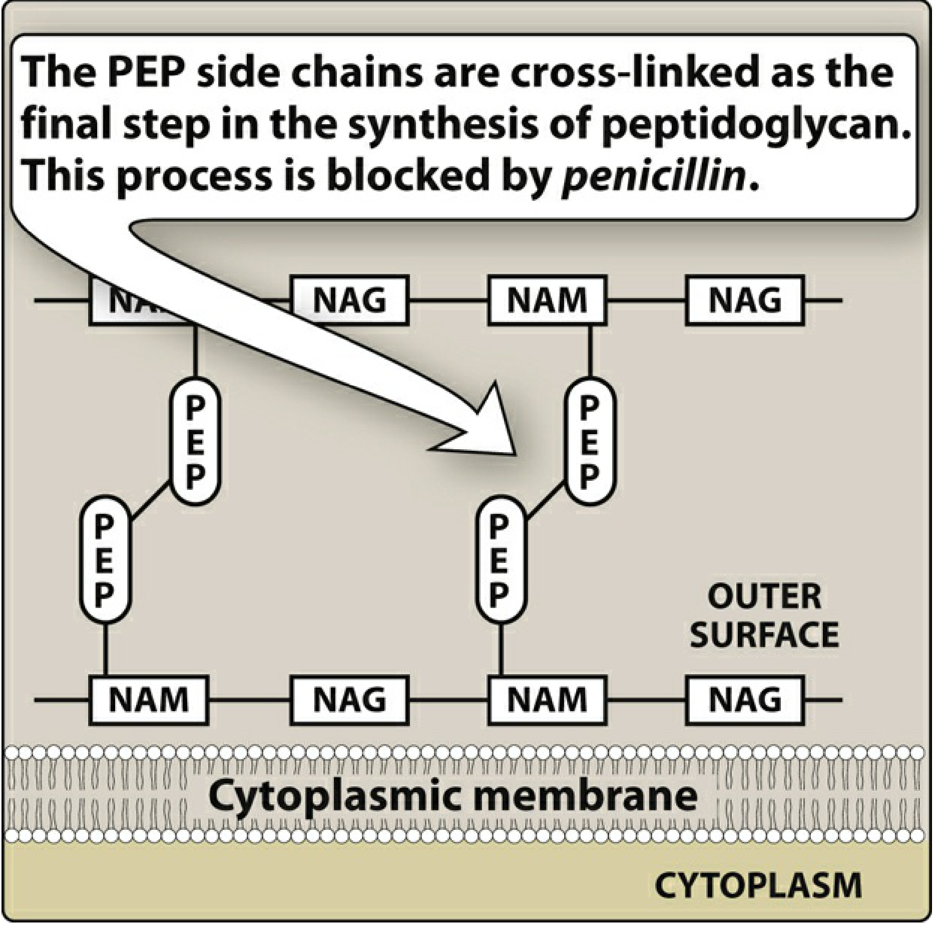
Fig. 1 - The PEP side chains of adjacent peptidoglycan strands must be cross-linked as the final step in cell wall synthesis. Penicillin blocks this step. (Lippincott Illustrated Reviews: Pharmacology, Fig. 29.3)
2. Structural Mimicry - The Key to Penicillin's Action
Penicillin's core structure - the 4-membered β-lactam ring fused to a thiazolidine ring - closely resembles the terminal D-Ala-D-Ala dipeptide of the peptidoglycan strand. This structural mimicry allows penicillin to act as a competitive, irreversible inhibitor of the enzymes that carry out transpeptidation.
3. Penicillin-Binding Proteins (PBPs)
The enzymes responsible for transpeptidation are membrane-anchored enzymes called Penicillin-Binding Proteins (PBPs). They sit in the periplasmic space, embedded in the cytoplasmic membrane. PBPs normally:
- Remove the terminal alanine from the D-Ala-D-Ala terminus
- Form a covalent cross-link between adjacent peptide side chains
When penicillin (a structural analog of D-Ala-D-Ala) enters the cell, it covalently binds to the active site of PBPs, permanently inactivating them. This halts the final cross-linking step of peptidoglycan synthesis.
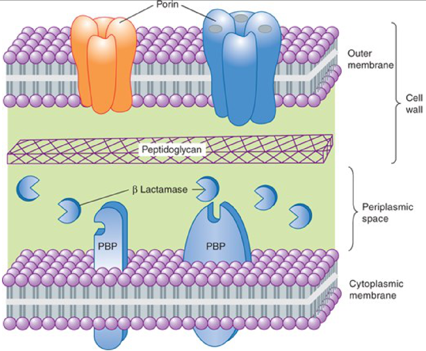
Fig. 2 - Cross-section of a gram-negative bacterial envelope. PBPs are located in the periplasmic space where they cross-link peptidoglycan. β-Lactamase, if present, also resides here and can destroy penicillin. (Katzung's Basic and Clinical Pharmacology, Fig. 43-3)
4. Consequences - Cell Lysis and Death
With PBPs inactivated:
- New peptidoglycan strands cannot be cross-linked
- The cell wall is weakened and loses structural integrity
- Bacteria also activate their own autolysins (enzymes that normally remodel the cell wall during growth)
- The combination of impaired wall synthesis + autolysin activity leads to cell lysis due to unopposed osmotic pressure
- The result: bactericidal killing
Because this mechanism targets an actively synthesizing cell wall, penicillin kills only actively growing and dividing bacteria. It has no effect on dormant or non-replicating cells.
5. Pharmacodynamics
Penicillin is time-dependent (not concentration-dependent) in its killing. Efficacy is maximized by keeping drug concentrations above the MIC (minimum inhibitory concentration) for as long as possible during the dosing interval - not by using very high peak concentrations.
6. Gram-Positive vs. Gram-Negative Bacteria
| Feature | Gram-Positive | Gram-Negative |
|---|---|---|
| Cell wall thickness | Thick peptidoglycan layer, near surface | Thin peptidoglycan, deeper |
| Outer membrane | Absent | Present (LPS bilayer) |
| Drug access to PBPs | Easy - penicillin diffuses freely | Must pass through porins (water-filled channels) |
| General susceptibility | High (in absence of resistance) | Variable; depends on porin availability |
7. Resistance Mechanisms
Three main ways bacteria evade penicillin:
-
β-Lactamase production - The most common mechanism. These enzymes hydrolyze the β-lactam ring, destroying penicillin's activity before it can reach PBPs. Gram-positive bacteria secrete them extracellularly; gram-negatives sequester them in the periplasmic space.
-
Altered PBPs - Mutations produce PBPs with low affinity for β-lactam antibiotics. MRSA (S. aureus) is the classic example: the mecA gene encodes a modified PBP2a that binds penicillin very poorly, conferring resistance to virtually all commercially available β-lactams.
-
Decreased permeability / efflux pumps - Gram-negative bacteria may lose or alter porins, reducing drug entry. Active efflux pumps (e.g., in Klebsiella pneumoniae, P. aeruginosa) can also pump the drug back out before it reaches PBPs.
Sources:
- Lippincott Illustrated Reviews: Pharmacology, 7th ed., Chapter 29 - β-Lactam Antibiotics, pp. 941-948
- Katzung's Basic and Clinical Pharmacology, 16th ed., Chapter 43 - Beta-Lactam & Other Cell Wall- & Membrane-Active Antibiotics, pp. 1239-1240
In short
Penicillin - Mechanism of Action (Short)
Penicillin inhibits bacterial cell wall synthesis by blocking the final step - transpeptidation (cross-linking of peptidoglycan strands).
How:
- Penicillin mimics the D-Ala-D-Ala terminus of peptidoglycan
- It covalently binds to Penicillin-Binding Proteins (PBPs) - the transpeptidase enzymes
- Cross-linking is blocked → cell wall weakens
- Autolysins degrade the defective wall → cell lysis and death
Key points:
- Bactericidal, time-dependent killing
- Works only on actively dividing bacteria
- Gram-positive organisms are more susceptible (thicker, accessible peptidoglycan)
- Main resistance: β-lactamase production (destroys the β-lactam ring)
This is a shared conversation. Sign in to Orris to start your own chat.