
Chicken poc treatment
chickenpox varicella rash skin lesions
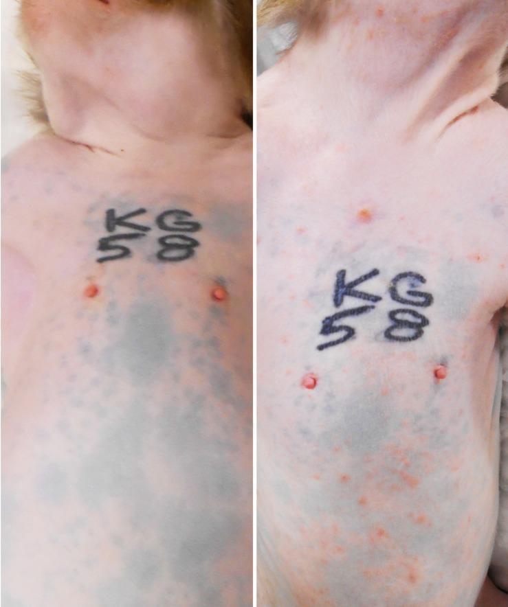
A comparative side-by-side clinical photograph illustrating the progression of primary varicella (chickenpox) rash in a rhesus macaque model. The left panel shows the baseline pre-inoculation state, displaying clear skin on the neck, torso, and abdomen with normal anatomical markings and identifying tattoos. The right panel, captured at 9 days post-inoculation (dpi), demonstrates a severe, generalized acute skin rash. The morphology of the lesions includes a dense distribution of small, erythematous (reddish/pinkish) macules and papules. The rash is widespread, involving the neck, shoulders, chest, and ventral abdomen. This comparison serves to highlight the clinical manifestation of Simian Varicella Virus (SVV), which mirrors the presentation of Varicella-Zoster Virus (VZV) in humans, focusing on the density, distribution, and characteristic appearance of primary varicella lesions.
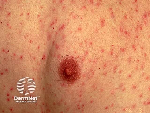
Clinical photography of skin lesions depicting a varicella (chickenpox)–like vesicular exanthem concentrated on the trunk with scattered papules and crusted lesions. The image shows numerous small erythematous papules with central vesiculation and crust formation on a backdrop of unremarkable skin. In classic varicella, lesions appear in crops, evolving from macules to vesicles (dew drops on a rose petal) and eventually crusting, often involving the trunk more than the face, and accompanied by pruritus, fever, and malaise. The depicted lesion is in a later crusted stage, while adjacent lesions are in various stages, reflecting the dynamic evolution of the eruption. Relevant imaging: not radiologic; this is dermatologic clinical photography. Diagnostic relevance: a hallmark varicella rash that aids rapid clinical diagnosis in outpatient or inpatient settings, allowing differential with disseminated herpes simplex, pityriasis rosea, or pityriasis versicolor; contagious until crusting. Clinically, management includes supportive care, antipyretics, antihistamines for itch, and antiviral therapy in high-risk patients; infection control measures to prevent transmission. Potential educational uses include dermatology training, telemedicine triage, clinical case discussions, and AI-based image retrieval for vesicular exanthems.
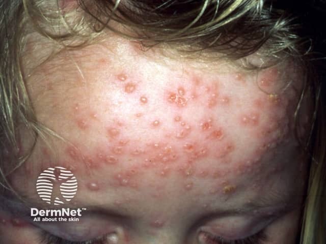
Clinical photography of active varicella (chickenpox) rash on the frontal scalp and forehead of a pediatric patient. Modality: digital color photograph taken under ambient lighting; standard clinical technique without dermoscopy or fluorescence. The image shows numerous small vesicles on an erythematous, slightly raised base distributed across the forehead and extending toward the temple. Lesions are in multiple evolutionary stages, with some vesicles capturing clear serous content, others showing crusted surfaces, and crusts around the margins. The pattern is characteristic of varicella: centripetal distribution with trunk and face involvement, crops of lesions appearing sequentially, and a tendency for vesicles to be more numerous on exposed skin areas. The epidermis and superficial dermis contain superficial fluid-filled vesicles, each about 2-4 mm in diameter, some conforming to the classic dew drop on a rose petal description. Facial involvement may accompany fever and malaise in pediatric patients. Although lesions may be pruritic, Nikolsky sign is typically absent; scarring is uncommon in children. Diagnosis is clinical, supported by history of varicella exposure or vaccination status; differential includes disseminated herpes zoster, pityriasis rosea, and vesicular dermatitis. Recognizing multistage lesions in a contagious exanthem is clinically significant for isolation, supportive therapy, and public health reporting as Varicella-zoster virus infection.
"varicella" AND "treatment" AND "acyclovir"
Chickenpox (Varicella) Treatment
1. Supportive Treatment (All Patients)
- Fever: Paracetamol (acetaminophen). Never aspirin in children - risk of Reye syndrome.
- Itch (pruritus): Calamine lotion, cool baths, antihistamines (e.g. diphenhydramine, cetirizine).
- Skin care: Keep nails cut short to reduce scratching and secondary bacterial skin infection.
- Isolation: Patients are contagious from 1-2 days before rash onset until ALL lesions are dried and crusted.
2. Antiviral Treatment - Acyclovir
Who does NOT routinely need antivirals:
- Otherwise healthy children with uncomplicated varicella - acyclovir is licensed but not routinely recommended because it only modestly decreases symptoms by about 1 day and is not cost-effective in this group.
Who SHOULD receive oral acyclovir (start within 24 hours of rash):
| Group | Regimen |
|---|---|
| Adolescents & adults | Acyclovir 800 mg 5x/day; or valacyclovir 1 g 3x/day (preferred) or famciclovir 500 mg 3x/day |
| Children with chronic pulmonary or skin disease | Acyclovir 20 mg/kg 4x/day (max 800 mg/dose) |
| Mildly immunocompromised | Same oral regimens |
| Pregnant women (especially 3rd trimester) | Consider acyclovir (IV if complications) |
| Neonates (mother had varicella near delivery) | Oral acyclovir |
| Household contacts at high risk | Consider acyclovir |
Note: Valacyclovir and famciclovir achieve higher antiviral drug levels than oral acyclovir and are preferred in nonpregnant adults. Valacyclovir (20 mg/kg 3x/day, max 1 g/dose) is also approved for children ages 2-18 years.
Who needs IV acyclovir (inpatient):
| Group | Regimen |
|---|---|
| Immunocompromised (all ages) | 500 mg/m² every 8 hours (children); 10 mg/kg every 8 hours (adults) for 7-10 days |
| Varicella pneumonia | IV acyclovir 10 mg/kg every 8 hours - best started within 36 hours of hospitalization |
| Severe/disseminated disease | IV acyclovir |
3. Special Situations
| Situation | Management |
|---|---|
| Acyclovir-resistant VZV | Foscarnet 40 mg/kg every 8 hours for 2 weeks or until all lesions crusted |
| HIV-infected adults | Individualized; evaluate for pneumonia or other organ involvement |
| Pregnancy with complications | IV acyclovir recommended |
| Congenital varicella | No controlled data that treatment affects congenital varicella syndrome |
4. Post-Exposure Prophylaxis
- Varicella vaccine given within 3-5 days of exposure can prevent or modify disease.
- Varicella-Zoster Immune Globulin (VariZIG) is given to high-risk exposed persons (immunocompromised, newborns, susceptible pregnant women) as soon as possible after exposure.
- Oral acyclovir post-exposure chemoprophylaxis in susceptible children has been shown to result in either no varicella or a milder modified illness.
5. Complications to Watch For
- Secondary bacterial skin infections (Staphylococcus, Streptococcus)
- Pneumonia (especially in adults and immunocompromised)
- Encephalitis/cerebellar ataxia
- Disseminated disease in immunocompromised