
Diagnosis of appendicitis
appendicitis diagnosis CT scan ultrasound
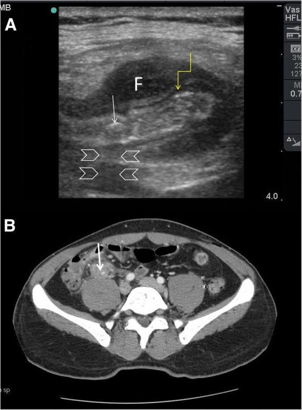
This composite figure demonstrates the diagnostic imaging of acute appendicitis via Point-of-Care Ultrasound (POCUS) and Computed Tomography (CT). Image A is a high-frequency linear probe ultrasound showing a cross-section of a thickened, non-compressible appendix. Key features include a hypoechoic, edematous muscularis propria (indicated by a yellow arrow) and an intraluminal hyperechoic appendicolith/faecolith (white arrow) exhibiting prominent posterior acoustic shadowing (arrowheads). A localized anechoic fluid collection (marked 'F') surrounds the inflamed appendix, consistent with periappendiceal fluid or a phlegmon. Image B is an axial CT scan of the abdomen and pelvis with intravenous contrast, confirming the ultrasound findings in the right iliac fossa. It shows a dilated appendix with wall enhancement and an associated hyperdense faecolith (white arrow) situated anterior to the psoas muscle. This comparison illustrates the utility of POCUS in identifying classic sonographic signs of appendicitis—wall thickening, fecaliths with shadowing, and periappendiceal fluid—and its correlation with definitive CT imaging in a clinical setting of fever and right lower quadrant pain.
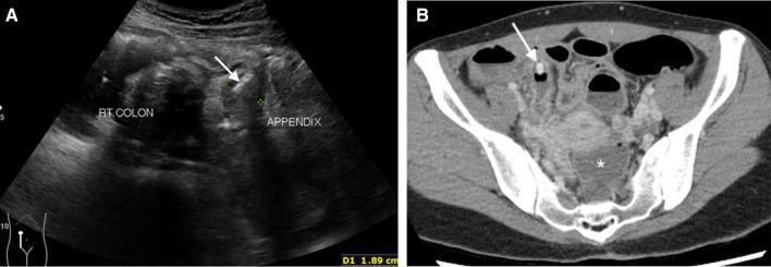
This dual-modality clinical image depicts acute perforated appendicitis in a 35-year-old female. Image A is a transverse abdominal ultrasound showing a dilated, non-compressible appendix adjacent to the right colon. A distinct hyperechoic focus with posterior acoustic shadowing is identified within the appendiceal lumen (arrow), characteristic of an intraluminal appendicolith. Image B is an axial contrast-enhanced CT scan of the pelvis, confirming the presence of a high-attenuation obstructive appendicolith (arrow). Key secondary findings of complicated appendicitis are present, including a significant fluid collection in the rectouterine pouch (cul-de-sac), marked with an asterisk, and generalized peritoneal enhancement. These findings are highly suggestive of appendiceal perforation and secondary peritonitis. The content is suitable for medical education regarding the radiological diagnosis of emergency gastrointestinal pathology, highlighting both sonographic and tomographic features of appendiceal obstruction and its complications.
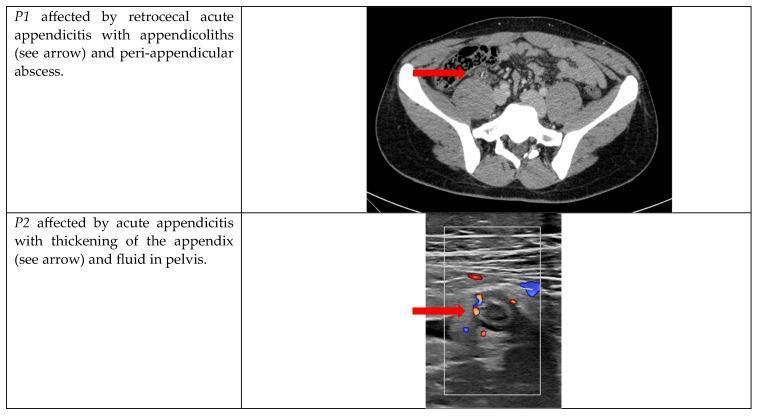
This composite educational graphic presents two diagnostic imaging modalities illustrating acute appendicitis. The top panel (P1) is a transverse axial CT scan of the lower abdomen and pelvis. A red arrow identifies signs of retrocecal acute appendicitis, characterized by the presence of hyperdense appendicoliths and an adjacent peri-appendicular abscess. Bony landmarks including the ilium and sacrum are visible. The bottom panel (P2) displays a Color Doppler ultrasound image in the transverse plane. A red arrow points to a thickened, non-compressible, blind-ended tubular structure consistent with an inflamed appendix. The Doppler overlay demonstrates increased peripheral vascularity (hyperemia) within the appendiceal wall, a hallmark of inflammation. These images serve to compare radiological findings across CT and ultrasonography in the context of complicated and uncomplicated appendicitis for medical training and surgical triage.
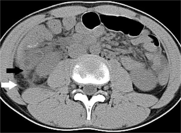
This transverse (axial) CT scan of a pediatric patient demonstrates a primary diagnosis of complicated appendicitis, characterized by an enlarged appendix and secondary inflammatory changes in the retroperitoneal space (RPS). The image highlights fluid collections and thickening across several interfascial planes. Specifically, fluid accumulation is visible in the lateral conal plane (indicated by a black arrow) and the subfascial plane (indicated by a white arrow). Additionally, pathology is noted in the retrorenal plane and surrounding the bridging septa. These findings manifest as regions of increased density and fluid tracks that disrupt the normal fat attenuation of the retroperitoneum. The scan serves as a clinical example of how inflammatory processes from the appendix can track into specific fascial compartments, which is a key radiological indicator for distinguishing between complicated and uncomplicated appendicitis in a clinical setting.
"appendicitis" AND "diagnosis"
Diagnosis of Appendicitis
1. Pathophysiology Basis for Symptoms
- Luminal distension and bacterial proliferation
- Visceral inflammation (causing diffuse, poorly localised pain)
- Progression to mural ischaemia and perforation if untreated
- Bailey and Love's Short Practice of Surgery, p. 1358
2. History and Symptoms
Classic sequence:
- Periumbilical or central colicky pain - the initial visceral pain from appendiceal distension (midgut origin - referred to the umbilicus via T10)
- Anorexia - present in virtually all cases; a highly constant feature
- Nausea and 1-2 episodes of vomiting - vomiting follows the onset of pain (important: pain before vomiting differentiates appendicitis from gastroenteritis)
- Pain migration to the right iliac fossa (RIF) - as parietal peritoneum becomes inflamed, pain shifts and becomes constant, more intense, and localised to the RIF
- Low-grade fever - temperature of 37.2-37.7°C with pulse 80-90 bpm; typically appears after the first 6 hours. In 20% of patients there is no pyrexia or tachycardia in early stages
-
The classic visceral-somatic pain sequence occurs in only ~50% of cases
-
Atypical presentation is more common in the elderly
-
A pelvic appendix may cause suprapubic pain, tenesmus, and urinary frequency without classical RIF tenderness
-
Retrocaecal appendix may present with loin pain, with absent anterior tenderness
-
Bailey and Love's Short Practice of Surgery, pp. 1360-1361
3. Physical Examination Signs
| Sign | Description |
|---|---|
| McBurney's point tenderness | Maximum tenderness at 1/3 of the way from the anterior superior iliac spine (ASIS) to the umbilicus; the most consistent sign |
| Guarding / rigidity | Involuntary muscle guarding over the RIF |
| Rebound tenderness (Blumberg) | Pain on release of deep pressure; indicates peritoneal irritation |
| Rovsing's sign | Palpation of the left iliac fossa produces pain in the RIF; positive in ~50% |
| Psoas sign | Patient lies with right hip flexed; pain on hip extension suggests retrocaecal appendix |
| Obturator sign (Cope's test) | Flexion and internal rotation of the right hip causes hypogastric pain; suggests pelvic appendix |
| Pointing sign | Patient points to the site of maximal pain |
Retrocaecal appendix: Rigidity may be absent; deep pressure may not reach the appendix (the "silent appendix"). Deep loin tenderness and quadratus lumborum rigidity may be more prominent.
Pelvic appendix: Complete absence of abdominal rigidity. Tenderness elicited on rectal examination (rectovesical pouch or pouch of Douglas). Rectal examination is mandatory in all patients with lower abdominal pain.
- Bailey and Love's Short Practice of Surgery, pp. 1360-1361
- Fischer's Mastery of Surgery, p. 7854
4. Laboratory Investigations
| Test | Findings & Utility |
|---|---|
| WBC count | Leukocytosis (>10,000/mm³) is the earliest marker; however, a normal WBC does not exclude appendicitis. Leukopenic presentations are documented. WBC does not distinguish simple from perforated appendicitis |
| C-reactive protein (CRP) | Elevated CRP supports diagnosis; CRP >10 mg/L in children <6 years may predict appendicitis. Used alone, insufficient sensitivity/specificity |
| Combined WBC + CRP | Combined sensitivity up to 98%. Both normal with low pretest probability makes appendicitis very unlikely (100% negative predictive value in one study of 98 patients) |
| ESR | Limited utility alone |
| Urinalysis | Essential to exclude UTI/renal colic. Sterile pyuria or microscopic haematuria can occur in appendicitis (from adjacent inflammation) |
| Pregnancy test (βhCG) | Mandatory in all females of reproductive age to exclude ectopic pregnancy |
Novel biomarkers (e.g. calprotectin, PCT) have not yet demonstrated sufficient utility for routine use.
- Tintinalli's Emergency Medicine, p. 565-566
- Fischer's Mastery of Surgery, p. 7854
5. Scoring Systems
Modified Alvarado Score (MANTRELS):
| Feature | Points |
|---|---|
| Migration of pain to RIF | 1 |
| Anorexia or urinary acetone | 1 |
| Nausea or vomiting | 1 |
| RLQ tenderness | 2 |
| Rebound tenderness | 1 |
| Elevated temperature | 1 |
| WBC >10,000/mm³ | 2 |
| Total | 9 |
- Score 1-4: Low risk
- Score 5-9: Possible or probable appendicitis
- Score ≥7: Probable appendicitis
Other Scoring Systems:
- Appendicitis Inflammatory Response (AIR) score - currently the best-performing in adults
- Adult Appendicitis Score (AAS) - comparable performance
- Pediatric Appendicitis Score (PAS) / Samuel score - used in children
- Tintinalli's Emergency Medicine, p. 565
- Current Surgical Therapy 14e, p. 3459
6. Imaging
Plain Radiography
- Generally not helpful; findings are nonspecific
- May show a non-specific bowel gas pattern, localised ileus, or an appendicolith (visible in up to 50% of children)
Ultrasound (US)
- Non-compressible appendix >6 mm in diameter
- Wall thickening and periappendiceal fluid
- Appendicolith with posterior acoustic shadowing
- Colour Doppler: periappendiceal hyperaemia
CT Abdomen/Pelvis
- Dilated appendix >6 mm with wall thickening
- Periappendiceal fat stranding
- Appendicolith
- Periappendiceal abscess or free fluid
- In perforation: loss of appendix wall continuity, pneumoperitoneum
MRI
- Graded-compression US first
- MRI if US non-diagnostic
- CT as last resort if MRI also non-diagnostic
- Tintinalli's Emergency Medicine, pp. 566-567
- Roberts and Hedges' Clinical Procedures, p. 1708
7. Differential Diagnosis
| Category | Conditions |
|---|---|
| GI | Mesenteric lymphadenitis, Meckel's diverticulitis, terminal ileitis/Crohn's, intussusception, epiploic appendagitis, bowel obstruction |
| GU | Renal colic, UTI/pyelonephritis, ovarian torsion, ectopic pregnancy, PID/tubo-ovarian abscess, testicular pain |
| Musculoskeletal | Psoas abscess, abdominal wall/rectus hematoma |
| Other | Mesenteric adenitis (in children), functional abdominal pain |
In females of reproductive age, PID and ovarian pathology must always be considered.
- Goldman-Cecil Medicine, p. 2191
8. Special Populations
Children
- Difficult to assess; presentation is often atypical especially in infants
- Perforation rate is much higher due to delayed diagnosis (thin omentum unable to wall off inflammation)
- US preferred as initial imaging (avoids radiation)
- PAS score used
Elderly
- Pain localisation to RIF is unusual
- Symptoms may be blunted; leukocytosis may be absent
- Higher perforation rate at presentation
Pregnant Patients
- Appendix displaced superiorly and laterally by gravid uterus
- Peritoneal signs may be reduced
- Most common non-obstetric surgical emergency in pregnancy
- Imaging algorithm: US → MRI → CT
9. Summary: Diagnostic Approach
Suspected Appendicitis
        │
        ▼
Clinical assessment (history + examination)
+ Lab work (WBC, CRP, urinalysis, βhCG in females)
+ Apply scoring system (AIR/Alvarado)
        │
   ┌────┴────┐
High suspicion    Diagnostic uncertainty
(adult, clear     (atypical, pregnancy,
 presentation)     child, obese)
        │                │
  Surgical              Imaging
consultation     ┌───────┴──────────┐
        │        US (first-line in   CT (adults,
        ▼        children/pregnant)  not pregnant)
   Appendectomy         │            │
                    Non-diagnostic   │
                        └─────────MRI/CT
Key principle: Clinical judgment of an experienced clinician remains the most accurate diagnostic tool. Scoring systems and imaging are adjuncts - they should not override strong clinical suspicion.
- Fischer's Mastery of Surgery, p. 7854
- Bailey and Love's Short Practice of Surgery, p. 3007
Treatment protocol
"appendicitis" AND "treatment" AND "antibiotics"
PMID: 38740595
PMID: 38682788
PMID: 39827891
PMID: 39826968
Treatment Protocol for Appendicitis
Step 1: Classify the Appendicitis
| Type | Definition |
|---|---|
| Uncomplicated | Non-perforated, no abscess, no phlegmon, no gangrene |
| Complicated | Perforation with peritonitis, gangrenous appendicitis, phlegmon, or abscess formation (2-10% of all cases) |
Step 2: Initial Resuscitation (all patients)
- IV fluid resuscitation (crystalloid)
- IV antibiotics - start promptly (preoperative dose; repeat intraoperatively if surgery is prolonged)
- Analgesia - opioid-sparing multimodal approach preferred
- NBM (nil by mouth)
- VTE prophylaxis for admitted patients
- Urinalysis, FBC, CRP, βhCG (if female)
- Surgical consultation
Step 3: Definitive Treatment - Decision Tree
Acute Appendicitis Confirmed
         │
    ┌────┴────────────────┐
    │                     │
UNCOMPLICATED         COMPLICATED
    │                     │
    ├── Operative          ├── With diffuse peritonitis/
    │   (standard)        │   sepsis → Emergency surgery
    │                     │
    └── Non-operative      └── With phlegmon/abscess →
        (select patients)      Non-operative first
A. UNCOMPLICATED APPENDICITIS
Option 1: Appendectomy (Standard of Care)
- Lower superficial wound infection rate (~50% lower)
- Shorter hospital stay (~1.1 days shorter)
- Less postoperative pain, faster return to normal activity
- Better for obese patients, young women, diagnostically uncertain cases
- Allows full abdominal survey
- Fischer's Mastery of Surgery, p. 7844
- Sleisenger and Fordtran's GI & Liver Disease, p. 2442
- Mulholland and Greenfield's Surgery, p. 458
Option 2: Non-operative Management (Antibiotics Alone)
Key Evidence
| Trial / Study | Key Finding |
|---|---|
| APPAC trial (Finland) | At 1 year, 72.7% of antibiotic-treated patients avoided appendectomy; at 5 years, 39.1% had undergone appendectomy |
| CODA trial (USA, n=1,552) | Antibiotics non-inferior to appendectomy at 90 days; ~40% required appendectomy at 1 year |
| Cochrane Review 2024 (13 RCTs, n=3,358) | ~69% avoided surgery at 1 year; antibiotic group had fewer wound infections but more recurrences |
| IPD Meta-analysis 2025 (Lancet Gastroenterol Hepatol) | Antibiotics safe alternative; ~66% avoided surgery at 1 year. CRITICAL: patients with appendicolith on imaging had significantly more complications with antibiotics (OR 2.82) and ~49% required appendectomy |
| Pediatric RCT 2025 (Lancet) | In children 5-16 years, antibiotic treatment was inferior to appendectomy by the pre-specified non-inferiority margin (34% vs. 7% failure); appendectomy remains preferred in children |
Antibiotic Regimen for Non-operative Management (APPAC-style):
- IV: Ertapenem 1g daily for 3 days (hospital)
- Oral step-down: Levofloxacin 500 mg daily + metronidazole 500 mg TDS for 7 days
Criteria for Non-operative Management (Uncomplicated Only):
- CT-confirmed uncomplicated appendicitis
- No appendicolith
- Clinically stable, no signs of peritonitis
- Able to comply with follow-up
- Patient preference after informed consent
- Appendicolith present (significantly higher failure/complication rate)
- Perforation, abscess, phlegmon
- Diffuse peritonitis
- Immunocompromised patients
- Children (antibiotics shown inferior in 2025 Lancet RCT)
- Failure to improve within 24-48h of antibiotics
-
~1 in 3 patients will still need appendectomy within 1 year
-
If appendicitis recurs, definitive surgery is recommended
-
Risk of delayed diagnosis of neoplasm (colonoscopy recommended in patients >40 years after non-operative management)
-
Fischer's Mastery of Surgery, p. 7854
-
Mulholland and Greenfield's Surgery, pp. 457-458
B. COMPLICATED APPENDICITIS
1. Perforated Appendicitis with Diffuse Peritonitis / Sepsis
- Urgent source control is mandatory
- Lavage and suction of the peritoneal cavity
- Intraoperative cultures
- IV antibiotics continued postoperatively for 4 days after source control
- Longer duration if source control is inadequate or patient is immunosuppressed
- Single agent: Piperacillin-tazobactam 4.5g IV Q8h OR Ertapenem 1g IV daily
- Combination: Cephalosporin (3rd-gen) or fluoroquinolone + metronidazole
- Avoid: Ampicillin-sulbactam (high E. coli resistance), tigecycline (increased mortality vs comparators)
-
Cefepime or ceftazidime + metronidazole OR
-
Piperacillin-tazobactam monotherapy OR
-
Imipenem / meropenem / doripenem
-
Add vancomycin if MRSA suspected
-
Alternative for beta-lactam allergy: Vancomycin + aztreonam + metronidazole
-
Current Surgical Therapy 14e, p. 1255
2. Appendiceal Abscess or Phlegmon (Without Diffuse Peritonitis)
- Bowel rest + IV fluids + IV antibiotics
- CT-guided percutaneous drainage if abscess ≥3 cm is present (single abscess, accessible)
- Monitor closely for clinical response (fever, WBC, pain trend)
- Step down to oral antibiotics when improving; typically 7-14 days total
- Interval appendectomy: NOT routinely recommended (recurrence risk only ~7.4%)
- Colonoscopy or repeat CT in patients >40 years to exclude underlying neoplasm (found in ~1.2-17% in some series, increasing with age)
-
Failure to respond within 48-72 h
-
Clinical deterioration
-
Rupture/spreading peritonitis
-
Mulholland and Greenfield's Surgery, pp. 458-459
C. SPECIAL POPULATIONS
Pregnancy
- Laparoscopic appendectomy is safe throughout pregnancy (including 2nd/3rd trimester)
- Recommended as first-line definitive treatment
- Second trimester preferred if timing allows
- Non-operative management may be considered but carries higher risk of fetal loss with perforation
- Open appendectomy as alternative; debate continues regarding which approach carries lower fetal risk in late pregnancy
- Postoperative tocolysis: only if uterine irritability is documented
Children
- Appendectomy remains the standard - 2025 Lancet RCT showed antibiotics were inferior to appendectomy (34% vs. 7% failure rate at 1 year)
- Laparoscopic approach preferred
- Thin, poorly developed omentum means faster progression to perforation
- Perforation rates up to 90% in infants <2 years
Elderly
- Higher perforation rate at presentation
- More comorbidities increase surgical risk - may favour initial non-operative management in select cases
- Lower threshold for broad-spectrum antibiotics
D. POST-APPENDECTOMY CARE
- Same-day discharge or next day
- Diet as tolerated post-operatively
- Oral analgesia (paracetamol ± NSAIDs ± short course opioid)
- Return to normal activity within 1-2 weeks
- Continue IV antibiotics for 4 days after source control
- Monitor for:
- Wound infection (most common complication) - open wound if superficial
- Intra-abdominal abscess (higher with laparoscopic approach) - usually requires percutaneous drainage
- Stump appendicitis (rare, 1/50,000) - re-operation required
- Enterocutaneous fistula (rare) - conservative initially; ileocecectomy if persistent
Summary Treatment Algorithm
Confirmed Appendicitis
        │
   Uncomplicated?
     YES    NO (complicated)
      │           │
   Surgical?  Peritonitis?
   preference   YES    NO
      │          │      │
 Laparoscopic  Urgent  Abscess/
 appendectomy surgery  phlegmon
  (preferred) (emer-     │
      │       gency)   Non-op
   OR if     + broad    Abx +
   patient    Abx      drain if
   prefers               ≥3cm
 Antibiotics-             │
 first if NO          Interval
 appendicolith     appendectomy
   + informed         NOT
   consent          routine
- 2024 Cochrane Review confirms antibiotics are a viable alternative to surgery for uncomplicated appendicitis in adults, with ~69% avoiding surgery at 1 year and fewer wound complications, but lower overall treatment success
- 2025 Lancet IPD meta-analysis shows appendicolith presence is a strong contraindication to antibiotic-first management (OR 2.82 for complications)
- 2024 SAGES guidelines give conditional recommendations for operative management as first-line but acknowledge antibiotics as an acceptable alternative with shared decision-making
- 2025 Lancet pediatric RCT definitively showed antibiotics inferior to appendectomy in children - appendectomy remains the standard in pediatric patients
Choice of oral medication and iv fluids in non complicated appendicitis
"appendicitis" AND "oral antibiotic" AND "nonoperative"
"appendicitis" AND "antibiotic" AND "nonoperative management"
PMID: 41604201
PMID: 39504344
uncomplicated appendicitis antibiotic regimen IV oral 2024 2025 guideline amoxicillin clavulanate ciprofloxacin metronidazole
Oral Medications and IV Fluids in Uncomplicated Appendicitis
- Pre-operative / perioperative (patient going to surgery)
- Non-operative management (antibiotics-first strategy, no surgery)
I. IV FLUIDS
Goal
Choice of IV Fluid
| Fluid | Recommendation |
|---|---|
| Lactated Ringer's (LR) | Preferred crystalloid - balanced electrolyte solution, more physiological, reduces hyperchloraemic acidosis |
| Normal Saline (0.9% NaCl) | Acceptable alternative; large volumes can cause hyperchloraemic metabolic acidosis |
| Hartmann's solution | Equivalent to LR, used interchangeably |
How Much?
- Give 500 mL - 1 L IV crystalloid bolus initially in a euvolaemic adult presenting with mild-moderate dehydration
- Reassess: HR, BP, urine output, mucous membranes
- Target urine output ≥0.5 mL/kg/hr
- Maintain IV fluids while NBM (nil by mouth)
- In uncomplicated appendicitis without sepsis, aggressive fluid loading is not required - the goal is simply euvolemia, not resuscitation
"Euvolemia should be established with IV access and crystalloid administration" before proceeding to surgery.
- Fischer's Mastery of Surgery, p. 7839
II. ANTIBIOTICS
Context A: Going to Surgery (Perioperative Prophylaxis)
Recommended IV Antibiotic (Single Preoperative Dose):
| Agent | Dose | Notes |
|---|---|---|
| Cefazolin (1st-gen cephalosporin) | 1-2 g IV (3 g if >120 kg) | First-line SSI prophylaxis; covers skin flora + some GI organisms |
| Cefoxitin (2nd-gen cephalosporin) | 1-2 g IV | Better anaerobic coverage; alternative |
| Ceftriaxone + metronidazole | 1-2 g IV + 500 mg IV | Good gram-negative + anaerobic cover |
| Piperacillin-tazobactam | 3.375 g IV | Broad-spectrum; useful if contamination expected |
- Ciprofloxacin 400 mg IV + metronidazole 500 mg IV
- Or clindamycin + gentamicin (less preferred; potential nephrotoxicity)
Duration: For confirmed uncomplicated (non-perforated) appendicitis, a single preoperative dose is sufficient. No prolonged postoperative antibiotics are needed.
- Fischer's Mastery of Surgery, p. 3412; Current Surgical Therapy 14e
Context B: Non-Operative Management (Antibiotics as Definitive Treatment)
Standard Two-Phase Regimen (APPAC Trial Protocol)
| Agent | Dose | Frequency |
|---|---|---|
| Ertapenem | 1 g IV | Once daily for 3 days |
Ertapenem is a carbapenem with excellent gram-negative and anaerobic coverage, once-daily dosing, and good tolerability. Used in APPAC (n=530) with 72.7% success at 1 year.
| Agent | Dose |
|---|---|
| Ceftriaxone 1-2 g IV Q24h + Metronidazole 500 mg IV Q8h (or 1.5 g Q24h) | Combined gram-neg + anaerobe cover |
| Piperacillin-tazobactam 4.5 g IV Q8h | Single-agent broad-spectrum |
| Ciprofloxacin 400 mg IV Q12h + Metronidazole 500 mg IV Q8h | Alternative; note increasing fluoroquinolone resistance |
| Regimen | Doses | Notes |
|---|---|---|
| Ciprofloxacin 500 mg BD + Metronidazole 500 mg TDS | 7 days | Most widely used; consistent with international guidelines; used in APPAC trial oral phase |
| Amoxicillin-clavulanate 875/125 mg BD | 7 days | Convenient single agent; first-line in many centres |
| Cephalexin 500 mg QDS + Metronidazole 400 mg TDS | 7 days | Alternative when amoxicillin resistance is a concern (avoids fluoroquinolone) |
| Levofloxacin 500 mg OD + Metronidazole 500 mg TDS | 7 days | Used in original APPAC oral phase |
- Ciprofloxacin 500 mg BD + Metronidazole 500 mg TDS for 7 days
Do NOT use:
- Ampicillin-sulbactam alone (high E. coli resistance)
- TMP-SMX alone (high local resistance to E. coli in many regions)
- Metronidazole alone (no gram-negative cover)
CODA Trial Regimen (USA, n=1,552)
- IV ertapenem for 24 hours (inpatient), then
- Oral ciprofloxacin + metronidazole to complete a 10-day total course
- Demonstrated antibiotics were non-inferior at 90 days; 40% required appendectomy at 1 year
Fully Oral Regimen (Outpatient-Only Protocol)
- Ciprofloxacin 500 mg BD + Metronidazole 500 mg TDS x 7 days (90% of patients in that series)
- 76% avoided appendectomy at 1 year
- Suitable only for clinically stable, reliable patients who can return immediately if symptoms worsen
III. SUMMARY TABLE
IV Phase (24-72 hours, inpatient)
| Scenario | First-Line IV | Alternative |
|---|---|---|
| Non-operative management | Ertapenem 1 g OD | Ceftriaxone 1-2 g OD + Metronidazole 500 mg Q8h |
| Preoperative prophylaxis (going to surgery) | Cefazolin 1-2 g (single dose) | Cefoxitin 1-2 g OR Ceftriaxone + Metronidazole |
| Penicillin/ceph allergy | Ciprofloxacin 400 mg Q12h + Metronidazole 500 mg Q8h | Aztreonam + Metronidazole |
Oral Step-Down Phase (7 days, outpatient)
| First-Line | Alternative | Allergy |
|---|---|---|
| Ciprofloxacin 500 mg BD + Metronidazole 500 mg TDS | Amoxicillin-clavulanate 875/125 mg BD | Ciprofloxacin + Metronidazole (if not already used) |
| OR Amoxicillin-clavulanate 875/125 mg BD | Cephalexin 500 mg QDS + Metronidazole 400 mg TDS |
IV Fluids
| Fluid | Rate / Volume |
|---|---|
| Lactated Ringer's (preferred) | 500 mL-1 L bolus to establish euvolemia; then 100-125 mL/hr while NBM |
| Normal Saline (alternative) | Same rate; watch for hyperchloraemia with large volumes |
IV. CRITERIA TO STEP DOWN FROM IV TO ORAL (before discharge)
- Afebrile (<38°C) for >24 hours
- Pain controlled without IV analgesia
- Return of bowel function / tolerating oral intake
- WBC trending down (<12,000)
- Clinically improving (reduced RIF tenderness)
If the patient fails to meet these criteria within 48-72 hours, re-evaluate - consider CT reassessment and escalation to surgery.