A 1-week-old newborn is seen for abnormal thyroid function tests that were performed as part of routine screening.  Serum thyroxine (T4) was low and serum TSH was normal.  The patient was born by spontaneous vaginal delivery at 40 weeks of gestation to a 28-year-old primigravida.  During the pregnancy, the mother took acetaminophen intermittently for low back pain.  Examination of the newborn shows no abnormal findings.  Repeat thyroid function test results are as follows: Free T4 1.2 μg/dL (normal: 0.8-1.95) Total T4 4 μg/dL TSH 3 μU/mL Which of the following is the most likely cause of the patient's abnormal thyroid function tests? A. Decreased T4 to triiodothyronine conversion [11%] B. Deficiency of thyroxine-binding globulin [56%] C. Hypoalbuminemia [7%] D. Hypopituitarism [4%] E. Thyroid dysgenesis [20%] Incorrect. Correct answer is B 56% answered correctly Explanation: This neonate with a low total thyroxine (T4) but normal free T4 and normal TSH likely has a deficiency of thyroxine-binding globulin (TBG).  Congenital TBG deficiency is a benign, X-linked disorder usually identified on newborn screening. In general, the majority of circulating T4 is bound to TBG, which then serves as a storage pool to replenish the biologically active free T4 that is continuously cleared by the kidneys.  Therefore, TBG helps maintain a constant free T4 level. When TBG is deficient, less T4 is bound and a greater proportion is allowed to freely circulate.  Total T4 is low due to the decrease in bound T4 fraction.  However, since physiologic feedback is intact, TSH and free T4 levels remain normal.  Because the levels of active (free) thyroid hormone are normal, patients are clinically euthyroid and asymptomatic, so no treatment is required. (Choice A)  Certain placentally transferred maternal medications (eg, propylthiouracil) decrease conversion of T4 to triiodothyronine (T3), causing transient neonatal hypothyroidism (ie, low T4, high TSH).  Acetaminophen does not interfere with thyroid hormone synthesis. (Choice C)  Because only a small percentage of T4 is bound to albumin, hypoalbuminia does not significantly affect thyroid hormone levels.  In addition, this patient has no risk factors for hypoalbuminemia (eg, malnutrition, sepsis, gastrointestinal/renal loss). (Choice D)  Hypopituitarism causes central hypothyroidism, which is characterized by low TSH, total T4, and free T4.  Other midline defects (eg, cleft lip) or hormonal imbalances (eg, hypoglycemia due to ACTH deficiency) are often present. (Choice E)  Thyroid dysgenesis (eg, ectopy, hypoplasia) is the most common cause of congenital hypothyroidism, which is usually asymptomatic at birth.  In contrast to this case, laboratory findings include high TSH and low total and free T4. Educational objective: Thyroxine-binding globulin deficiency is a benign disorder characterized by low total thyroxine (T4) and normal free T4 and TSH levels.  Patients are euthyroid and do not require treatment. Time spent:176 Seconds QID:19746

The explanation here is clear and the correct answer is well-justified. Let me give you a concise, reinforced summary of the key concepts.
Reading File
Finding Sources
Reading File
Reading File
Reading File
Here is a detailed breakdown of this question and the underlying physiology, drawn from authoritative textbook sources.

Thyroxine-Binding Globulin (TBG) Deficiency - Concept Breakdown

The Core Logic: Free vs. Total T4

This question hinges on understanding the difference between total T4 and free T4:
Hormone FractionWhat it MeasuresClinical Relevance
Total T4Bound + Free T4Affected by binding protein levels
Free T4Unbound, biologically active T4True reflection of thyroid status
TSHPituitary feedback hormoneMost sensitive indicator of thyroid function
The key principle, from Katzung's Basic & Clinical Pharmacology:
"Only about 0.04% of total T4 and 0.4% of T3 exist in the free form. Many physiologic and pathologic states and drugs affect T4, T3, and thyroid transport. However, the actual levels of free hormone generally remain normal, reflecting feedback control."

Thyroid Hormone Binding Proteins

From Tietz Textbook of Laboratory Medicine (Table 57.2), T4 is distributed across three binding proteins:
Protein% of T4 CarriedNotes
TBG75%Dominant carrier - highest affinity
Transthyretin (TTR/prealbumin)~20%Lower affinity
Albumin~5%Very low affinity, but huge capacity
TBG carries the vast majority of circulating T4. Its functions are:
  1. Limiting urinary loss of thyroid hormones
  2. Buffering fluctuations in thyroid output so free hormone stays constant
  3. Acting as a reservoir to maintain steady free hormone levels

What Happens in TBG Deficiency?

When TBG is absent or reduced:
↓ TBG
  → Less T4 bound to protein
  → Total T4 falls (because most T4 was bound to TBG)
  → Transiently, free T4 might rise slightly
  → Pituitary senses normal/slightly elevated free T4
  → TSH remains suppressed/normal
  → Thyroid adjusts output downward
  → New steady state: LOW total T4, NORMAL free T4, NORMAL TSH
The patient's labs fit this pattern perfectly:
  • Free T4: 1.2 ng/dL - normal
  • Total T4: 4 μg/dL - low (but actually within range for a 1-week-old on prematurity scale)
  • TSH: 3 μU/mL - normal
The patient is clinically euthyroid because the biologically active (free) hormone is normal.

Why the Other Choices Fail

A. Decreased T4 to T3 conversion Drugs that inhibit 5'-deiodinase (amiodarone, PTU, beta-blockers, iodinated contrast, corticosteroids) cause low T3 and high reverse T3. If anything, you'd see compensatory TSH rise. Acetaminophen has no effect on thyroid metabolism - confirmed in Katzung.
C. Hypoalbuminemia From Tietz's table above, albumin carries only ~5% of circulating T4. Even severe hypoalbuminemia would minimally affect total T4 levels. This patient also has no risk factors for hypoalbuminemia (no malnutrition, sepsis, or protein-losing disease).
D. Hypopituitarism Central (secondary/tertiary) hypothyroidism = low TSH + low free T4 + low total T4. TSH here is normal at 3 μU/mL, ruling this out entirely. Hypopituitarism also commonly presents with midline defects or hypoglycemia (from ACTH deficiency).
E. Thyroid dysgenesis This is the most common cause of congenital hypothyroidism (ectopic or hypoplastic thyroid). The pattern would be:
  • High TSH (pituitary sensing hormone deficiency)
  • Low total T4
  • Low free T4
Here, free T4 is normal - this rules out primary hypothyroidism of any cause.

Congenital TBG Deficiency - Key Facts

  • Genetics: X-linked recessive (gene on Xq22); affects ~1 in 5,000-15,000 newborns
  • Expression: Males fully affected (hemizygous); females are carriers with partial deficiency
  • Detection: Often caught on newborn screening (low total T4 triggers reflex testing)
  • Treatment: None required - patients are euthyroid with normal free T4 and TSH
  • Pitfall: Never treat based on total T4 alone without checking free T4 and TSH

Summary Table: Differential Diagnosis of Abnormal TFTs in Neonates

ConditionTotal T4Free T4TSHTreatment
TBG deficiencyLowNormalNormalNone
Thyroid dysgenesisLowLowHighLevothyroxine
Central hypothyroidismLowLowLow/normalLevothyroxine
Decreased T4→T3 conversionVariableLowHighStop offending drug
HypoalbuminemiaMinimally affectedNormalNormalTreat cause
The pattern of low total T4 with normal free T4 and normal TSH is the hallmark of a binding protein deficiency - and TBG (carrying 75% of T4) is by far the most impactful when deficient.
  • Katzung's Basic and Clinical Pharmacology, 16th Ed, p. 1075
  • Tietz Textbook of Laboratory Medicine, 7th Ed - Circulating Thyroid Hormone Binding Proteins
  • The Harriet Lane Handbook, 23rd Ed - Age-based thyroid function reference ranges

Explain me core concept that was due to which my question get wrong

The One Core Concept You Missed

Looking at the answer distribution, 20% chose Thyroid dysgenesis (E) - which is the most common wrong answer. That's likely where you went.
Here is the single concept that separates the right answer from the wrong one:

Free T4 vs. Total T4 - They Are NOT the Same Thing

Most students instinctively think:
"Low T4 = hypothyroidism = something wrong with the thyroid"
That mental shortcut is wrong and is exactly what this question is testing.

The Analogy That Makes This Click

Think of thyroid hormone like cash in a wallet vs. cash in a bank:
AnalogyThyroid
Free T4Cash in your wallet (immediately usable)Biologically active hormone
Bound T4Cash locked in a bank (stored, not usable right now)Attached to TBG - inactive
Total T4Wallet + Bank combinedFree + Bound together
TBG is the bank. If the bank closes (TBG deficiency), your total assets (total T4) look low - but your spendable cash (free T4) is perfectly fine.
The body only "feels" free T4. TSH responds only to free T4. So:
  • Free T4 normal → pituitary happy → TSH normal → patient euthyroid
  • Total T4 low → just means less is stored in the "bank" (TBG)

The Two Patterns You Must Memorize

Pattern 1 - TRUE Hypothyroidism (thyroid dysgenesis, Hashimoto's, etc.):
Total T4 ↓   +   Free T4 ↓   +   TSH ↑
The thyroid cannot make enough hormone. Both the bank AND the wallet are empty. The pituitary screams for more (TSH rises).
Pattern 2 - Binding Protein Problem (TBG deficiency):
Total T4 ↓   +   Free T4 NORMAL   +   TSH NORMAL
The bank is gone, but the wallet is fine. The pituitary sees normal free T4, so TSH stays quiet. Patient feels nothing.

Why Thyroid Dysgenesis Was Tempting

The trap was:
  1. Newborn screening → low T4 flagged
  2. Most common cause of congenital hypothyroidism = thyroid dysgenesis
  3. You picked the "most common" without checking if the free T4 and TSH fit
Thyroid dysgenesis would give you high TSH because the thyroid is not producing enough hormone, so the pituitary compensates. Here, TSH was 3 - perfectly normal.
The question gave you the diagnosis in the labs. You just needed the framework to read it.

The 10-Second Decision Rule for Thyroid Questions

Step 1: Is free T4 low?
    YES → True hypothyroidism → Check TSH
              TSH high → Primary (thyroid problem)
              TSH low/normal → Central (pituitary/hypothalamus problem)
    NO → Free T4 is normal → NOT hypothyroidism
              Total T4 low + Free T4 normal → Binding protein deficiency (TBG)
              Total T4 high + Free T4 normal → Binding protein excess
In this case: Free T4 = 1.2 (normal) → stop here → NOT true hypothyroidism → answer is B.

Bottom Line

You likely knew thyroid dysgenesis is common in newborns and pattern-matched to that. The question was designed to punish exactly that reflex. The discriminating detail was the normal free T4 - which immediately rules out any true hypothyroid state and points to a binding protein problem. Always check free T4 before concluding a patient is hypothyroid.

Explain pelvic organ prolapse

Pelvic organ prolapse (POP) occurs when the pelvic floor muscles and connective tissue weaken, allowing pelvic organs (bladder, uterus, rectum) to descend into or outside the vaginal canal. It's common in older women, especially after vaginal childbirth.
Searching sources...
Finding Sources
Reading File
Reading File
Reading File
Reading File
Reading File
Reading File
Finding Sources
Finding Sources
Reading File
Finding Sources
Reading File
Loading Image
Loading Image
Here is a complete, structured explanation of pelvic organ prolapse (POP) from authoritative textbook sources.

Pelvic Organ Prolapse (POP)

Definition

Pelvic organ prolapse is the descent of one or more pelvic organs (bladder, uterus, small bowel, rectum) through the vaginal canal due to failure of the supporting structures of the pelvic floor. The vagina serves as the common pathway through which all pelvic organs can herniate.

Anatomy of Pelvic Support - The Foundation

Understanding POP requires knowing the three-layer support system:

1. Levator Ani Muscle Complex

The most important active support structure. It creates the levator hiatus - the opening through which the urethra, vagina, and rectum pass. Under normal resting tone, this muscle keeps the urogenital hiatus closed, compressing the vagina against the pubic bone.
"The urogenital hiatus is kept closed by the tonic activity of the levator muscles, which compresses the urethra, vagina, and rectum against the pubic bone and therefore closes the lumen of the vagina." - Campbell-Walsh-Wein Urology

2. Endopelvic Fascia and Ligaments

When the levator muscles are intact, fascia bears minimal load. But once muscle tone is lost, all pelvic support falls on the ligaments - which then stretch and fail over time.
Key ligaments involved:
  • Uterosacral ligaments - suspend the uterus/vaginal apex to the sacrum
  • Cardinal ligaments - lateral supports for the uterus and cervix
  • Pubocervical fascia - supports the anterior vaginal wall (bladder)
  • Rectovaginal septum - supports the posterior vaginal wall (rectum)

3. DeLancey's Three Levels of Vaginal Support

A useful anatomical framework:
LevelStructuresWhat it Supports
I (Apical)Uterosacral-cardinal ligament complexUterus, vaginal vault
II (Lateral)Arcus tendineus fascia pelvisMid-vagina, bladder, rectum
III (Distal)Perineal membrane, perineal bodyLower vagina, urethra

Pathophysiology - Why Prolapse Happens

Three mechanisms working together:

A. Muscle Injury

Vaginal delivery is the #1 cause. MRI studies show up to 20% of primiparous women have visible levator ani defects after vaginal delivery - defects never seen in nulliparous women. When the levator muscle tears or weakens, the urogenital hiatus widens, and prolapse begins.

B. Connective Tissue Failure

Collagen metabolism is significantly altered in women with POP. An abnormal ratio of collagen type I : type III weakens the endopelvic fascia. This is why conditions like Ehlers-Danlos syndrome and Marfan syndrome strongly predispose to POP.

C. Neuropathic Injury

The pudendal nerve (S2-S4) and levator ani nerve (S3-S5) can be stretched or compressed during vaginal delivery. Studies show electromyographic evidence of neuropathy in 24-29% of women 6 weeks after delivery, contributing to progressive muscle atrophy and support failure.

Types of Prolapse (by Compartment)

The pelvic floor is divided into three compartments. Prolapse is named by which organ herniates through which compartment wall.
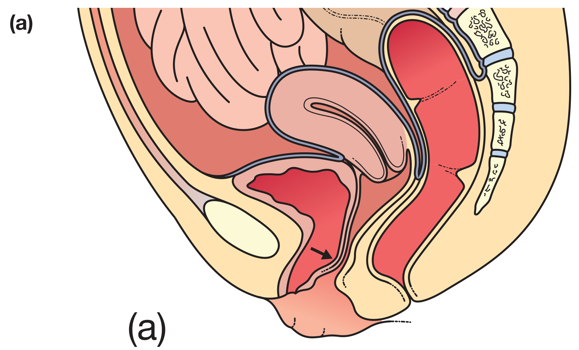
Urethrocele/cystocele (anterior compartment prolapse)
Figure (a): Urethrocele/cystocele - anterior wall descent with bladder herniation
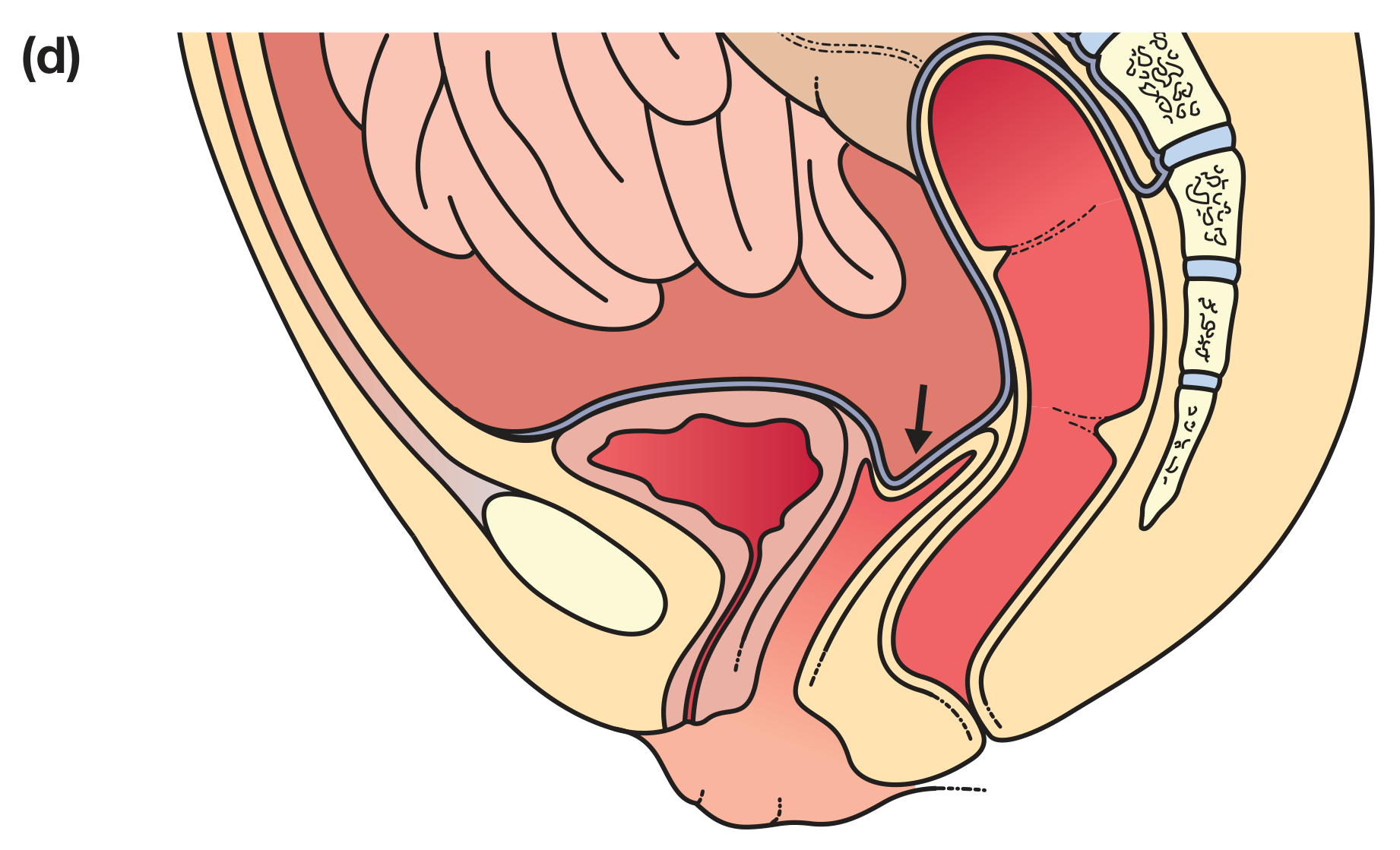
Vaginal vault prolapse (apical compartment prolapse)
Figure (d): Vaginal vault prolapse - apical descent post-hysterectomy

Anterior Compartment

TypeOrgan HerniatedMechanism
UrethroceleUrethraDefect in pubocervical fascia (distal)
CystoceleBladderDefect in pubocervical fascia (central or lateral)
CystourethroceleBladder + urethraCombined anterior wall defect
Symptoms: Vaginal bulge, urinary urgency, frequency, recurrent UTIs, incomplete bladder emptying. Note: stress urinary incontinence (SUI) can paradoxically decrease as the cystocele enlarges (due to urethral kinking causing obstruction).

Apical Compartment

TypeDescription
Uterine prolapseDescent of the uterus and cervix into or beyond the vagina
ProcidentiaComplete uterine prolapse - entire uterus protrudes outside the vagina
Vaginal vault prolapsePost-hysterectomy descent of the vaginal cuff
EnteroceleSmall bowel herniation through the pelvic floor (between vaginal cuff and rectum)
Symptoms: Dragging sensation, lump in vagina, vaginal discharge, ulceration/bleeding in procidentia.

Posterior Compartment

TypeOrgan HerniatedMechanism
RectoceleRectumDefect in the rectovaginal septum/fibromuscular layer
EnteroceleBowel + peritoneumCan occur posteriorly, especially post-hysterectomy
Symptoms: Difficulty with defecation, incomplete emptying, needing to splint (manually push on vaginal wall to defecate), pelvic pressure.
Key point: Apical prolapse is almost always present when there is significant anterior or posterior prolapse. Studies show clinically significant apical prolapse in 42%, 85%, and 100% of patients with stage 2, 3, and 4 anterior wall prolapse respectively. Surgeons must always assess the apex.

Risk Factors

CategorySpecific Factors
ObstetricVaginal delivery, increasing parity, prolonged labor, instrumental delivery, large baby
HormonalEstrogen deficiency (postmenopause weakens collagen and epithelium)
AnatomicalPrior hysterectomy (removes uterosacral/cardinal ligament complex)
LifestyleObesity, chronic constipation, chronic cough, heavy lifting
GeneticFamily history, Ehlers-Danlos, Marfan syndrome
AgeProgressive loss of muscle tone and collagen quality
Epidemiology: Only 24% of women seeking routine gynecologic care have NO prolapse at all. Stage 1-2 prolapse is so common it is considered part of the normal spectrum in adult women. Symptoms typically only appear when prolapse reaches the hymenal ring.

Symptoms

The most specific symptom for POP is a sensation of a vaginal bulge that can be seen or felt. Other symptoms include:
  • Pelvic pressure or heaviness
  • Low backache
  • Urinary dysfunction (frequency, nocturia, incomplete emptying, urgency)
  • Bowel dysfunction (constipation, incomplete emptying, need to splint)
  • Sexual dysfunction (body image, dyspareunia)
  • Discharge, ulceration (advanced cases)
Important caveat: Urinary and bowel symptoms correlate poorly with prolapse severity. Constipation alone without vaginal bulge sensation should not prompt surgical repair. The exception is a rectocele where the patient reports stool getting "caught in a pocket" requiring manual reduction.

Staging - POP-Q System

The Pelvic Organ Prolapse Quantification (POP-Q) system is the current gold standard, using the hymen as a fixed reference point (negative = above hymen, positive = below hymen):
StageCriteria
0No prolapse; all points well above hymen
ILeading edge > 1 cm above the hymen
IILeading edge within 1 cm above or below the hymen
IIILeading edge > 1 cm below hymen but not complete eversion
IVComplete eversion; vagina fully everted (procidentia)

Treatment

Conservative (First Line)

  • Pelvic floor muscle training (Kegel exercises) - strengthens levator ani; effective for mild-moderate prolapse
  • Lifestyle modification - weight loss, treat constipation, avoid heavy lifting
  • Topical estrogen - improves vaginal epithelial quality, eases pessary use in postmenopausal women
  • Pessary - a mechanical silicone device inserted into the vagina to physically support prolapsed organs. First-line for surgical poor candidates, those wishing to avoid surgery, or as a diagnostic trial. Very useful to confirm if symptoms are POP-related.

Surgical (Definitive)

Surgical approach depends on compartment and whether uterine preservation is desired:
CompartmentProcedure
AnteriorAnterior colporrhaphy (cystocele repair), paravaginal repair
Apical (vaginal)Uterosacral ligament suspension, sacrospinous fixation
Apical (abdominal/laparoscopic)Sacrocolpopexy (mesh) - gold standard for vault prolapse
PosteriorPosterior colporrhaphy (rectocele repair)
ObliterativeColpocleisis - surgical closure of vagina (for elderly non-sexually active women)
Posterior colporrhaphy outcomes: Anatomic cure in 76-96%, but significant rate of de novo dyspareunia (7-26%), especially when levator plication is performed. Modern techniques avoid levator plication to reduce dyspareunia.

High-Yield Summary Table

POP TypeOrganWallKey Symptom
CystoceleBladderAnteriorUrinary urgency, incomplete emptying
UrethroceleUrethraAnteriorStress incontinence
Uterine prolapseUterusApicalDragging, lump, procidentia
Vault prolapseVaginal cuffApicalPost-hysterectomy bulge
EnteroceleSmall bowelApical/PosteriorPelvic pressure
RectoceleRectumPosteriorIncomplete defecation, splinting
  • Campbell-Walsh-Wein Urology, Pathophysiology of POP
  • Berek & Novak's Gynecology, Chapter 31 - Pelvic Organ Prolapse
  • Bailey & Love's Short Practice of Surgery, 28th Ed, Chapter 87

Explain topic

Which topic would you like me to explain?
Asking for Preferences

Which topic would you like me to explain?

Asking for Preferences

Which aspect of POP would you like to go deeper on?

Searching sources...
Finding Sources
Finding Sources
Finding Sources

Send whole topic of pelvic organ prolapse again

Here is the complete topic of Pelvic Organ Prolapse:

Pelvic Organ Prolapse (POP)

Definition

Pelvic organ prolapse is the descent of one or more pelvic organs (bladder, uterus, small bowel, rectum) through the vaginal canal due to failure of the supporting structures of the pelvic floor. The vagina serves as the common pathway through which all pelvic organs can herniate.

Anatomy of Pelvic Support - The Foundation

Understanding POP requires knowing the three-layer support system:

1. Levator Ani Muscle Complex

The most important active support structure. It creates the levator hiatus - the opening through which the urethra, vagina, and rectum pass. Under normal resting tone, this muscle keeps the urogenital hiatus closed, compressing the vagina against the pubic bone.
"The urogenital hiatus is kept closed by the tonic activity of the levator muscles, which compresses the urethra, vagina, and rectum against the pubic bone and therefore closes the lumen of the vagina." - Campbell-Walsh-Wein Urology

2. Endopelvic Fascia and Ligaments

When levator muscles are intact, fascia bears minimal load. Once muscle tone is lost, all pelvic support falls on the ligaments - which then stretch and eventually fail.
Key ligaments:
  • Uterosacral ligaments - suspend the uterus/vaginal apex to the sacrum
  • Cardinal ligaments - lateral supports for the uterus and cervix
  • Pubocervical fascia - supports the anterior vaginal wall (bladder)
  • Rectovaginal septum - supports the posterior vaginal wall (rectum)

3. DeLancey's Three Levels of Vaginal Support

LevelStructuresWhat it Supports
I (Apical)Uterosacral-cardinal ligament complexUterus, vaginal vault
II (Lateral)Arcus tendineus fascia pelvisMid-vagina, bladder, rectum
III (Distal)Perineal membrane, perineal bodyLower vagina, urethra

Pathophysiology - Why Prolapse Happens

Three mechanisms work together:

A. Muscle Injury

Vaginal delivery is the #1 cause. MRI shows up to 20% of primiparous women have visible levator ani muscle defects after vaginal delivery - never seen in nulliparous women. When the levator tears or weakens, the urogenital hiatus widens and prolapse begins.

B. Connective Tissue Failure

Collagen metabolism is significantly altered in women with POP. An abnormal ratio of collagen type I : type III weakens the endopelvic fascia. This is why connective tissue disorders like Ehlers-Danlos syndrome and Marfan syndrome strongly predispose to POP.

C. Neuropathic Injury

The pudendal nerve (S2-S4) and levator ani nerve (S3-S5) can be stretched during vaginal delivery. EMG studies show neuropathy evidence in 24-29% of women at 6 weeks postpartum, contributing to progressive muscle atrophy and support failure.

Types of Prolapse (by Compartment)

Cystocele - anterior compartment prolapse
Figure (a): Urethrocele/Cystocele - anterior wall descent with bladder herniation
Vaginal vault prolapse - apical compartment
Figure (d): Vaginal vault prolapse - apical descent post-hysterectomy

Anterior Compartment

TypeOrgan HerniatedKey Feature
UrethroceleUrethraDistal defect in pubocervical fascia
CystoceleBladderCentral or lateral fascial defect
CystourethroceleBladder + urethraCombined anterior wall defect
Symptoms: Vaginal bulge, urinary urgency, frequency, recurrent UTIs, incomplete bladder emptying.
  • Important: Stress urinary incontinence (SUI) can paradoxically decrease as a cystocele enlarges, because the prolapsing bladder kinks the urethra creating obstruction. Occult SUI may unmask after surgical repair - always assess preoperatively.

Apical Compartment

TypeDescription
Uterine prolapseDescent of uterus and cervix into or beyond the vagina
ProcidentiaComplete prolapse - entire uterus protrudes outside the vagina; associated with vaginal ulceration, discharge, bleeding
Vaginal vault prolapsePost-hysterectomy descent of the vaginal cuff
EnteroceleSmall bowel + peritoneum herniation through the pelvic floor, between vaginal cuff and rectum; more common after hysterectomy
Symptoms: Dragging sensation, lump in vagina, pelvic pressure, vaginal discharge, bleeding/ulceration in advanced cases.

Posterior Compartment

TypeOrgan HerniatedKey Feature
RectoceleRectumDefect in rectovaginal septum/fibromuscular layer
EnteroceleSmall bowelCan occur posteriorly, especially post-hysterectomy
Descending perineum syndromeEntire perineumInferior detachment of rectovaginal septum from perineal body; causes pudendal neuropathy from stretch
Symptoms: Incomplete defecation, needing to splint (manually push on the posterior vaginal wall to defecate), constipation, pelvic pressure.
Critical rule: Apical prolapse is almost always present alongside significant anterior/posterior prolapse. Studies confirm clinically significant apical prolapse in 42%, 85%, and 100% of patients with stage 2, 3, and 4 anterior wall prolapse respectively. Always assess and address the apex surgically.

Risk Factors

CategorySpecific Factors
ObstetricVaginal delivery, increasing parity, prolonged labor, instrumental delivery, large baby
HormonalEstrogen deficiency (postmenopause weakens collagen and epithelium)
Prior surgeryHysterectomy (removes uterosacral/cardinal ligament complex), retropubic urethropexy
LifestyleObesity, chronic constipation, chronic cough, heavy lifting
GeneticFamily history, Ehlers-Danlos, Marfan syndrome
AgeProgressive loss of muscle tone and collagen quality
Epidemiology:
  • Only 24% of women seeking routine gynecologic care have NO prolapse
  • Stage 1-2 prolapse is so common it is part of the normal spectrum in adult women
  • Anterior wall prolapse is most common (34%), followed by posterior (19%), then uterine (14%)
  • Symptoms typically appear only when prolapse reaches or crosses the hymenal ring

Symptoms

The most specific symptom is a sensation or sight of a vaginal bulge. Other symptoms:
  • Pelvic pressure or heaviness, low backache
  • Urinary dysfunction: frequency, nocturia, urgency, incomplete emptying, recurrent UTIs
  • Bowel dysfunction: constipation, incomplete emptying, straining, need to splint
  • Sexual dysfunction: dyspareunia, body image concerns
  • Vaginal discharge, ulceration, bleeding (in advanced/procidentia cases)
Caveat: Urinary and bowel symptoms correlate poorly with prolapse severity. Constipation alone without a bulge sensation should not prompt surgery. A pessary trial is very useful - if symptoms improve with a pessary, they are likely POP-related and will improve with surgical repair.

Staging - POP-Q System

The Pelvic Organ Prolapse Quantification (POP-Q) is the gold standard. Nine anatomical points are measured during Valsalva, referenced to the hymen (negative = above hymen, positive = below):
Six vaginal measurement points: Aa, Ba (anterior wall), C, D (apex), Ap, Bp (posterior wall) Three additional measures: Genital hiatus (gh), perineal body (pb), total vaginal length (tvl)
StageCriteriaClinical Meaning
0No prolapse; all points well above hymenNormal
ILeading edge > 1 cm above hymenMild; usually asymptomatic
IILeading edge within 1 cm above or below hymenModerate; symptoms begin
IIILeading edge > 1 cm below hymen, not complete eversionSignificant; usually symptomatic
IVComplete vaginal eversion (procidentia)Severe
Baden-Walker system (older, simpler): Grades 0-3 using hymen as reference, still used in some centers.

Evaluation

History

  • Duration and progression of symptoms
  • Obstetric history (parity, mode of delivery, birth weights)
  • Prior pelvic surgeries
  • Bowel, bladder, and sexual function
  • Desire for future fertility
  • Desire for uterine preservation

Physical Examination

  • Performed with patient straining/Valsalva and standing if lying exam is unrevealing
  • Assess each compartment separately using a split speculum
  • Evaluate pelvic floor muscle strength
  • Document leading edge of prolapse relative to the hymen

Additional Workup

  • Urodynamics - mandatory before surgery; identifies occult SUI, detrusor overactivity, voiding dysfunction
  • Cystoscopy - if bladder involvement suspected
  • Defecating proctography / MRI defecography - for complex posterior compartment or rectal intussusception
  • Pelvic MRI - best for levator ani muscle defects

Treatment

Conservative (First-Line for Most Patients)

Pelvic Floor Muscle Training (PFMT / Kegel Exercises)

  • Strengthens levator ani, reduces prolapse symptoms
  • Effective for stage I-II; can slow progression
  • Requires consistent, supervised technique for best results

Lifestyle Modifications

  • Weight loss, treat chronic constipation, avoid heavy lifting, manage chronic cough (treat underlying cause)

Topical Estrogen (Postmenopausal Women)

  • Improves vaginal epithelial quality and collagen
  • Facilitates pessary fitting and reduces complications
  • Does not reverse prolapse itself

Pessary - Mechanical Support Device

A silicone device inserted vaginally to mechanically support the prolapsed organs.
Pessary TypeBest For
Ring pessaryMild-moderate prolapse; women with good sphincter tone
GehrungCystocele, rectocele
GellhornAdvanced prolapse; best anatomic support
Donut/CubeAdvanced prolapse when other types fail
ShaatzVaginal vault prolapse
Indications for pessary:
  • Patient prefers non-surgical management
  • Awaiting surgery
  • Poor surgical candidate (elderly, comorbidities)
  • Pregnancy
  • Diagnostic trial to confirm symptoms are POP-related
Complications: Vaginal discharge, odor, erosion, infection, urinary retention (if too large), de novo SUI (if kinking was masking it).

Surgical Management

Surgery is indicated when prolapse is symptomatic, impacts quality of life, and conservative measures have failed.
Two broad approaches: Vaginal vs. Abdominal (open/laparoscopic/robotic)

ANTERIOR COMPARTMENT REPAIR

Anterior Colporrhaphy (Cystocele Repair)

  • Most common procedure for anterior wall prolapse
  • Vaginal approach: plication of the pubocervical fascia in the midline
  • Native tissue repair (no mesh)
  • Anatomic success: 70-80% at 1-2 years
  • Recurrence rates higher than abdominal mesh-augmented repair

Paravaginal Repair

  • Repairs lateral detachment of the pubocervical fascia from the arcus tendineus
  • Can be done vaginally or abdominally (laparoscopic)
  • Used when cystocele is due to lateral (paravaginal) defect rather than central defect

APICAL COMPARTMENT REPAIR

The apex is the keystone of pelvic floor support. Apical repair is the most important component - anterior/posterior repairs fail long-term without concurrent apical fixation.

Vaginal Approaches

1. Uterosacral Ligament Suspension (USLS)
  • Vaginal cuff or cervix sutured to the uterosacral ligaments bilaterally
  • Good anatomic support; preserves vaginal length and axis
  • Risk: ureteral kinking (2-11%) - must confirm ureteral patency with cystoscopy intraoperatively
  • Native tissue; no mesh
2. Sacrospinous Ligament Fixation (SSLF)
  • Vaginal apex sutured to the sacrospinous ligament (usually right side)
  • Deflects vaginal axis posteriorly - higher risk of anterior wall recurrence
  • Risk: gluteal/posterior thigh pain (injury to pudendal nerve or vessels)
  • Native tissue; no mesh
3. Iliococcygeus Fixation
  • Vaginal apex sutured to iliococcygeus fascia bilaterally
  • Less commonly used; good for patients with attenuated sacrospinous ligaments

Abdominal/Laparoscopic/Robotic Approaches

Abdominal Sacrocolpopexy - Gold Standard for Vault Prolapse
  • Mesh (polypropylene) sutured to anterior and posterior vaginal wall, then fixed to the sacral promontory (L5/S1)
  • Can be done open, laparoscopic, or robotic
  • Superior long-term anatomic outcomes vs. vaginal native tissue (80-95% success)
  • Preserves vaginal length and normal axis
  • Risks: mesh erosion into vagina (3-7%), mesh-related infection, bowel/vessel injury, prolonged recovery
  • Preferred for: younger active women, large vault prolapse, prior failed vaginal repair
Laparoscopic/Robotic Sacrocolpopexy
  • Same principles as open; less blood loss, faster recovery
  • Robotic approach facilitates complex suturing in deep pelvis
  • Outcomes comparable to open sacrocolpopexy
Uterine Preservation Options (Hysteropexy)
  • For women who wish to keep the uterus
  • Sacrohysteropexy - mesh from cervix to sacrum (laparoscopic)
  • Manchester procedure - amputation of cervix + cardinal/uterosacral ligament shortening (vaginal)
  • Sacrospinous hysteropexy - vaginal fixation of cervix to sacrospinous ligament

POSTERIOR COMPARTMENT REPAIR

Posterior Colporrhaphy (Rectocele Repair)

  • Plication of the rectovaginal septum (fibromuscular layer) in the midline
  • Usually includes perineorrhaphy (reconstruction of the perineal body)
  • Anatomic cure: 76-96%
  • Key concern: De novo dyspareunia in 7-26% (up to 50% historically when levator plication was performed)
  • Modern technique: fascial plication without levator plication dramatically reduces dyspareunia risk
  • Functional symptom relief (constipation, splinting) is modest and inconsistent

Site-Specific Rectocele Repair

  • Identifies and repairs discrete breaks in the rectovaginal septum rather than plication
  • Theoretical advantage: restores normal anatomy; less distortion
  • Similar anatomic outcomes to traditional posterior colporrhaphy

OBLITERATIVE PROCEDURES

Colpocleisis (LeFort Procedure / Total Colpocleisis)

  • Surgical closure of the vaginal canal
  • For elderly women with advanced prolapse who are not sexually active and have high surgical risk
  • Most effective prolapse surgery with lowest morbidity
  • Irreversible - must counsel thoroughly; small risk of regret
  • Concurrent anti-incontinence procedure often performed as occult SUI may unmask

MESH CONSIDERATIONS

The FDA issued warnings in 2008 and 2011 regarding transvaginal mesh for POP repair:
  • No evidence of increased effectiveness over native tissue repair for anterior/posterior compartments
  • Significant mesh-related complications: erosion/extrusion, pain, dyspareunia, need for reoperation
  • Transvaginal mesh kits for POP largely abandoned in current practice
  • Abdominal sacrocolpopexy mesh (placed abdominally, not transvaginally) is still the gold standard for vault prolapse - different risk profile
Mesh complication assessment (IUGA/ICS 2011 classification):
  • Evaluate for location, size, and degree of mesh extrusion
  • Even absence of visible mesh can be associated with pelvic pain

CONCURRENT PROCEDURES

Anti-incontinence surgery with POP repair:
  • Occult SUI is common - masked by urethral kinking from prolapse
  • As prolapse is reduced, SUI often unmasks
  • Options: midurethral sling (tension-free vaginal tape/TVT, transobturator tape/TOT) at time of POP repair
  • The CARE trial showed prophylactic Burch colposuspension at sacrocolpopexy reduced postoperative SUI

Comparison of Approaches

FeatureVaginal Native TissueAbdominal Sacrocolpopexy
ApproachVaginalAbdominal/laparoscopic
RecoveryFasterLonger
Mesh usedNoYes
Anatomic successModerate (70-85%)High (80-95%)
Vaginal axisMay be alteredPreserved
Mesh complicationsNoneErosion 3-7%
Best forOlder/frail women, uterine prolapseYounger women, vault prolapse, recurrences
Ureteral risk (USLS)Yes (2-11%)No

High-Yield Summary

POP TypeOrganCompartmentKey SymptomSurgery
CystoceleBladderAnteriorUrinary urgency, incomplete emptyingAnterior colporrhaphy
UrethroceleUrethraAnteriorStress incontinenceAnterior colporrhaphy + sling
Uterine prolapseUterusApicalDragging, lump, procidentiaHysterectomy + apical suspension or hysteropexy
Vault prolapseVaginal cuffApicalPost-hysterectomy bulgeSacrocolpopexy (gold standard)
EnteroceleSmall bowelApical/PosteriorPelvic pressureEnterocele repair + apical suspension
RectoceleRectumPosteriorSplinting, incomplete defecationPosterior colporrhaphy

Key Principles to Remember

  1. Always assess the apex - anterior/posterior repairs fail without apical support
  2. Free T4 = Free Pelvic Floor - symptoms come from what's functionally compromised, not just what anatomically descends
  3. Treat the patient, not the prolapse - stage II prolapse without symptoms needs no treatment
  4. Pessary first - offers diagnosis + treatment; confirms symptoms are prolapse-related
  5. Occult SUI - always assess and counsel before repair; fixing prolapse may unmask hidden incontinence
  6. Mesh = abdominally placed only for modern POP surgery; transvaginal mesh kits are largely obsolete
  • Berek & Novak's Gynecology, Chapter 31
  • Campbell-Walsh-Wein Urology, Chapters 62, 68, 71
  • Bailey & Love's Short Practice of Surgery, 28th Ed
  • Sabiston Textbook of Surgery, Chapter 29
This is a shared conversation. Sign in to Orris to start your own chat.