
Walls of Middle ear
Reading File
The middle ear has 6 walls: roof (tegmental), floor (jugular), lateral (membranous), medial (labyrinthine), anterior (carotid/tubal), and posterior (mastoid). Let me pull the full textbook detail.
Searching sources...
Finding Sources
Finding Sources
Gray's Anatomy for Students has dedicated sections on each wall. Let me read them all now.
Reading File
Reading File
Loading Image
Loading Image
All the content is now in. Here is the complete answer.
Walls of the Middle Ear
The middle ear (tympanic cavity) is a box-like space with 6 walls. A helpful mnemonic is "TAMIL R" (Tegmental, Anterior, Mastoid, Internal/labyrinthine, Lateral/membranous, Roof-floor pair) -- or just remember them by direction.
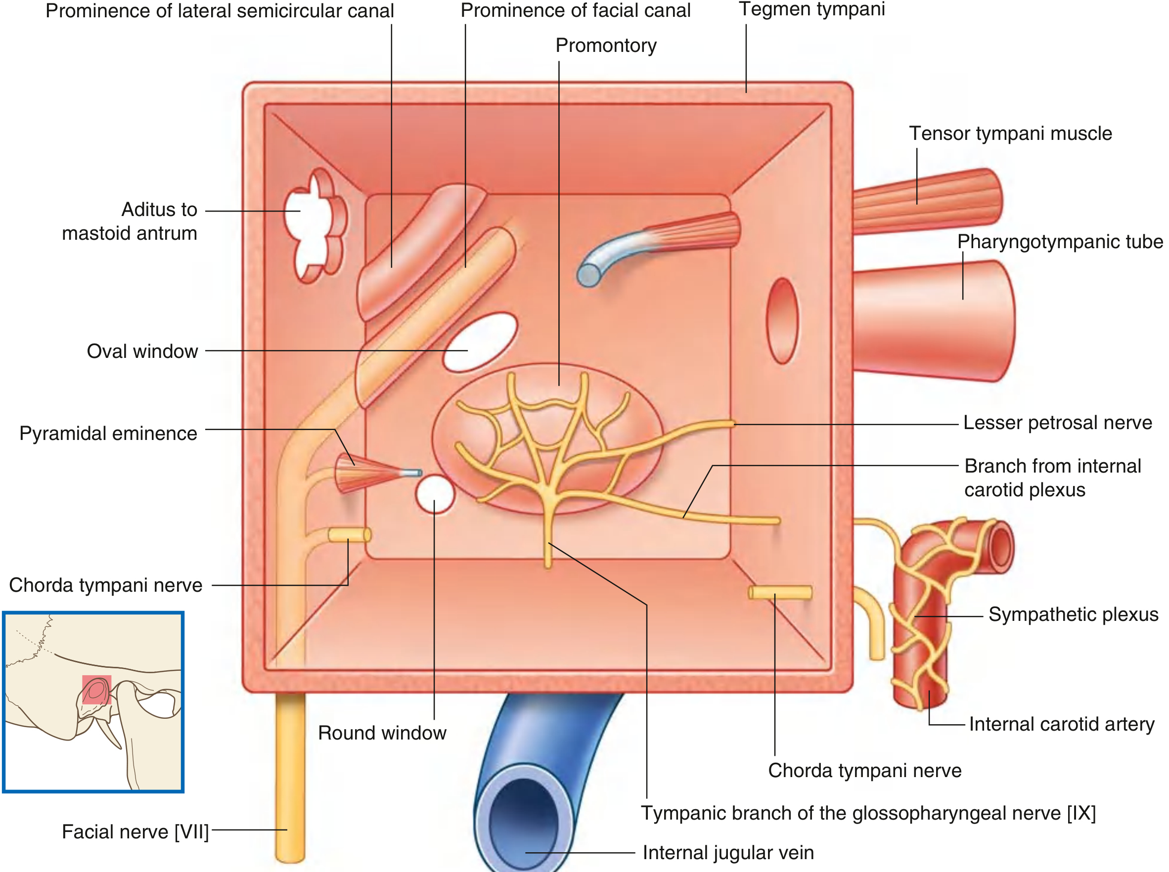
1. Tegmental Wall (Roof)
- Also called: Roof
- Bone: Tegmen tympani - a thin plate of bone on the anterior surface of the petrous part of the temporal bone
- Separates the middle ear from the middle cranial fossa
- Clinical note: Infection can spread superiorly through this thin plate to cause extradural abscess or meningitis
2. Jugular Wall (Floor)
- Also called: Floor
- Separates the tympanic cavity from the internal jugular vein (bulb of the jugular vein)
- May be thickened by mastoid air cells
- Contains a small aperture near the medial border through which the tympanic branch of CN IX (Jacobson's nerve) enters the middle ear
- Clinical note: A high-riding jugular bulb can protrude into the floor and appear as a bluish mass behind the tympanic membrane
3. Membranous Wall (Lateral Wall)
- Also called: Lateral wall
- Formed almost entirely by the tympanic membrane
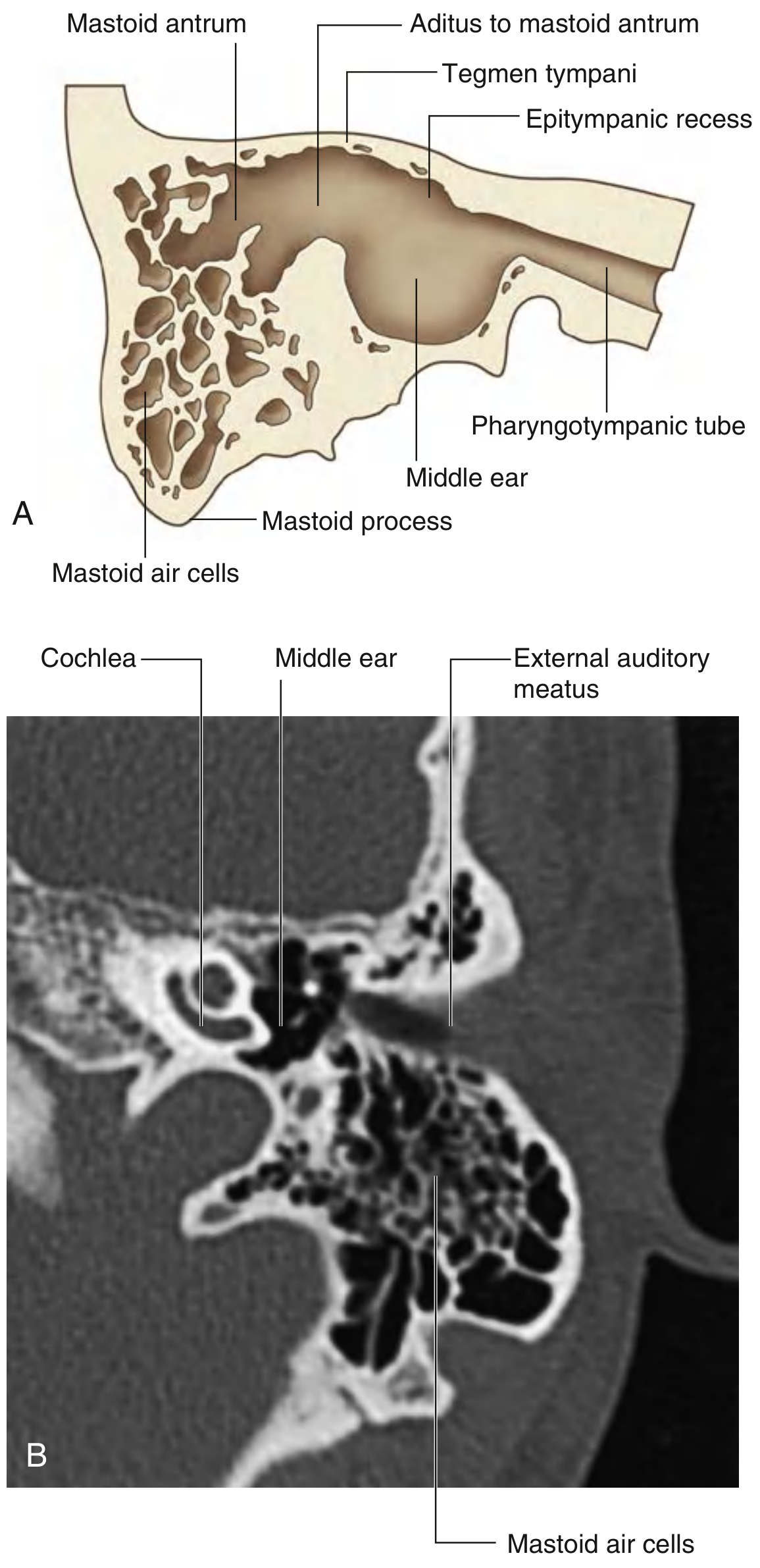
- The tympanic membrane does not extend superiorly into the epitympanic recess, so the upper part of this wall is formed by the bony lateral wall of the epitympanic recess
4. Mastoid Wall (Posterior Wall)
- Also called: Posterior wall
- Only partially complete
- Lower part: bony partition between tympanic cavity and mastoid air cells
- Upper part: the epitympanic recess is continuous with the aditus to the mastoid antrum
Key structures associated with this wall:
| Structure | Significance |
|---|---|
| Pyramidal eminence | Small bony projection through which the tendon of stapedius muscle enters the middle ear |
| Chorda tympani nerve | Branch of CN VII; enters the middle ear through an opening in this wall, crosses the handle of the malleus, exits via the anterior wall |
5. Anterior Wall (Carotid/Tubal Wall)
- Also called: Carotid wall or Tubal wall
- Only partially complete
- Lower part: thin layer of bone separating the tympanic cavity from the internal carotid artery
- Superior part is deficient, containing two large openings:
- Large opening for the pharyngotympanic tube (Eustachian tube) entering the middle ear
- Smaller opening for the canal containing the tensor tympani muscle
- Also contains the foramen for exit of the chorda tympani nerve from the middle ear
6. Labyrinthine Wall (Medial Wall)
- Also called: Medial wall or Promontory wall
- Simultaneously forms the lateral wall of the internal ear
- The most complex and clinically important wall
Key features:
| Feature | Details |
|---|---|
| Promontory | Rounded bulge produced by the basal coil of the cochlea; covered by the tympanic plexus (CN IX + sympathetic branches from internal carotid plexus) |
| Oval window (fenestra vestibuli) | Posterosuperior to the promontory; closed by the footplate of the stapes; transmits vibrations to the perilymph of the vestibule |
| Round window (fenestra cochleae) | Posteroinferior to the promontory; closed by the secondary tympanic membrane; acts as a pressure release valve for cochlear fluid |
| Prominence of the facial canal | Ridge of bone posterior and superior to the oval window, produced by CN VII in its bony canal - clinically important as it can dehisce |
| Prominence of the lateral semicircular canal | Broader ridge just above and posterior to the facial canal prominence |
The tympanic plexus on the promontory gives off the lesser petrosal nerve, which travels across the petrous temporal bone, exits via the foramen ovale, and enters the otic ganglion (supplying the parotid gland).
Summary Table
| Wall | Other Name | Key Relations / Contents |
|---|---|---|
| Roof | Tegmental | Tegmen tympani / Middle cranial fossa |
| Floor | Jugular | Internal jugular vein; tympanic branch of CN IX |
| Lateral | Membranous | Tympanic membrane; bony wall of epitympanic recess |
| Posterior | Mastoid | Aditus to mastoid antrum; pyramidal eminence (stapedius); chorda tympani entry |
| Anterior | Carotid/Tubal | Internal carotid artery; pharyngotympanic tube; tensor tympani canal; chorda tympani exit |
| Medial | Labyrinthine | Promontory (cochlea); oval window (stapes footplate); round window; facial canal; lateral SCC prominence |
Source: Gray's Anatomy for Students, pp. 1099-1100
This is a shared conversation. Sign in to Orris to start your own chat.