
Pneumoconiosis
pneumoconiosis lung pathology histology
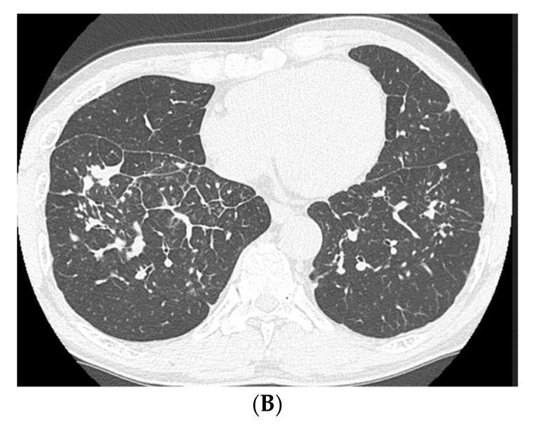
This diagnostic image is an axial slice from a High-Resolution Computed Tomography (HRCT) scan of the chest. It demonstrates bilateral lung parenchyma with a focus on the middle and lower lobes. The primary pathology consists of multiple, well-defined nodular shadows with a centrilobular and perilobular distribution. There is visible thickening of the intralobular septa, creating a reticulonodular pattern characteristic of interstitial lung disease or pneumoconiosis. The nodules are scattered throughout both lung fields but show higher density in the posterior segments. Key anatomical landmarks visible include the heart in the anterior mediastinum, the descending aorta, the main bronchi, and the ribs forming the thoracic cage. The image serves as a clinical illustration of disease progression and regression in a patient with pneumoconiosis and a sarcoid-like reaction, highlighting the importance of nodular morphology and septal thickening in diagnosing occupational lung diseases.
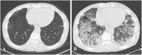
This diagnostic image consists of two axial low-dose chest computed tomography (LDCT) scans comparing chronic baseline lung disease with an acute pulmonary event. Image A (baseline) shows numerous, diffuse, small calcified nodular infiltrations distributed throughout both lung fields, characteristic of pneumoconiosis. Additional findings include minimal subpleural septal thickening and fine reticulations in the right middle lobe and both lower lobes, alongside calcified lymphadenopathy in the mediastinal and hilar regions. Image B (at admission) demonstrates a significant progression in pathology, featuring multifocal patchy areas of consolidation and diffuse ground-glass opacities (GGO) bilaterally. These acute findings are clinically suggestive of extensive pneumonia, pulmonary edema, or alveolar hemorrhage superimposed on the underlying chronic interstitial disease. The comparison highlights the transition from a stable nodular pattern to acute, severe parenchymal opacification, relevant for pulmonology and radiology education regarding complications in occupational lung disease.
This composite educational graphic presents a comparison of lung pathology and histology in a porcine model of acute respiratory distress syndrome (ARDS), contrasting two ventilation protocols: ARDSnet (low tidal volume) and time-controlled adaptive ventilation (TCAV). Top Panels (a-d): Gross pathology at necropsy with lungs inflated to 25 cmH2O. Panel (a) shows the ARDSnet lung with significant dark red consolidation, inflammation, and incomplete inflation. Panel (b) reveals a cut surface with diffuse inflammation and copious white edema foam in the airways. In contrast, panel (c) displays a TCAV-treated lung that appears healthy, pink, and fully inflated, with the cut surface in panel (d) showing dry, aerated tissue without edema. Bottom Panels (e-f): H&E stained histology (50 μm scale). Panel (e) demonstrates classic ARDS features: marked alveolar wall thickening (arrows), significant vessel congestion (arrowheads), and inflammatory infiltration. Panel (f) shows the TCAV group with preserved architecture, thin alveolar septa, and clear airspaces, illustrating the educational concept of ventilator-induced lung injury (VILI) prevention through lung stabilization.
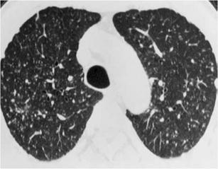
**Imaging Modality:** High-resolution computed tomography (HRCT) of the chest in axial plane, lung window. **Anatomical Region:** Upper lobes of the lungs at the level of the trachea. **Observed Pathology:** Coal workers' pneumoconiosis (CWP) presenting as simple pneumoconiosis. **Characteristic Visual Features:** * **Nodular Pattern:** Diffuse, bilateral distribution of small, ill-defined centrilobular nodules. * **Distribution:** Marked predominance in the upper lobes and posterior segments of the lungs. * **Nodule Morphology:** The nodules vary slightly in size and lack the "tree-in-bud" configuration, indicating an interstitial rather than an endobronchial inflammatory process. * **Lung Parenchyma:** Background lung attenuation appears largely preserved without significant evidence of conglomerate masses or progressive massive fibrosis (PMF) in this specific section. **Differentiating Features:** The absence of branching linear opacities (tree-in-bud) helps distinguish this occupational dust inhalation pattern from infectious bronchiolitis. The posterior and upper lobe distribution is characteristic of mineral dust exposure, such as coal dust or silica.
pneumoconiosis occupational lung disease
silicosis nodules chest X-ray radiology
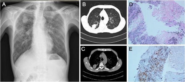
This composite image illustrates the radiological and histopathological presentation of pulmonary silicosis. Panel A is a posterior-anterior chest X-ray showing diffuse, small micronodular opacities throughout both lung fields and a prominent mass-like opacity in the right upper zone. Panels B and C are axial chest CT scans in lung and mediastinal windows, respectively, identifying a 25×33 mm consolidated nodule in the right upper lobe alongside widespread perilymphatic micronodularity. Panel D presents a Hematoxylin and Eosin (H&E) stained histopathological section from the biopsied lung nodule, demonstrating silicotic nodules characterized by dense fibrous tissue hyperplasia, hyaline degeneration, chronic inflammatory cell infiltration, and prominent black carbon (anthracotic) pigment deposits. Panel E shows immunohistochemical (IHC) staining, positive for CD163 (a histiocytic marker) and negative for CK (cytokeratin), supporting the diagnosis of a reactive/fibrotic process over malignancy. These findings are clinically relevant for diagnosing occupational pneumoconiosis, specifically silicosis with progressive massive fibrosis (PMF).
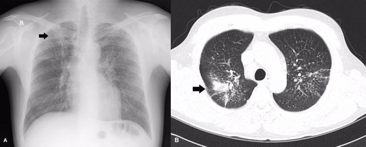
This composite medical image displays a chest radiograph (A) and an axial thin-section computed tomography (CT) scan (B) demonstrating occupational lung disease, specifically silicosis. (A) The posterior-anterior chest X-ray reveals diffuse, bilateral, ill-defined small nodular opacities with a predilection for the upper and middle lung zones. A prominent large opacity is noted in the right upper lobe (black arrow), suggesting Progressive Massive Fibrosis (PMF). (B) The axial CT scan at the level of the upper lobes confirms the presence of diffuse, multiple rounded centrilobular nodules. Significant findings include a large conglomerated mass in the right upper lobe (black arrow) and smaller areas of coalescing nodules in the left upper lobe. These radiological features are characteristic of complicated silicosis, where individual silicotic nodules merge into larger masses of fibrous tissue. The distribution is typically upper-lobe dominant, reflecting the common pattern seen in pneumoconiosis and sarcoidosis.
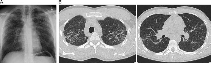
This diagnostic imaging set consists of a posteroanterior chest X-ray (A) and two axial high-resolution computed tomography (HRCT) slices (B) of the thorax, demonstrating characteristic findings of chronic interstitial lung disease. Panel A shows a projectional view of the chest with bilateral hazy opacities predominantly in the upper lobes, associated with a loss of lung volume suggestive of fibrotic changes. Panel B provides cross-sectional detail, revealing a diffuse reticulonodular pattern throughout the lung parenchyma. This is characterized by fine linear densities and small, ill-defined nodules, which are most concentrated in the upper lobes of both lungs. The CT scans also demonstrate signs of parenchymal architectural distortion and fibrosis. These radiological features are consistent with silicosis or other occupational pneumoconioses. The images are designed for medical education regarding thoracic radiology, specifically illustrating the transition from screening with conventional X-ray to the detailed characterization of interstitial patterns using HRCT.
asbestosis pleural plaques mesothelioma
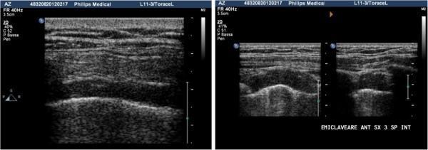
This diagnostic image consists of two thoracic ultrasound panels illustrating various forms of pleural pathology. The left panel shows a B-mode ultrasound scan of the pleura in a patient with asbestosis. It demonstrates focal pleural thickening characterized by a well-demarcated hypo-anechoic area that creates a 'doubling' of the pleural line. This represents a classic pleural plaque. The right panel displays two side-by-side scans (transverse and longitudinal views) of a more advanced pleural lesion. This plaque exhibits loss of the distinct tissue interface and shows direct infiltration into the adjacent chest wall muscle layers, evidenced by the disruption of normal muscular bands. In a clinical context, such infiltrative behavior is a critical sonographic sign suggestive of neoplastic transformation, such as malignant mesothelioma. These images are essential for medical students and radiologists in learning to differentiate benign asbestos-related plaques from malignant pleural disease using high-frequency linear probes.
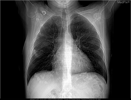
**Imaging Modality:** Posteroanterior (PA) chest radiograph (topogram/scout view). **Anatomical Region:** Thorax, including the lungs, mediastinum, and diaphragmatic surface. **Observed Pathology:** Multiple, discrete, calcified pleural plaques consistent with asbestos-related pleural disease. **Characteristic Visual Features:** - **Calcification:** High-attenuation, radio-opaque opacities are noted along the bilateral diaphragmatic pleura and the lateral chest walls. - **Distribution:** The lesions are multifocal and characteristically concentrated in the mid-to-lower thoracic zones. - **Morphology:** The plaques exhibit a "holly leaf" appearance in some areas, representing the irregular, jagged margins of calcified pleural thickening viewed en face. - **Sparing:** There is relative sparing of the lung apices and the costophrenic angles. **Relevant Clinical Context:** These findings are pathognomonic for chronic asbestos exposure. While the lung parenchyma appears relatively clear in this view, the presence of such extensive pleural calcification indicates a risk for underlying asbestosis (interstitial fibrosis) and increased susceptibility to bronchogenic carcinoma or malignant mesothelioma. **Key Diagnostic Features:** Bilateral, calcified diaphragmatic and parietal pleural plaques; absence of acute pleural effusion.
Pneumoconiosis
- Park's Textbook of Preventive and Social Medicine, p. 4635
- Robbins & Kumar Basic Pathology, p. 458
Classification
| Type | Causative Dust | Main Industries |
|---|---|---|
| Silicosis | Crystalline silica (SiO₂) | Mining, sandblasting, quarrying, pottery |
| Coal Workers' Pneumoconiosis (CWP) | Coal dust | Coal mining (especially Appalachia) |
| Asbestosis | Asbestos fibers | Shipyards, construction, plumbing |
| Siderosis | Iron dust | Iron/steel workers |
| Stannosis | Tin dust | Tin workers |
| Byssinosis | Cotton dust | Textile industry |
| Bagassosis | Sugarcane bagasse | Sugar mills |
| Berylliosis | Beryllium | Aerospace, nuclear industry |
1. Silicosis
Etiology & Pathogenesis
- Caused by inhalation of crystalline silica (quartz, cristobalite, or tridymite). Quartz is the most commonly implicated form.
- Silica particles are ingested by alveolar macrophages → lysosomal damage → inflammasome activation → release of IL-1, TNF, oxygen free radicals, and fibrogenic cytokines → progressive nodular fibrosis.
Types
| Form | Latency | Features |
|---|---|---|
| Chronic (Classic) | >10 years of low-level exposure | Most common; simple silicosis or PMF |
| Accelerated | 5-10 years of high-level exposure | Resembles chronic PMF |
| Acute (silicoproteinosis) | Weeks to few years of very high exposure | Rare; rapidly progressive; poor prognosis |
Morphology
- Early: tiny discrete silicotic nodules in upper lung zones
- Microscopically: concentrically arranged hyalinized collagen fibers in a whorled pattern - pathognomonic
- Polarized microscopy reveals weakly birefringent silica particles in nodule centers
- Advanced: nodules coalesce → Progressive Massive Fibrosis (PMF) → honeycombing
Clinical Features
- Insidious onset; early symptoms: irritant cough, exertional dyspnea, chest pain
- CXR: "eggshell" calcification of hilar nodes; "snowstorm" appearance of lung fields in nodular stage
- HRCT: upper lobe predominant, well-defined centrilobular nodules
- Late complications: PMF, pulmonary hypertension, cor pulmonale
- Silico-tuberculosis: silica inhibits macrophage killing of mycobacteria → markedly increased susceptibility to TB
- Associated with modest increased risk of lung cancer
2. Coal Workers' Pneumoconiosis (CWP) - "Black Lung"
Pathogenesis
Spectrum of Disease
- Anthracosis: Carbon pigment in macrophages, no tissue reaction - seen in urban dwellers and smokers too; benign
- Simple CWP: Coal macules (dust-laden macrophages + delicate collagen network) and coal nodules in upper/upper-lower lobes; minimal lung dysfunction; centrilobular emphysema may develop
- Complicated CWP / PMF: Coalescence of nodules → black scars >2 cm (sometimes up to 10 cm) of dense collagen + pigment; impaired lung function; less than 10% of simple CWP progresses to PMF
Clinical Features
- Simple CWP is usually benign with little functional impairment
- PMF causes increasing pulmonary dysfunction, pulmonary hypertension, and cor pulmonale
- Usually presents >10 years after initial exposure
- HRCT: upper lobe predominant small nodules; in complicated CWP, large conglomerate opacities
- Robbins & Kumar Basic Pathology, pp. 458-460
3. Asbestosis & Asbestos-Related Disease
Pathogenesis
Asbestos-Related Conditions
| Condition | Notes |
|---|---|
| Asbestosis (parenchymal fibrosis) | Diffuse interstitial fibrosis, lower lobe predominant, subpleural |
| Pleural plaques | Most common manifestation; well-circumscribed dense collagen; often calcified; bilateral parietal pleura + diaphragm |
| Pleural effusion | May be the first manifestation |
| Diffuse pleural fibrosis | Uncommon |
| Lung carcinoma | Risk markedly amplified by smoking |
| Mesothelioma | Rare; strongly linked to crocidolite; latency 25-45 years |
| Laryngeal carcinoma | Increased risk |
Morphology
- Asbestos bodies: golden-brown, fusiform or beaded rods with translucent center - asbestos fibers coated with iron-containing proteinaceous material (ferritin) - pathognomonic
- Fibrosis begins in lower lobes and subpleurally (contrast to silicosis: upper lobe, nodular)
- Progression: honeycombing; visceral pleural adhesions; pulmonary artery narrowing
- Pleural plaques on anterior/posterolateral parietal pleura and diaphragmatic domes; often calcified ("holly leaf" appearance on CXR)
Clinical Features
- Dyspnea 10-20 years after exposure, with cough and sputum
- Bilateral basal crackles
- CXR: lower zone reticular opacities; calcified pleural plaques
- May progress to cor pulmonale and death
- Pleural plaques are usually asymptomatic
- Robbins & Kumar Basic Pathology, pp. 460-461
Key Comparisons: Silicosis vs. CWP vs. Asbestosis
| Feature | Silicosis | CWP | Asbestosis |
|---|---|---|---|
| Causative dust | Crystalline silica | Coal dust | Asbestos fibers |
| Lung zone | Upper lobes | Upper lobes | Lower lobes (subpleural) |
| Pattern | Nodular fibrosis | Macules/nodules → PMF | Diffuse interstitial fibrosis |
| CXR finding | Snowstorm; eggshell nodes | Small opacities | Lower zone reticulations; pleural plaques |
| Pathognomonic | Whorled hyaline nodule | Black scars | Asbestos bodies (ferruginous bodies) |
| TB risk | High (silico-TB) | Moderate | Not prominent |
| Malignancy | Lung cancer (modest) | Lung cancer | Mesothelioma, lung cancer |
| Latency | Chronic: >10 yrs | >10 years | 10-20 yrs (parenchymal); 25-45 yrs (mesothelioma) |
Other Important Pneumoconioses
Byssinosis
- Cotton, flax, or hemp dust inhalation (textile workers)
- Unique feature: symptoms worse on Monday (after weekend rest), improving through the week
- Mechanism: bronchoconstriction from substances in the cotton bract
- Chronic exposure: irreversible COPD-like picture
Bagassosis
- Inhalation of bagasse (sugarcane residue dust)
- Caused by Thermoactinomyces sacchari - a form of hypersensitivity pneumonitis
- Acute phase: acute diffuse bronchiolitis; skiagram shows mottling
- Chronic phase: diffuse fibrosis, emphysema, bronchiectasis
- Prevention: keep moisture content >20% and spray with 2% propionic acid
Farmer's Lung
- Hypersensitivity pneumonitis from moldy hay (Micropolyspora faeni, Thermoactinomyces vulgaris)
- Type III/IV immunological reaction
Prevention Principles (Applicable to All Pneumoconioses)
- Dust control: wet drilling, enclosed apparatus, exhaust ventilation, substitution
- Personal protection: mechanical filter masks, respirators with air supply
- Medical control: pre-employment and periodic medical examination + chest X-rays
- Regulatory control: Silicosis is a notifiable disease under the Factories Act 1948 and Mines Act 1952 (India)
Recent Evidence
- A dose-response meta-analysis confirms a clear cumulative silica exposure-silicosis relationship (PMID 39107111, Thorax 2024).
- A systematic review and meta-analysis confirms significantly elevated TB risk in silicosis patients (PMID 37349198, Pulmonology 2025).
- A 2025 meta-analysis in Thorax links inhalational exposures broadly with interstitial lung disease risk (PMID 40518258).
Mental health warning sign and causes
mental health warning signs and causes WHO 2024
https://www.who.int/news-room/fact-sheets/detail/mental-diso…
"mental health" AND "risk factors" AND prevention
mental health biopsychosocial model diagram
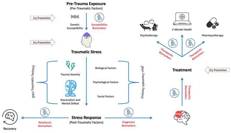
A comprehensive flowchart outlining the biopsychosocial model of Post-Traumatic Stress Disorder (PTSD) progression and management. The diagram follows a logical sequence from 'Pre-Trauma Exposure' to 'Recovery.' The first stage, Pre-Trauma, highlights genetic susceptibility and susceptibility biomarkers as targets for primary prevention. The central 'Traumatic Stress' phase identifies peri-traumatic factors, including trauma severity, dissociation, mental defeat, and biological, psychological, and social variables, marking the stage for secondary prevention. The 'Stress Response' (post-traumatic factors) leads to either 'Recovery'—facilitated by resilience biomarkers—or to a 'Treatment' phase. Treatment is categorized into three modalities: Psychotherapy (symbolized by figures in session), E-Mental Health (laptop icon), and Pharmacotherapy (pill icon), all of which are mediated by therapeutic biomarkers and represent tertiary prevention. Diagnostic biomarkers are positioned between the stress response and treatment stages. The infographic effectively illustrates the integration of biomarkers and multi-level prevention strategies (1ry, 2ry, 3ry) within the clinical trajectory of traumatic stress disorders.

An educational infographic depicting a collaborative learner model for mental health literacy. The diagram is organized into two horizontal tiers. The top tier illustrates the integration of 'Popular representations' (laypeople, symbolized by an group icon and a lightbulb) and 'Professional representations' (experts, symbolized by a brain profile with gears) into a central node labeled 'Increased knowledge (literacy),' shown as a head with a complex internal gear system. The bottom tier, connected via a vertical bidirectional arrow to the knowledge node, outlines the 'Use in mental health' through four radiating domains: 1) Social (graduation cap and globe), 2) Political (figures at podiums), 3) Medical (stethoscope), and 4) Prevention/stigma (person protected by open hands). This pathophysiology-related diagram emphasizes the synthesis of community beliefs and clinical expertise to improve public health outcomes, specifically in the context of neurodevelopmental conditions like ASD. It serves as a visual framework for medical sociology and psychiatric health education.
Mental Health: Warning Signs & Causes
What is Mental Health?
Causes & Risk Factors
1. Biological Factors
| Factor | Examples |
|---|---|
| Genetics | Family history; heritability of schizophrenia, bipolar disorder, depression. Genomic risk is polygenic (many common alleles each with small effect). A key theme: individual risk genes show phenotypic pleiotropy - they contribute to multiple disorders |
| Neurochemistry | Imbalances in serotonin, dopamine, norepinephrine, GABA; dysregulated HPA (stress-cortisol) axis |
| Brain structure/function | Altered prefrontal-limbic connectivity; hippocampal volume reduction in depression/PTSD |
| Medical conditions | Thyroid disorders, Parkinson's, epilepsy, TBI, autoimmune encephalitis |
| Substance use | Alcohol, cannabis, stimulants, opioids can precipitate or worsen disorders |
| Prenatal factors | Maternal stress, infections, nutritional deficiencies during pregnancy |
- Harrison's Principles of Internal Medicine 22E, p. 2801-2804; Kaplan & Sadock's Comprehensive Textbook of Psychiatry
2. Psychological Factors
| Factor | Examples |
|---|---|
| Early adversity | Childhood trauma, abuse, neglect, loss of a parent |
| Attachment disruption | Insecure attachment in early relationships; harsh parenting, physical punishment |
| Personality traits | High neuroticism, low emotional resilience, rigid thinking styles |
| Cognitive patterns | Negative attribution style; learned helplessness; rumination |
| Coping deficits | Maladaptive coping mechanisms (avoidance, dissociation) |
| Trauma | PTSD can develop from any extremely threatening/horrific event |
3. Social & Environmental Factors
| Factor | Examples |
|---|---|
| Poverty & inequality | Financial insecurity is one of the strongest predictors |
| Violence & conflict | Domestic violence, war, forced displacement |
| Bullying & social exclusion | A leading risk factor, especially in adolescents |
| Social isolation | Loneliness dramatically increases depression and anxiety risk |
| Environmental deprivation | Lack of access to education, safe housing, decent work |
| Global threats | Economic downturns, pandemics, climate change, humanitarian emergencies |
"Risks can emerge at any stage of life, but those occurring during sensitive developmental periods, especially early childhood, are particularly harmful." - WHO Fact Sheet on Mental Health
Warning Signs by Domain
Emotional / Mood Warning Signs
- Persistent sadness, emptiness, or hopelessness (nearly every day for 2+ weeks)
- Unexplained irritability, anger, or mood swings
- Excessive fear, worry, or dread that is disproportionate and uncontrollable
- Feeling emotionally numb or disconnected from reality (depersonalization/derealization)
- Extreme mood shifts between highs and lows (possible bipolar)
- Persistent feelings of worthlessness, excessive guilt, or shame
Cognitive Warning Signs
- Difficulty concentrating, remembering, or making decisions
- Confused thinking or disorganized speech
- False beliefs with no basis in reality (delusions) - e.g., feeling persecuted, grandiose
- Hearing, seeing, or feeling things that others do not (hallucinations)
- Racing thoughts or unusually rapid speech
- Persistent negative thoughts about self, world, or future
Behavioral Warning Signs
- Withdrawal from friends, family, and previously enjoyed activities
- Neglecting personal hygiene or self-care
- Drastic changes in sleep (insomnia, hypersomnia, irregular patterns)
- Significant changes in appetite or weight (increase or decrease)
- Declining performance at work, school, or in responsibilities
- Increased use of alcohol or substances to cope
- Risky, reckless, or impulsive behavior
- Self-harm behaviors (cutting, burning)
- Expressing thoughts of death, dying, or suicide
Physical Warning Signs
- Unexplained physical complaints (headaches, fatigue, gastrointestinal problems) with no organic cause
- Persistent low energy or fatigue unrelated to physical illness
- Psychomotor agitation (can't sit still) or retardation (slowed movement/speech)
- Panic attacks: sudden intense fear with palpitations, shortness of breath, dizziness, sweating
Warning Signs by Condition (Quick Reference)
| Condition | Key Warning Signs |
|---|---|
| Depression | Persistent low mood, anhedonia (loss of pleasure), fatigue, sleep changes, appetite changes, worthlessness, thoughts of death; for ≥2 weeks |
| Anxiety Disorders | Excessive worry, restlessness, muscle tension, sleep difficulty, concentration problems, avoidance behaviors |
| Bipolar Disorder | Episodes of extreme elevated/irritable mood (mania), decreased need for sleep, grandiosity, alternating with depressive episodes |
| Schizophrenia / Psychosis | Delusions, hallucinations, disorganized thinking, social withdrawal, flat affect, declining self-care (often insidious onset) |
| PTSD | Re-experiencing trauma (flashbacks, nightmares), avoidance, hypervigilance, negative mood/cognition - after a traumatic event |
| Eating Disorders | Distorted body image, restriction of food, binge-purge behaviors, extreme preoccupation with food/weight |
| OCD | Intrusive unwanted thoughts (obsessions) + repetitive behaviors to neutralize anxiety (compulsions) |
| ADHD | Inattention, impulsivity, hyperactivity - impairing multiple domains of life |
Who is Most Vulnerable?
- Children and adolescents: Bullying, academic pressure, social media exposure, adverse childhood experiences; depression is the #2 cause of inpatient hospitalization in under-18s
- Elderly: Social isolation, bereavement, chronic illness, cognitive decline
- Women: Higher rates of depression and anxiety (hormonal, social, and trauma-related factors)
- People in poverty: Chronic stress, insecurity, reduced access to care
- Refugees and displaced persons: Compound trauma and loss
- LGBTQ+ individuals: Minority stress, discrimination, family rejection
- Frontline/healthcare workers: Burnout, vicarious trauma, high-demand environments
Protective Factors (What Builds Resilience)
- Strong social support and community ties
- Access to quality education and decent work
- Emotional intelligence and coping skills
- Safe, stable, nurturing relationships in early childhood
- Physical activity and adequate sleep
- Access to mental health care and reduction of stigma
- Sense of purpose and meaning
When to Seek Help Immediately
- Expresses suicidal thoughts or intent to harm others
- Is experiencing severe psychosis (cannot distinguish reality)
- Has harmed or is about to harm themselves
- Is unable to care for themselves due to mental illness