
Give details on the main topic "Surgical diseases of liver and pancreas". Focus on the following topic learning outcomes: 1. The basic surgical anatomy and main functions of the liver 2. List common clinical presentations of liver disease such as jaundice, hepatomegaly, and right upper quadrant pain 3. Identify important laboratory and imaging investigations used in evaluating liver diseases 4. Outline the main causes and differential diagnoses of jaundice 5. Interpret basic investigation results to differentiate medical from surgical causes of liver disease 6. Describe the anatomy and main functions of the pancreas 7. Recognise common clinical features of pancreatic disease such as pancreatitis and pancreatic masses 8. Identify appropriate laboratory and imaging investigations for suspected pancreatic disease 9. Outline the main differential diagnoses for pancreatic pathology based on clinical and investigation findings 10. Describe, in simple terms, the difference between benign and malignant pancreatic conditions. Use Bailey and Love Short Practice of Surgery 28th Edition, Sabiston Textbook of Surgery 21st Edition, and Shwartz's Principles of Surgery 11th Edition.
Couinaud liver segments surgical anatomy
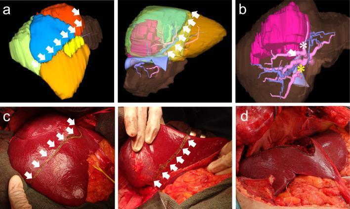
This composite educational graphic illustrates the preoperative planning and intraoperative execution of an anatomical liver resection (bisegmentectomy 7 and 8) for hepatocellular carcinoma. Panel (a) presents 3D CT volumetric reconstructions of the liver from anterior and posterior perspectives. Couinaud segments are color-coded for surgical mapping: Segment 5 (yellow), Segment 6 (orange), Segment 7 (green), and Segment 8, which is subdivided into dorsal (blue) and ventral (red) portions. White arrows delineate the planned virtual resection line. Panel (b) focuses on the portal vascular anatomy, highlighting the portal veins (P5, dorsal P8, and P6) using asterisks and arrows to guide anatomical dissection. Panel (c) contains two clinical photographs showing the intraoperative liver surface after portal staining with indigo carmine. The demarcation line, representing the planned transection boundary between the stained and unstained segments, is marked with cautery (indicated by white arrows). Panel (d) shows the final surgical site after the completion of the bisegmentectomy, displaying the exposed resection surface of the remnant liver. This series demonstrates the integration of 3D diagnostic imaging with real-time surgical navigation and staining techniques for precise oncological resection.
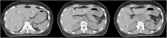
This diagnostic image consists of three axial contrast-enhanced CT scans of the upper abdomen, specifically illustrating hepatic segmental anatomy. The images utilize nine numbered circular markers to identify liver segments based on the Couinaud classification system. The first (superior) slice identifies the caudate lobe (Point 1), the superior left lateral segment (Point 2), the superior left medial segment (Point 4a), and the superior right segments (Points 7 and 8). Subsequent inferior slices identify the inferior left lateral segment (Point 3), inferior left medial segment (Point 4b), and the right lobe segments (Points 5 and 6). Key anatomical landmarks are visible for orientation, including the vertebral body and spinal canal posteriorly, the kidneys, the spleen in the left upper quadrant, and the fluid-filled stomach. The liver parenchyma appears homogeneous, and major vascular structures such as the inferior vena cava and portal venous branches are discernible. This visual serves as a topographical guide for identifying functional liver segments in clinical radiology and surgical planning.
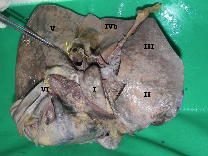
A clinical photograph of the inferior (visceral) surface of an explanted cadaveric human liver, positioned to demonstrate gross anatomical structures and Couinaud's segmental anatomy. The organ displays a mottled brownish-gray parenchymal texture. Key anatomical regions are labeled with Roman numerals: Segment I (caudate lobe), Segment II and III (left lateral section), Segment IVb (quadrate lobe), and Segments V and VI (right lobe). A yellow arrow indicates the lateral extent of the transverse fissure (porta hepatis). The image highlights an accessory inferior hepatic sulcus (AIS) extending postero-laterally from the transverse fissure into the right liver, being probed by surgical dissecting forceps. Visible landmarks include the gallbladder fossa and major vascular structures at the hilum. This image serves as an educational tool for surgical anatomy, hepatic lobectomy planning, and identifying variations in liver surface morphology, specifically the accessory sulci that may relate to the underlying portal venous branches.
pancreatitis acute CT scan pancreas
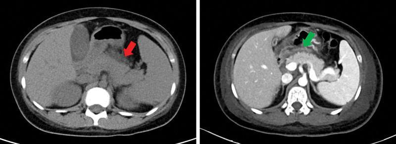
This abdominal computerized tomography (CT) scan consists of two axial views demonstrating features of acute pancreatitis and associated complications. The image on the left, a non-contrast CT, features a red arrow indicating a swollen, heterogeneously dense pancreas with peripancreatic fat stranding and indistinct margins, signs characteristic of acute inflammation. The image on the right is a contrast-enhanced CT where the green arrow points to a significant collection of low-density fluid in the anterior pararenal space and abdominal cavity, consistent with hydrops abdominis or peripancreatic fluid collection. Anatomical landmarks include the liver on the right side of the image, the spleen on the left, and the stomach located anteriorly to the pancreas. The contrast scan clearly delineates the vasculature and reinforces the presence of extensive peritoneal fluid and inflammatory changes surrounding the pancreatic head and body. These findings are clinically relevant for diagnosing systemic complications of autoimmune conditions like Systemic Lupus Erythematosus (SLE) affecting the gastrointestinal system.
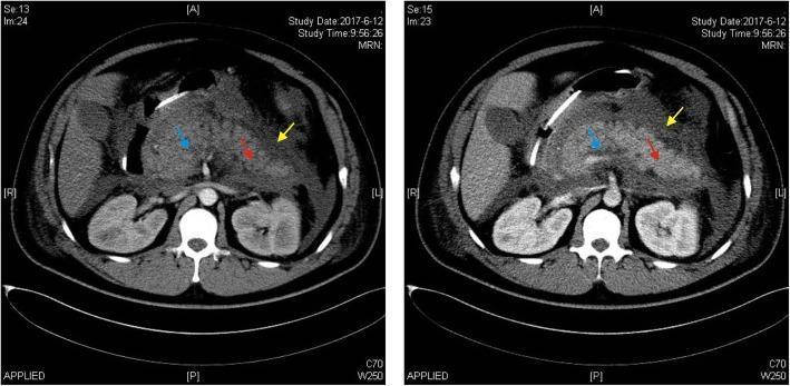
This diagnostic image consists of two side-by-side axial abdominal CT scans demonstrating features of severe acute pancreatitis. The left panel shows a non-contrast (plain) CT, while the right panel shows a contrast-enhanced CT. The pancreas is diffusely enlarged (edematous) with a significant loss of the normal sharp margins and disappearance of clear peripancreatic adipose tissue planes. Colored arrowheads on the enhanced scan identify key pathological findings: the blue arrowhead points to a hypo-enhancing region representing an acute necrotic collection; the red arrowhead indicates areas of pancreatic edema; and the yellow arrowhead highlights an acute peripancreatic fluid collection (APFC). The contrast-enhanced image is critical for distinguishing between viable, enhancing pancreatic parenchyma and non-enhancing necrotic tissue. There is a visible water-density shadow in the peripancreatic space, characteristic of inflammatory exudate. This comparison is used in medical education to illustrate the diagnostic criteria and complications of pancreatitis according to the Atlanta consensus.
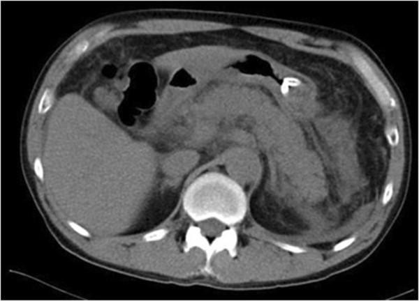
This diagnostic axial abdominal computed tomography (CT) scan illustrates hallmarks of acute pancreatitis. The central feature is significant pancreatomegaly, where the pancreas appears diffusely enlarged and swollen. Surrounding the organ, there is a notable increase in peripancreatic fat stranding and inflammation, evidenced by increased density and blurred margins between the pancreas and adjacent retroperitoneal fat. Anatomical landmarks include the liver on the left of the image (right anatomical side), the vertebral column posteriorly, and gas-filled bowel loops appearing as low-attenuation (dark) structures anteriorly and laterally. The image demonstrates soft tissue density variations across abdominal organs and subcutaneous fat at the periphery. This scan is educationally significant for demonstrating the radiological presentation of drug-induced or idiopathic acute pancreatitis, focusing on organ morphology and peripancreatic fluid/fat changes essential for diagnostic classification in gastroenterology and radiology.
Surgical Diseases of the Liver and Pancreas
PART I: THE LIVER
LO1 - Surgical Anatomy and Main Functions of the Liver
Gross and Segmental Anatomy
- Falciform ligament - connects the liver to the anterior abdominal wall and carries the ligamentum teres (obliterated left umbilical vein)
- Coronary ligaments - form the boundaries of the bare area (where the liver is in direct contact with the diaphragm)
- Left and right triangular ligaments - lateral extensions of the coronary ligaments
- Hepatoduodenal ligament - the free edge of the lesser omentum, carrying the portal triad: portal vein, hepatic artery, and common bile duct (the porta hepatis or hepatic hilum)
Couinaud Segmental Classification
- Portal venous branch
- Hepatic arterial branch
- Biliary drainage
- Hepatic venous outflow
- Segment I: Caudate lobe (posterior, drains directly into IVC)
- Segments II, III: Left lateral section (left of falciform ligament)
- Segment IV (IVa, IVb): Left medial section (quadrate lobe)
- Segments V, VI: Right lobe inferior
- Segments VII, VIII: Right lobe superior
"There are two anatomical lobes with a separate blood supply, bile duct and venous drainage... Resection is based on anatomical lines to preserve maximal functioning liver and blood supply." - BL, p. 1512
Blood Supply - Dual Hepatic Circulation
- Portal vein (80% of hepatic blood flow, ~60% of oxygen): carries nutrient-rich blood from the gut and spleen
- Hepatic artery (20% of flow, ~40% of oxygen): branch of the celiac axis, carries oxygenated blood
Ligament of Pringle / Hepatoduodenal Ligament
Main Functions of the Liver
| Category | Functions |
|---|---|
| Metabolic | Glucose metabolism (glycolysis, gluconeogenesis, glycogen storage); urea formation from protein catabolism; lipid metabolism |
| Synthetic | Albumin (transport of fatty acids, steroids, waste); coagulation factors (I, II, V, VII, VIII, IX, X, XI); complement proteins; angiotensin |
| Excretory | Bilirubin conjugation and excretion in bile; bile salt synthesis |
| Detoxification | Drug and hormone metabolism (Cytochrome P450 system); removal of gut endotoxins and foreign antigens; alcohol metabolism |
| Storage | Vitamins A, D, E, K, and B12; glycogen; iron and copper |
| Immunological | Kupffer cells (part of mononuclear phagocyte system) remove bacteria and debris from portal blood |
| Temperature | Maintains core body temperature via heat production |
| Acid-base | pH balance and correction of lactic acidosis |
Regeneration
LO2 - Common Clinical Presentations of Liver Disease
Jaundice
Hepatomegaly
- Congestive cardiac failure (tender, pulsatile in tricuspid regurgitation)
- Hepatitis (viral, alcoholic - tender)
- Cirrhosis (initially enlarged, later shrunken)
- Primary or secondary liver tumours (irregular, hard surface)
- Hepatic cysts, abscesses
- Infiltrative disorders (fatty liver, amyloidosis)
Right Upper Quadrant (RUQ) Pain
- Acute hepatitis - dull RUQ ache with tender hepatomegaly
- Liver abscess - severe RUQ pain with swinging fever
- Biliary colic - severe, colicky RUQ/epigastric pain radiating to right shoulder (from gallstones)
- Liver tumours - dragging/aching pain from capsular distension
Other Clinical Signs and Symptoms
- Spider naevi (>5 is pathological)
- Palmar erythema
- Leukonychia (white nails)
- Clubbing
- Dupuytren's contracture
- Gynaecomastia, testicular atrophy (in alcohol-related disease)
- Caput medusae (distended periumbilical veins - portal hypertension)
- Ascites (shifting dullness, fluid thrill)
- Peripheral oedema
- Splenomegaly (portal hypertension)
- Flapping tremor / asterixis (hepatic encephalopathy)
LO3 - Laboratory and Imaging Investigations for Liver Disease
Laboratory Investigations
| Test | Normal Range | Clinical Significance |
|---|---|---|
| Bilirubin (total) | 5-17 µmol/L (0.3-1.2 mg/dL) | Elevated in haemolysis, hepatocellular dysfunction, or biliary obstruction |
| Conjugated (direct) bilirubin | <5 µmol/L | Elevated specifically in intrahepatic/posthepatic (surgical) jaundice |
| Alkaline Phosphatase (ALP) | 30-140 IU/L | Particularly elevated in cholestatic disease / biliary obstruction |
| Alanine Transaminase (ALT) | 5-40 IU/L | Hepatocellular damage marker (more liver-specific than AST) |
| Aspartate Transaminase (AST) | 5-40 IU/L | Hepatocellular damage; also elevated in cardiac/muscle injury |
| Gamma-Glutamyl Transpeptidase (GGT) | 10-48 IU/L | Sensitive marker of alcohol intake; elevated in cholestatic disease |
| Albumin | 35-50 g/L | Synthetic function marker; falls in chronic liver disease |
| Prothrombin Time (PT) / INR | 12-16 s | Coagulation factor synthesis; prolonged in hepatocellular failure |
| Total Protein | 60-85 g/L |
- Full Blood Count (FBC): anaemia (chronic disease/haemolysis), thrombocytopenia (portal hypertension), macrocytosis (alcohol)
- Hepatitis serology: HBsAg, anti-HCV, anti-HAV, anti-HEV
- Auto-antibodies: ANA, ASMA (autoimmune hepatitis); AMA (primary biliary cholangitis)
- Ferritin, transferrin saturation: haemochromatosis
- Caeruloplasmin, serum copper: Wilson's disease
- Alpha-fetoprotein (AFP): hepatocellular carcinoma (HCC) tumour marker
- CEA, CA 19-9: metastatic disease surveillance
Imaging Investigations
- First-line imaging modality for the liver
- Inexpensive, no radiation, widely available
- Excellent for: biliary pathology (gallstones, duct dilatation), focal liver lesions, ascites, portal vein assessment with Doppler
- Limitations: operator-dependent, poor visualisation through ribs or bowel gas, obesity
- Contrast-enhanced US (CEUS) improves lesion characterisation
"Ultrasonography is a useful initial imaging test of the liver because it is inexpensive, widely available, involves no radiation exposure, and is well tolerated by patients. It is excellent for diagnosing biliary pathology and focal liver lesions." - SPS, p. 1383
- Triple-phase CT (non-contrast, arterial, portal venous): gold standard for characterising focal liver lesions
- HCC: arterial enhancement with "washout" in portal phase
- Liver metastases: typically hypodense on portal phase
- Assesses liver volume, vascular anatomy, and extrahepatic disease
- Excellent for biliary anatomy and staging of malignancy
- Superior soft tissue resolution
- MRI with hepatocyte-specific contrast (e.g. Primovist/gadoxetate): excellent for characterising small lesions, detecting metastases, differentiating HCC from regenerative nodules
- MRCP (Magnetic Resonance Cholangiopancreatography): non-invasive, no radiation, excellent visualisation of biliary tree and pancreatic duct - the investigation of choice for suspected biliary obstruction
- MR Elastography: assesses liver stiffness/fibrosis non-invasively
"MRCP provides visualisation of dilated bile ducts, and the high spatial and contrast resolution often enables accurate assessment of the level of occlusion in the biliary tree." - SPS, p. 1387
- Functional imaging using FDG (18F-fluorodeoxyglucose)
- Used for staging malignancy, detecting occult metastases, and confirming lymph node involvement
- FDG uptake correlates with metabolic activity - high-grade HCC is PET-avid, low-grade HCC may be PET-cold
- BL, p. 1857-1861; SPS, p. 1385-1387
- ERCP (Endoscopic Retrograde Cholangiopancreatography): diagnostic and therapeutic for biliary obstruction; allows stenting, stone extraction, biopsy
- PTC (Percutaneous Transhepatic Cholangiography): when ERCP is not possible; allows biliary drainage
- Liver Biopsy: for histological diagnosis of parenchymal disease; guided by US or CT
- Laparoscopy + Laparoscopic Ultrasonography: staging of HPB cancers, detects peritoneal metastases, avoids unnecessary laparotomy
- Angiography: for therapeutic embolisation, TACE (transarterial chemoembolisation)
- Octreotide scan (Scintigraphy): for somatostatin receptor-positive neuroendocrine tumours
LO4 - Causes and Differential Diagnoses of Jaundice
"Workup of a patient with jaundice is simplified by organizing the possible causes of the disorder into groups based on the location of bilirubin metabolism... prehepatic, intrahepatic, and posthepatic." - SPS, p. 1381
Prehepatic Jaundice (Unconjugated Hyperbilirubinaemia)
- Haemolytic anaemia: hereditary spherocytosis, sickle cell disease, G6PD deficiency, thalassaemia
- Acquired haemolysis: autoimmune haemolytic anaemia (Coombs test positive), microangiopathic haemolytic anaemia, drug-induced, transfusion reactions
- Ineffective erythropoiesis: megaloblastic anaemia
- Impaired albumin transport: severe malnutrition, burns (reduced carrier protein)
- Gilbert's syndrome: benign, common (~4-7% of population); reduced glucuronyltransferase activity causing mild unconjugated hyperbilirubinaemia during fasting or stress
- Crigler-Najjar syndrome: rare neonatal condition; severe absence of glucuronyltransferase; can cause bilirubin encephalopathy
Intrahepatic Jaundice (Medical / Hepatocellular Causes)
- Viral hepatitis: Hepatitis A, B, C, D, E
- Alcoholic hepatitis
- Drug-induced liver injury (DILI): paracetamol (acetaminophen) overdose, halothane, isoniazid-rifampicin, NSAIDs, antibiotics, antidepressants, valproic acid
- Autoimmune hepatitis
- Cirrhosis (any cause: alcohol, viral, metabolic, autoimmune)
- Non-alcoholic fatty liver disease (NAFLD/NASH)
- Ischaemic hepatitis ("shock liver"): from any cause of reduced hepatic perfusion
- Sepsis
- Intrahepatic cholestasis: primary biliary cholangitis (PBC), primary sclerosing cholangitis (PSC), drugs
- Hereditary excretion defects: Dubin-Johnson syndrome, Rotor's syndrome (impaired secretion of conjugated bilirubin)
- Pregnancy-related: intrahepatic cholestasis of pregnancy
- Wilson's disease, haemochromatosis, alpha-1 antitrypsin deficiency
Posthepatic (Obstructive) Jaundice - SURGICAL Causes
- Choledocholithiasis (common bile duct stones) - most common surgical cause
- Blood clot (haemobilia)
- Parasites (Ascaris, Clonorchis)
- Bile duct strictures: benign post-surgical (post-cholecystectomy), post-inflammatory (PSC, chronic pancreatitis)
- Cholangiocarcinoma (bile duct cancer): can be intrahepatic, perihilar (Klatskin tumour - most common), or distal
- Ampullary carcinoma
- Carcinoma of the head of pancreas - the most common cause of painless progressive obstructive jaundice in the elderly; "Courvoisier's law" - if a palpable, non-tender gallbladder accompanies jaundice, the cause is unlikely to be gallstones (as chronic gallstone disease scars the gallbladder wall, preventing distension)
- Enlarged lymph nodes at the porta hepatis (lymphoma, metastases - Mirizzi syndrome can mimic this)
- Pancreatitis (head oedema compressing distal bile duct)
- Hepatic metastases / HCC compressing intrahepatic ducts
| Feature | Prehepatic | Intrahepatic | Posthepatic (Obstructive) |
|---|---|---|---|
| Bilirubin type | Unconjugated | Mixed | Conjugated (direct) |
| Urine colour | Normal | Dark | Dark (bilirubinuria) |
| Stool colour | Normal | Normal or pale | Pale/clay |
| Pruritis | Absent | Variable | Present |
| ALP | Normal | Mildly raised | Markedly raised |
| AST/ALT | Normal | Markedly raised | Mildly raised |
| US finding | Normal | Normal/diffuse | Dilated bile ducts |
LO5 - Interpreting Investigations: Medical vs Surgical Causes of Liver Disease
Step 1 - History clues
- Surgical: painless progressive jaundice + weight loss (pancreatic cancer); colicky RUQ pain + fever + jaundice (Charcot's triad of cholangitis from choledocholithiasis); palpable gallbladder (Courvoisier's sign)
- Medical: viral prodrome, contacts, IV drug use, alcohol history, medications, autoimmune features
Step 2 - LFT pattern
- Obstructive pattern: ALP disproportionately elevated (e.g. ALP 600 IU/L, ALT 80 IU/L); conjugated bilirubin predominates; prolonged PT may occur late
- Hepatocellular pattern: ALT/AST disproportionately elevated (e.g. ALT 1200 IU/L, ALP 150 IU/L); both conjugated and unconjugated bilirubin elevated; PT may be significantly prolonged early
Step 3 - Bilirubin fractionation
- Predominantly conjugated (direct) bilirubin = hepatocellular or obstructive (requires further differentiation by imaging)
- Predominantly unconjugated (indirect) bilirubin = prehepatic or defective conjugation
Step 4 - Ultrasound
- Dilated intra- or extrahepatic bile ducts = obstructive/surgical cause until proven otherwise
- Common bile duct (CBD) >8 mm diameter is abnormal (>10 mm post-cholecystectomy)
- May reveal the cause: gallstones in CBD, pancreatic mass, liver lesion
- No duct dilatation = medical/hepatocellular cause more likely (biopsy if indicated)
Step 5 - Further imaging
- CT/MRCP: define the level and cause of obstruction; assess resectability
- ERCP: if therapeutic intervention needed (stenting, stone extraction)
Summary Algorithm
Jaundice
  ├── Unconjugated bilirubin dominant → Prehepatic (haemolysis, Gilbert's)
  └── Conjugated bilirubin dominant
        ├── ALT/AST markedly raised → Hepatocellular (hepatitis, DILI)
        ├── ALP markedly raised → Obstructive
        │     └── Ultrasound → Dilated ducts?
        │           ├── YES → SURGICAL cause → CT/MRCP → define level and cause
        │           └── NO → Intrahepatic cholestasis (PBC, PSC, drugs) → Biopsy
PART II: THE PANCREAS
LO6 - Anatomy and Main Functions of the Pancreas
Gross Anatomy
- Head: lies in the C-loop of the duodenum; the uncinate process is a downward and medial projection behind the superior mesenteric vessels
- Neck: lies directly over the superior mesenteric vein (SMV) and portal vein confluence - important surgical landmark
- Body: crosses the spine; the splenic vein runs along its posterior surface
- Tail: extends to the hilum of the spleen; lies within the splenorenal ligament
Ductal Anatomy
- Main pancreatic duct (duct of Wirsung): runs the entire length of the pancreas and joins the common bile duct to form the ampulla of Vater (hepatopancreatic ampulla), which opens into the second part of the duodenum at the major papilla. Controlled by the sphincter of Oddi
- Accessory duct (duct of Santorini): drains the upper head and opens separately at the minor papilla (2 cm proximal to major papilla)
- Pancreas divisum: failure of fusion of ventral and dorsal pancreatic ducts (5-10% of population); can cause recurrent pancreatitis
Relations and Surgical Importance
- Anterior: lesser sac (epiploic foramen gives access)
- Posterior: aorta, IVC, left renal vein, both adrenals, splenic vein, superior mesenteric vessels
- The bile duct passes through or posterior to the head of the pancreas - pancreatic head pathology commonly causes obstructive jaundice
- The duodenum shares a blood supply with the pancreatic head (pancreaticoduodenal arteries) - hence they are resected together in a Whipple procedure
Blood Supply
- Pancreatic head: superior and inferior pancreaticoduodenal arteries (from gastroduodenal artery and SMA respectively)
- Body and tail: splenic artery branches (dorsal pancreatic, great pancreatic artery)
- Venous drainage is to the portal vein via splenic and SMV
Functions of the Pancreas
- Produces 1-2 litres/day of pancreatic juice (pH 8.0-8.3, rich in bicarbonate from ductal cells)
- Digestive enzymes (secreted as inactive zymogens to prevent autodigestion):
- Proteases: trypsinogen (activated by enterokinase in duodenum to trypsin), chymotrypsinogen, proelastase, procarboxypeptidase
- Lipase and colipase: fat digestion
- Amylase: starch digestion
- Phospholipase A2, DNase, RNase
- Regulated by: secretin (stimulates bicarbonate), cholecystokinin (CCK) (stimulates enzyme secretion)
- Beta (β) cells (70%): insulin - lowers blood glucose
- Alpha (α) cells (20%): glucagon - raises blood glucose
- Delta (δ) cells: somatostatin - inhibits insulin and glucagon
- PP cells: pancreatic polypeptide
LO7 - Common Clinical Features of Pancreatic Disease
Acute Pancreatitis
"The underlying mechanism of injury in pancreatitis is thought to be premature activation of pancreatic enzymes within the pancreas, leading to a process of autodigestion." - BL, p. 7597
- Gallstones (most common in UK/Ireland - 40-50%)
- Ethanol/alcohol (30-35%)
- Trauma
- Steroids
- Mumps (and other viral infections)
- Autoimmune (IgG4-related)
- Scorpion venom
- Hyperlipidaemia / Hypercalcaemia
- ERCP (post-procedural)
- Drugs (azathioprine, thiazides, furosemide, oestrogens, tetracyclines)
- Pain: severe, constant epigastric/upper abdominal pain, radiating to the back; may be relieved by sitting forward; builds over minutes to hours
- Nausea and vomiting
- Fever (low-grade early; high fever suggests secondary infection/cholangitis)
- Tachycardia, hypotension (in severe disease - systemic inflammatory response)
- Abdominal tenderness: epigastric, often with guarding; periumbilical bruising (Cullen's sign) and flank bruising (Grey Turner's sign) - indicate retroperitoneal haemorrhage, appear 24-48 hours after onset
- Ileus - absent bowel sounds, abdominal distension
- Jaundice - if gallstone impacted at ampulla or oedema of head compressing CBD
- Respiratory: basal crackles, pleural effusion (especially left-sided), in severe disease - ARDS
- Mild (interstitial oedematous pancreatitis): most cases (80%); no organ failure, no necrosis; mortality <1%
- Moderate: transient organ failure (<48 hrs) or local complications
- Severe (necrotising pancreatitis): persistent organ failure (>48 hrs) and/or infected necrosis; mortality 15-30%
- Ranson's criteria (11 parameters at admission and 48 hours)
- APACHE II score
- Glasgow/Imrie criteria (8 parameters at 48 hours) - score ≥3 indicates severe attack
- CT Severity Index (Balthazar score): grades A-E based on CT findings of necrosis and fluid collections; used after 72 hours
Chronic Pancreatitis
- Pain: recurrent or persistent epigastric pain radiating to the back - the dominant symptom
- Exocrine insufficiency: steatorrhoea (pale, fatty, offensive stools, floating), malabsorption, weight loss - occurs when >90% of acinar function is lost
- Endocrine insufficiency: diabetes mellitus ("pancreatogenic diabetes") - occurs late
- Obstructive jaundice: from fibrosis of the head of pancreas compressing the bile duct
- Duodenal obstruction: from fibrosis in the pancreatic head
- Pancreatic pseudocyst formation
Pancreatic Masses
- Painless, progressive obstructive jaundice (head of pancreas cancer pressing on CBD) - classic presentation; Courvoisier's gallbladder may be palpable
- Epigastric/back pain: visceral pain from retroperitoneal invasion; often described as a boring, persistent ache; worse at night; improved by sitting forward
- Weight loss and cachexia: from malabsorption and systemic tumour effects
- New-onset diabetes mellitus in an older patient (destruction of islets; or paraneoplastic effect)
- Steatorrhoea: from pancreatic duct obstruction
- Trousseau's syndrome: migratory thrombophlebitis (venous thrombosis) associated with pancreatic adenocarcinoma
- Palpable epigastric mass: usually advanced disease
- Gastric outlet obstruction: duodenal invasion by head of pancreas tumour
LO8 - Laboratory and Imaging Investigations for Pancreatic Disease
Laboratory Investigations
| Test | Finding |
|---|---|
| Serum amylase | >3× upper limit of normal (diagnostic); peaks early (6-12 hrs), returns to normal in 3-5 days |
| Serum lipase | More sensitive and specific than amylase; remains elevated longer (7-14 days); preferred by many centres |
| FBC | Leukocytosis (inflammation/infection); haematocrit rise (haemoconcentration - severity marker) |
| U&E, creatinine | Renal function; markers of severity |
| LFTs | ALP/bilirubin raised in gallstone pancreatitis; ALT >150 IU/L strongly suggests gallstone aetiology |
| Calcium | Hypocalcaemia (severity marker; saponification with free fatty acids); or hypercalcaemia (cause) |
| Glucose | Hyperglycaemia (severity marker) |
| CRP | CRP >150 mg/L at 48 hours is a marker of severity/necrosis |
| Triglycerides | If hypertriglyceridaemia is the suspected cause (>1000 mg/dL) |
| LDH | Raised in severe pancreatitis (Ranson criteria) |
- CA 19-9 (carbohydrate antigen 19-9): tumour marker for pancreatic adenocarcinoma; elevated in ~80% of cases; not diagnostic alone (elevated in benign biliary disease too); useful for monitoring treatment response and recurrence
- CEA: less specific
- Faecal elastase-1: marker of exocrine insufficiency (<200 µg/g stool = significant insufficiency)
- 72-hour faecal fat test: gold standard for malabsorption but rarely used clinically
- HbA1c, fasting glucose: assess endocrine insufficiency
- IgG4 levels: elevated in autoimmune pancreatitis
- PRSS1, SPINK1, CFTR genetic testing: if hereditary pancreatitis suspected
Imaging Investigations
- First-line for suspected pancreatitis
- Detects gallstones (cause of pancreatitis) - sensitivity >95% for gallbladder stones but poor for CBD stones
- Detects pancreatic oedema, peripancreatic fluid
- Detects masses, duct dilatation, and vascular involvement (with Doppler)
- Often limited by overlying bowel gas
- Gold standard for:
- Confirming diagnosis when in doubt
- Assessing severity of acute pancreatitis (Balthazar scoring/CT Severity Index) - best performed 72 hours after onset to allow full evolution of necrosis
- Detecting and characterising pancreatic masses
- Staging pancreatic cancer (assessing vascular involvement, metastases, resectability)
- Detecting complications (pseudocysts, vascular complications, abscesses)
- BL, p. 1691: "CT remains the mainstay of accurate diagnosis"
- Superior to CT for soft tissue characterisation of pancreatic lesions
- MRCP: non-invasive ductal imaging - excellent for pancreatic duct changes in chronic pancreatitis (irregular "chain of lakes" appearance), ductal stones, ampullary pathology
- Better than CT for detecting small liver metastases
- Essential for evaluating cystic pancreatic lesions (IPMN, MCN vs pseudocyst)
- Highly sensitive (>90%) for pancreatic masses, especially lesions <2 cm
- Allows fine needle aspiration (FNA)/biopsy for histological diagnosis
- Best modality for staging local vascular invasion (portal vein, SMA, SMV)
- Used to evaluate cystic lesions and obtain fluid for CEA, amylase, and cytology
- Detects CBD stones with high sensitivity (better than CT/US)
- Diagnostic and therapeutic for biliary and pancreatic ductal pathology
- Detects CBD stones, allows stenting of biliary obstruction (e.g. cancer, strictures)
- "Chain of lakes" appearance in main duct of chronic pancreatitis
- Thick mucus extruding from ampulla is diagnostic of main duct IPMN
- Carries risk of post-ERCP pancreatitis (2-5%)
- Largely replaced for diagnosis by MRCP; retained for therapy
- For staging of pancreatic cancer, detecting distant metastases
- Differentiating between autoimmune pancreatitis (which can mimic cancer) and malignancy
- Staging of pancreatic cancer prior to laparotomy - detects small peritoneal metastases and liver metastases not seen on CT
LO9 - Differential Diagnoses of Pancreatic Pathology
For Epigastric Pain / Acute Abdominal Presentation:
- Acute pancreatitis (gallstones, alcohol) - most common
- Peptic ulcer disease (duodenal/gastric ulcer) or perforation
- Biliary colic / cholecystitis / cholangitis
- Acute mesenteric ischaemia
- Perforated hollow viscus
- Aortic aneurysm (ruptured or dissection)
- Inferior myocardial infarction (can mimic epigastric pain)
- Pancreatitis from other causes (post-ERCP, drug-induced, hypercalcaemia)
For Chronic Pancreatic Pain:
- Chronic pancreatitis (alcohol, idiopathic, hereditary)
- Pancreatic cancer (must always be excluded)
- Autoimmune pancreatitis (IgG4)
- Pancreatic pseudocyst
- Peptic ulcer disease
- Functional dyspepsia / irritable bowel syndrome
For Painless Obstructive Jaundice + Weight Loss:
- Carcinoma of the head of the pancreas (most important to exclude)
- Cholangiocarcinoma (distal CBD / Klatskin tumour)
- Ampullary carcinoma (periampullary cancer)
- Duodenal carcinoma
- Autoimmune pancreatitis (mimics cancer on imaging)
- Sclerosing cholangitis (PSC)
- Chronic pancreatitis (inflammatory head mass)
- Choledocholithiasis (CBD stones - usually associated with pain/cholangitis)
- Metastatic lymphadenopathy at the hepatoduodenal ligament
- Lymphoma
For Pancreatic Cystic Lesion on Imaging:
- Pseudocyst (most common; post-pancreatitis; high amylase in cyst fluid, low CEA)
- Serous cystadenoma (benign; older women; multiple small cysts)
- Mucinous cystic neoplasm (MCN) (premalignant; perimenopausal women; body/tail; ovarian-type stroma; thick-walled)
- Intraductal papillary mucinous neoplasm (IPMN) (premalignant to malignant; head of pancreas; main duct IPMNs are high risk)
- Solid pseudopapillary neoplasm (SPN): rare; young women; part-solid, part-cystic
- Cystic neuroendocrine tumour
"It is important to differentiate a pseudocyst from an APFC... a cystic neoplasm may be confused with a chronic pseudocyst. EUS and aspiration of the cyst fluid are very useful." - BL, p. 8203-8216
"Fluid from a pseudocyst typically has a low CEA level, and levels above 400 ng/mL are suggestive of a mucinous neoplasm." - BL, p. 8211
LO10 - Benign vs Malignant Pancreatic Conditions
Benign Pancreatic Conditions
- An inflammatory (not neoplastic) condition; the vast majority of cases resolve completely
- Most patients (80%) have mild disease with mortality <1%
- However, severe necrotising pancreatitis carries significant mortality (15-30%)
- Does not progress to cancer (though chronic pancreatitis is a risk factor)
- Irreversible but benign inflammatory/fibrotic condition
- Causes permanent exocrine and endocrine insufficiency
- An independent risk factor for pancreatic adenocarcinoma (but not inevitably malignant)
- Management is medical/endoscopic/surgical symptom control
- A benign fluid collection following pancreatitis or trauma; enclosed by fibrous and granulation tissue (not a true epithelial-lined cyst)
- Typically benign and many resolve spontaneously
- Requires intervention only if symptomatic, large (>6 cm), infected, or to exclude malignancy
- "Multiple small cysts, almost like bubble wrap" - BL, p. 8735
- Benign - almost zero malignant potential
- Typically in older women
- Can be observed safely if asymptomatic and diagnosis is certain
- Benign, steroid-responsive condition caused by IgG4-related disease
- Critically important because it mimics pancreatic cancer on imaging and clinically
- Distinguishing features: diffuse enlargement ("sausage pancreas"), halo sign on CT, elevated serum IgG4, rapid response to steroids, associated with other IgG4-related diseases (cholangitis, sialadenitis)
- Arise from islet cells; include insulinoma, gastrinoma, glucagonoma, VIPoma
- Many are functional (produce hormones causing clinical syndromes)
- Most insulinomas are benign (90%)
- Gastrinomas have higher malignant potential (~60% malignant)
Malignant Pancreatic Conditions
- The most common and most lethal primary pancreatic malignancy (~85% of all pancreatic cancers)
- Arises from ductal cells; most commonly in the head of the pancreas (60-70%)
- Prognosis is very poor: overall 5-year survival <10%; only ~20% of patients are resectable at presentation
- Risk factors: smoking, chronic pancreatitis, diabetes mellitus, obesity, family history, BRCA2/PALB2/ATM gene mutations
- Pathology: dense desmoplastic stroma; early perineural invasion, lymphovascular invasion, and distant metastases
- Tumour markers: CA 19-9 elevated in ~80%; used for monitoring, not diagnosis
- Treatment: surgical resection (Whipple's pancreaticoduodenectomy for head lesions; distal pancreatectomy for body/tail) is the only curative option; adjuvant chemotherapy (FOLFIRINOX, gemcitabine/capecitabine) improves survival
- Mucinous Cystic Neoplasm (MCN): carries significant risk of malignant transformation; all MCNs in fit patients should be resected - BL, p. 8737
- Intraductal Papillary Mucinous Neoplasm (IPMN):
- Main duct IPMN: high risk of malignancy (~60-70%); resection recommended
- Branch duct IPMN: lower risk; management depends on size, mural nodules, symptoms
- Pancreatic Intraepithelial Neoplasia (PanIN): microscopic precursor to PDAC (PanIN-1, -2, -3)
- Arises at the ampulla of Vater
- Generally better prognosis than PDAC (5-year survival ~40%) due to earlier presentation (obstructive jaundice when small)
- Treated by Whipple's procedure
- Malignant NETs (e.g. malignant gastrinoma, nonfunctioning NETs) tend to be slow-growing and have far better prognosis than PDAC
- Even with liver metastases, survival can be measured in years with appropriate treatment
Simple Comparison: Benign vs Malignant Pancreatic Conditions
| Feature | Benign (e.g. Pancreatitis, Pseudocyst) | Malignant (e.g. PDAC) |
|---|---|---|
| Nature | Inflammatory or benign neoplastic | Malignant neoplasm |
| Course | Often self-limiting or treatable | Progressive, often fatal |
| Spread | Does not metastasise | Spreads to lymph nodes, liver, peritoneum |
| Pain | Yes (from inflammation) | Yes (from invasion, neuropathy) |
| CA 19-9 | May be mildly elevated | Often markedly elevated |
| CT appearance | Diffuse swelling, fluid | Mass lesion (often hypodense), duct cutoff |
| Resectability | N/A (surgical drainage if needed) | Only 20% resectable at presentation |
| Prognosis | Usually good | Very poor (5-year survival <10%) |
| Treatment goal | Symptom control, address cause | Curative resection if possible; palliation if not |
Summary of Key Points
- The liver has 8 Couinaud segments, each functionally independent, enabling anatomical resection
- Dual blood supply (80% portal, 20% hepatic artery); regenerates to original volume after resection
- Key functions: synthesis (albumin, clotting factors), metabolism (bilirubin, drugs), storage (vitamins, glycogen)
- Clinical presentations: jaundice, hepatomegaly, RUQ pain, ascites, encephalopathy
- Investigations: LFTs pattern (obstructive vs hepatocellular), ultrasound (dilated ducts vs no dilatation), CT/MRCP for level and cause
- Jaundice classification - prehepatic (unconjugated), intrahepatic (mixed), posthepatic/obstructive (conjugated); the last is the surgical cause
- Retroperitoneal organ; head lies in duodenal C-loop; shares blood supply with duodenum
- Exocrine (digestive enzymes, bicarbonate) + endocrine (insulin, glucagon) functions
- Acute pancreatitis: severe epigastric pain, amylase/lipase elevation; causes include gallstones and alcohol
- Pancreatic cancer: painless progressive jaundice, weight loss, back pain; CA 19-9 elevated; poor prognosis
- Investigations: US (first-line), CECT (severity/staging), MRCP (ductal anatomy), EUS-FNA (tissue diagnosis)
- Benign conditions (pancreatitis, pseudocyst, serous cystadenoma) are treatable and non-lethal; malignant (PDAC) has very poor prognosis with <10% 5-year survival
- Bailey and Love's Short Practice of Surgery, 28th Edition (BL) - Chapters 69, 72 (pp. 1512-1862, 7376-8750)
- Schwartz's Principles of Surgery, 11th Edition (SPS) - Chapter 31 (pp. 1381-1390)
- Sabiston Textbook of Surgery, 21st Edition (SAB) - Chapters on liver and pancreas