I now have enough information from the textbooks to write comprehensive answers. Let me compile everything.---
LONG ANSWER QUESTIONS (15 Marks Each)
LONG Q1 — Urea Cycle: Detailed Note with Reactions & Major Metabolic Disorders
Introduction
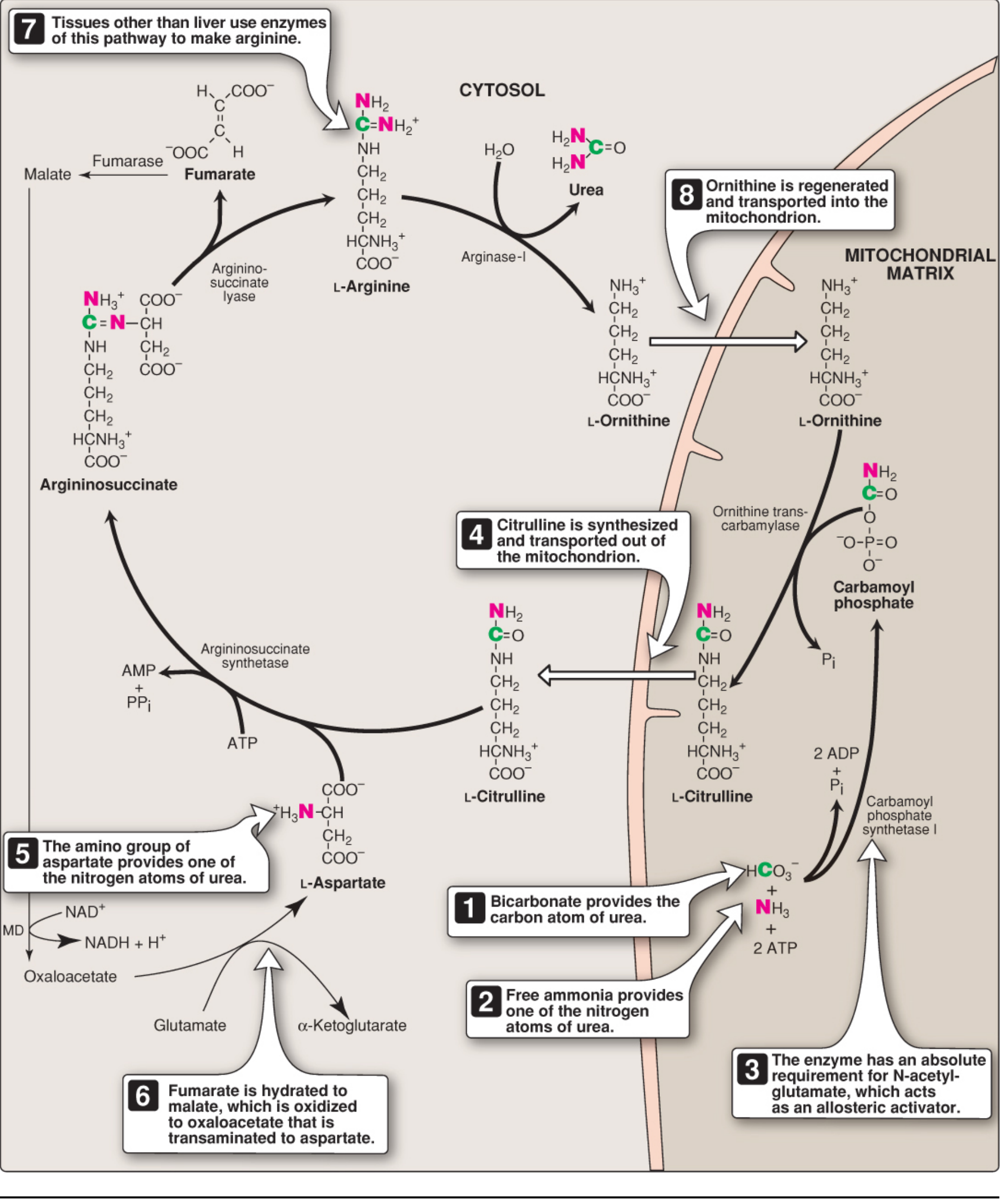
Urea is the major disposal form of amino groups derived from amino acids and accounts for ~90% of the nitrogen-containing components of urine. One nitrogen of the urea molecule is supplied by free ammonia (NH₄⁺) and the other by aspartate. The carbon and oxygen of urea are derived from CO₂ (as HCO₃⁻). Urea is produced exclusively in the liver and transported in the blood (measured as Blood Urea Nitrogen, BUN) to the kidneys for excretion. The urea cycle was proposed in 1932 by Hans Krebs and Kurt Henseleit and was originally called the Krebs–Henseleit cycle.
Normal blood ammonia: 30–60 µM. Urea is synthesized to detoxify this, since ammonia is especially toxic to the brain and CNS.
Reactions of the Urea Cycle
The first two reactions occur in the mitochondrial matrix; the remaining three occur in the cytosol.
Step 1 — Synthesis of Carbamoyl Phosphate (Mitochondria)
NH₄⁺ + HCO₃⁻ + 2 ATP → Carbamoyl Phosphate + 2 ADP + Pᵢ
Enzyme: Carbamoyl Phosphate Synthetase I (CPS I)
- Located in mitochondria of liver and intestine
- Requires N-acetylglutamate (NAG) as an obligatory allosteric activator
- NAG is synthesized by N-acetylglutamate synthase (NAGS) from acetyl-CoA + glutamate; arginine is an activator of NAGS
- Consumes 2 ATP
(Note: CPS II — cytosolic, glutamine as N-source, for pyrimidine synthesis; no NAG needed)
Step 2 — Formation of Citrulline (Mitochondria)
Carbamoyl Phosphate + Ornithine → Citrulline + Pᵢ
Enzyme: Ornithine Transcarbamoylase (OTC)
- Citrulline is transported to the cytosol via an antiporter (citrulline out, ornithine in)
Step 3 — Formation of Argininosuccinate (Cytosol)
Citrulline + Aspartate + ATP → Argininosuccinate + AMP + PPᵢ
Enzyme: Argininosuccinate Synthetase
- The α-amino group of aspartate provides the second nitrogen of urea
- Driven by cleavage of ATP → AMP + PPᵢ (equivalent to 2 ATP used)
Step 4 — Cleavage of Argininosuccinate (Cytosol)
Argininosuccinate → Arginine + Fumarate
Enzyme: Argininosuccinate Lyase
- Fumarate enters the TCA cycle or is converted to malate → oxaloacetate → re-transaminated to aspartate (links urea cycle to TCA cycle)
- Arginine is the immediate precursor of urea
Step 5 — Hydrolysis of Arginine (Cytosol)
Arginine + H₂O → Ornithine + Urea
Enzyme: Arginase I (virtually exclusive to liver)
- Regenerates ornithine, completing the cycle
- Urea diffuses to blood → kidneys → excreted in urine
Overall Stoichiometry
Aspartate + NH₄⁺ + HCO₃⁻ + 3 ATP + H₂O → Urea + Fumarate + 2 ADP + AMP + 2Pᵢ + PPᵢ
4 high-energy phosphate bonds consumed per molecule of urea synthesized.
Regulation of the Urea Cycle
- NAG is an essential activator of CPS I (rate-limiting enzyme)
- Arginine activates NAGS → more NAG → more CPS I activity (positive feedback)
- Short-term: substrate availability
- Long-term: enzyme induction (e.g., high-protein diet induces urea cycle enzymes)
Major Metabolic Disorders of the Urea Cycle
Deficiencies in each enzyme lead to hyperammonemia. The most severe manifestation is cerebral toxicity leading to intellectual disability or death.
| Disorder | Enzyme Deficient | Key Features |
|---|
| CPS I Deficiency | CPS I | Hyperammonemia, no orotic aciduria |
| OTC Deficiency (most common) | Ornithine Transcarbamoylase | X-linked; hyperammonemia + orotic aciduria (carbamoyl phosphate overflows into pyrimidine pathway) |
| Citrullinemia (Type I) | Argininosuccinate Synthetase | Elevated citrulline in blood/urine |
| Argininosuccinic Aciduria | Argininosuccinate Lyase | Argininosuccinate in blood/urine/CSF; neurological features |
| Arginase Deficiency | Arginase I | Elevated blood arginine; spastic paraplegia, mental retardation |
| NAGS Deficiency | N-acetylglutamate Synthase | Like CPS I deficiency; responds to N-carbamylglutamate |
OTC deficiency is the most common (1 in 80,000); it is X-linked. A hallmark is orotic aciduria (excess carbamoyl phosphate floods the pyrimidine pathway → orotic acid excreted in urine).
Treatment principles:
- Low-protein diet to reduce nitrogen load
- Benzoic acid + glycine → hippuric acid (excreted; removes 1 N)
- Phenylbutyrate/Phenylacetate + glutamine → phenylacetylglutamine (excreted; removes 2 N per molecule)
- Arginine supplementation (for defects after argininosuccinate synthesis)
- Hemodialysis in acute crises
LONG Q2 — Transamination Reaction in Catabolism of Amino Acids
Definition
Transamination is the transfer of the α-amino group from an amino acid to an α-keto acid, producing a new amino acid and a new keto acid. It is catalyzed by enzymes called transaminases (aminotransferases) and requires pyridoxal phosphate (PLP) — a derivative of vitamin B₆ — as a coenzyme.
General Reaction
Amino acid₁ + α-Keto acid₂ ⇌ α-Keto acid₁ + Amino acid₂
This is a reversible reaction with ΔG ≈ 0.
Most Important Transamination Reactions
1. Alanine Aminotransferase (ALT / GPT)
L-Alanine + α-Ketoglutarate ⇌ Pyruvate + L-Glutamate
2. Aspartate Aminotransferase (AST / GOT)
L-Aspartate + α-Ketoglutarate ⇌ Oxaloacetate + L-Glutamate
Glutamate is the central collector of amino groups — most amino acids donate their nitrogen to α-ketoglutarate, forming glutamate.
Role of PLP (Pyridoxal Phosphate)
PLP acts as a prosthetic group (covalently bound to enzyme via Schiff base). It shuttles between pyridoxal phosphate (acceptor of amino group → forms pyridoxamine phosphate) and back, acting as the true amino group carrier. This is called the ping-pong (double displacement) mechanism.
Role in Amino Acid Catabolism
Transamination initiates catabolism of most amino acids:
- The amino group is removed first, leaving a carbon skeleton (α-keto acid)
- The carbon skeleton enters amphibolic pathways (TCA cycle intermediates) as:
- Pyruvate → glucogenic
- Acetyl-CoA / Acetoacetyl-CoA → ketogenic
- Oxaloacetate, α-Ketoglutarate, Succinyl-CoA, Fumarate → glucogenic
| Amino Acid | Product of Transamination | Metabolic fate |
|---|
| Alanine | Pyruvate | Gluconeogenesis |
| Aspartate | Oxaloacetate | TCA/gluconeogenesis |
| Glutamate | α-Ketoglutarate | TCA cycle |
| Leucine | α-Keto-isocaproate | Ketogenesis |
Exceptions — amino acids that do NOT transaminate first: Proline, Hydroxyproline, Threonine, Lysine (their α-amino groups do not participate in transamination).
Glucose-Alanine Cycle
In muscle, pyruvate receives the amino group from glutamate → alanine is released to the blood → taken up by liver → re-transaminated to pyruvate → used for gluconeogenesis. This is a key mechanism of inter-organ nitrogen transport.
Link to Urea Synthesis
Glutamate formed by transamination undergoes oxidative deamination (by glutamate dehydrogenase) → liberates NH₄⁺ → enters the urea cycle. Glutamate is therefore the immediate precursor of the ammonia that enters the urea cycle.
LONG Q3 — Bile Pigments: Names, Formation, and Associated Diseases
What are Bile Pigments?
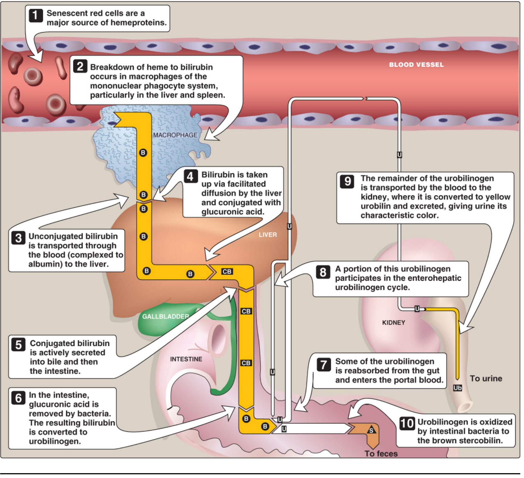
Bile pigments are colored compounds derived from the catabolism of heme. Collectively, they include biliverdin, bilirubin, urobilinogen, urobilin, and stercobilin. They are end products of heme degradation and are excreted primarily via bile.
Formation of Bile Pigments
Step 1 — Heme release:
After ~120 days, senescent RBCs are phagocytosed by the mononuclear phagocyte system (MPS) in the liver, spleen, and bone marrow. ~85% of heme comes from RBC hemoglobin.
Step 2 — Biliverdin formation:
Heme → (Heme oxygenase, requires NADPH + O₂) → Biliverdin (green) + CO + Fe²⁺
Step 3 — Bilirubin formation:
Biliverdin → (Biliverdin reductase, NADPH) → Bilirubin (red-orange)
Bilirubin is water-insoluble (unconjugated/indirect bilirubin); transported in blood bound to albumin.
Step 4 — Hepatic uptake and conjugation:
- Bilirubin dissociates from albumin, enters hepatocyte, binds to ligandin
- Two molecules of glucuronic acid are added by bilirubin UDP-glucuronosyltransferase (UGT)
- → Bilirubin diglucuronide (conjugated/direct bilirubin) — water soluble
- Actively secreted into bile canaliculi (rate-limiting step)
Step 5 — Intestinal metabolism:
- Intestinal bacteria deconjugate CB → reduce it to urobilinogen (colorless)
- Urobilinogen is partly reabsorbed (enterohepatic circulation) → re-excreted in bile or appears in urine as urobilin (yellow)
- The remainder is oxidized in the colon to stercobilin (brown color of feces)
Normal Values
- Total plasma bilirubin: ≤ 1 mg/dL
- Jaundice appears at: ≥ 2–3 mg/dL
Diseases Associated with Bile Pigment Metabolism
| Disease | Mechanism | Bilirubin Type Elevated |
|---|
| Hemolytic Jaundice | Excess RBC destruction → excess bilirubin | Unconjugated (indirect) |
| Hepatitis / Cirrhosis | Liver cell damage → impaired conjugation + secretion | Both UCB and CB |
| Obstructive (Posthepatic) Jaundice | Bile duct obstruction (gallstone, tumor) | Conjugated (direct); dark urine, pale stools |
| Gilbert Syndrome | Mild UGT deficiency (AD); benign; UCB mildly elevated | Unconjugated |
| Crigler-Najjar Syndrome I | Complete absence of UGT; severe UCB → kernicterus | Unconjugated |
| Crigler-Najjar Syndrome II | Partial UGT deficiency; less severe | Unconjugated |
| Dubin–Johnson Syndrome | Defect in canalicular transport of CB | Conjugated |
| Rotor Syndrome | Impaired hepatic uptake/storage of bilirubin | Conjugated |
| Neonatal (Physiologic) Jaundice | Immature UGT at birth; resolves in 4 weeks | Unconjugated → treated with phototherapy |
| Kernicterus | UCB (exceeds albumin binding) crosses BBB → basal ganglia damage | Unconjugated |
LONG Q4 — Transaminases: Definition and Diagnostic Importance
Definition
Transaminases (aminotransferases) are enzymes that catalyze transamination — transfer of an amino group from an amino acid to an α-keto acid. All require pyridoxal phosphate (PLP) as coenzyme.
The two clinically most important transaminases are:
-
ALT (Alanine Aminotransferase) — formerly called GPT (Glutamate Pyruvate Transaminase)
- Reaction: L-Alanine + α-Ketoglutarate ⇌ Pyruvate + L-Glutamate
- Predominantly in liver cytosol; highly specific for liver
-
AST (Aspartate Aminotransferase) — formerly called GOT (Glutamate Oxaloacetate Transaminase)
- Reaction: L-Aspartate + α-Ketoglutarate ⇌ Oxaloacetate + L-Glutamate
- Found in liver, heart, skeletal muscle, kidney, brain (less specific for liver)
Normal Serum Values
- ALT: 7–56 U/L
- AST: 10–40 U/L
Diagnostic Importance of Transaminases
When cells are damaged or necrotic, intracellular transaminases leak into the bloodstream → elevated serum levels. This makes them excellent markers of cellular injury.
1. Liver Diseases
- Viral hepatitis (A, B, C): Very high ALT and AST (often 10–100× normal); ALT > AST
- Alcoholic hepatitis: AST > ALT (typically AST:ALT ratio > 2:1) — "De Ritis ratio"
- Cirrhosis: Moderate elevation
- Non-alcoholic fatty liver disease (NAFLD): Mild-moderate elevation of ALT
- Drug-induced hepatotoxicity: Marked elevation (e.g., paracetamol overdose)
- Obstructive jaundice: Mild elevation; alkaline phosphatase more prominent
2. Myocardial Infarction (Heart Attack)
- AST rises within 6–12 hrs, peaks at 24–36 hrs, returns to normal by 3–5 days
- Now largely replaced by troponin and CK-MB for cardiac diagnosis, but AST is still informative
- ALT is NOT elevated in uncomplicated MI → helps differentiate from liver disease
3. Skeletal Muscle Diseases
- Muscular dystrophies, rhabdomyolysis → elevated AST and CK
4. De Ritis Ratio (AST:ALT)
| Ratio | Interpretation |
|---|
| > 2:1 | Alcoholic liver disease |
| < 1 (ALT > AST) | Viral hepatitis / NAFLD |
| > 1 | Cirrhosis, liver fibrosis |
5. Other Clinical Applications
- Pulmonary embolism: Mild AST elevation
- Hemolytic anemia: Mild elevation
- Monitoring liver toxicity of drugs (e.g., statins, antitubercular drugs, methotrexate)
- Preoperative assessment
- Pregnancy: Serial transaminase monitoring in obstetric cholestasis, HELLP syndrome
LONG Q5 — Porphyrins: Types and Clinical Importance of Porphyria
What are Porphyrins?
Porphyrins are cyclic compounds formed by the linkage of four pyrrole rings through methine bridges (=CH–). They are the structural backbone of heme (iron-containing), chlorophyll (magnesium-containing), and vitamin B₁₂ (cobalt-containing).
In humans, heme (protoporphyrin IX + Fe²⁺) is the functional porphyrin — it is the prosthetic group of:
- Hemoglobin and myoglobin (oxygen transport)
- Cytochromes (electron transport)
- Catalase and peroxidase
- Tryptophan pyrrolase
Heme Synthesis Pathway (Brief Overview)
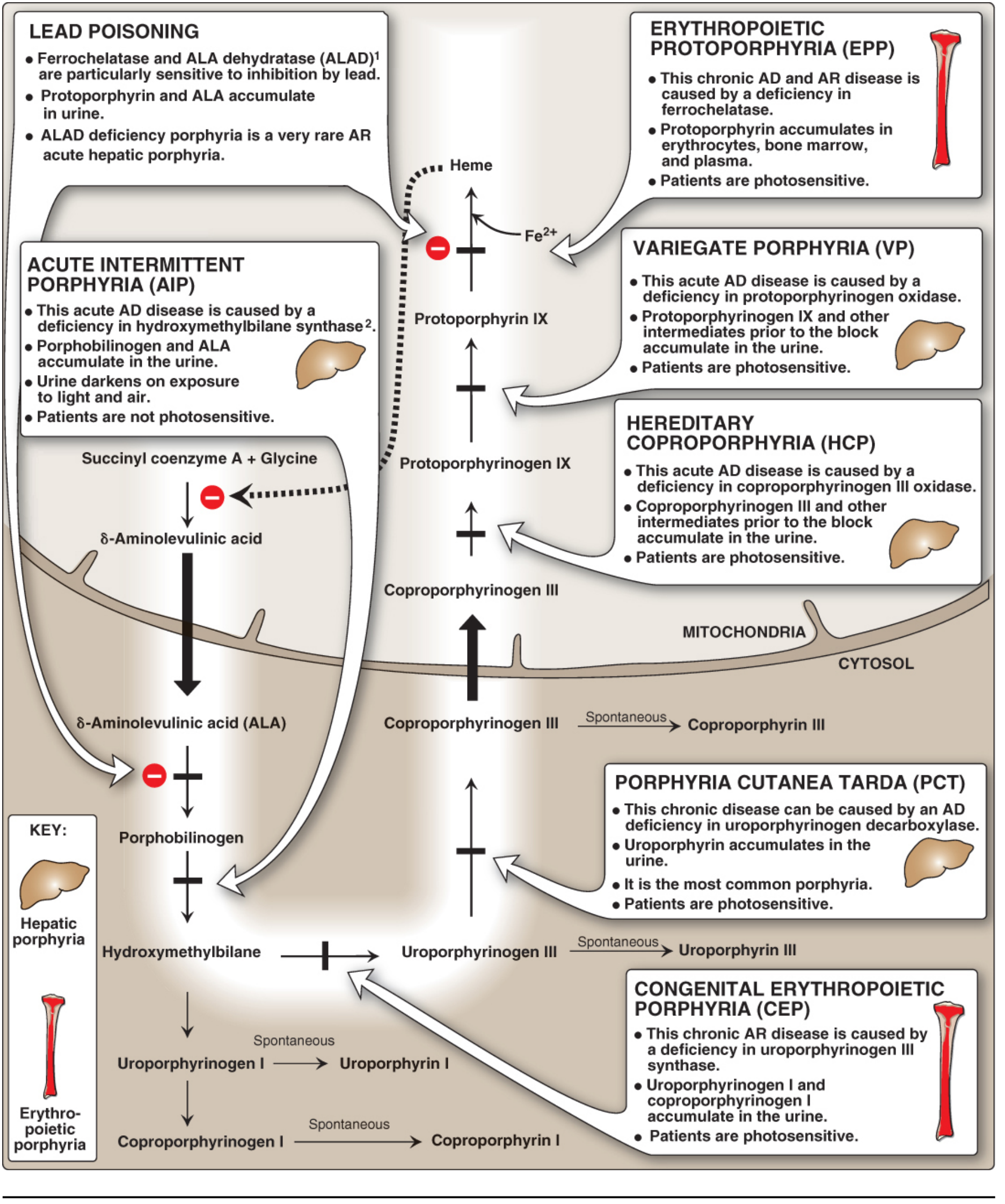
- Glycine + Succinyl-CoA → δ-Aminolevulinic acid (ALA) — catalyzed by ALA synthase (ALAS), the rate-limiting enzyme; in mitochondria
- 2 ALA → Porphobilinogen (PBG) (by ALA dehydratase / PBG synthase)
- 4 PBG → Hydroxymethylbilane (by PBG deaminase / uroporphyrinogen I synthase)
- → Uroporphyrinogen III (by uroporphyrinogen III cosynthase)
- → Coproporphyrinogen III (by uroporphyrinogen decarboxylase, UROD)
- → Protoporphyrinogen IX (by coproporphyrinogen oxidase)
- → Protoporphyrin IX (by protoporphyrinogen oxidase)
- Protoporphyrin IX + Fe²⁺ → Heme (by ferrochelatase)
What is Porphyria?
Porphyrias are rare inherited (occasionally acquired) disorders resulting from deficiency of specific enzymes in the heme synthesis pathway. Each type leads to accumulation of a unique pattern of porphyrin intermediates.
The word "porphyria" derives from the Greek porphyra (purple), referring to the red-blue color of porphyrin-laden urine.
Classification of Porphyrias
A. By Site of Enzyme Defect:
| Classification | Site |
|---|
| Erythropoietic | Bone marrow (erythroid cells) |
| Hepatic | Liver |
B. By Clinical Presentation:
| Classification | Features |
|---|
| Acute (neurovisceral) | Abdominal pain, neuropsychiatric symptoms, motor neuropathy |
| Chronic (cutaneous) | Photosensitivity, skin fragility, blistering |
Types of Porphyria and Clinical Features
1. Acute Intermittent Porphyria (AIP) — Most Common Acute Porphyria
- Enzyme deficient: PBG deaminase (hydroxymethylbilane synthase)
- Inheritance: Autosomal dominant (AD)
- Presentation: Acute attacks of severe colicky abdominal pain, vomiting, constipation, peripheral neuropathy, psychiatric symptoms (anxiety, hallucinations), autonomic dysfunction (hypertension, tachycardia)
- Urine: Dark red/port-wine urine; elevated ALA and PBG
- Triggers: Drugs (barbiturates, sulfonamides, oral contraceptives), fasting, alcohol, infection
- No photosensitivity (accumulation is before tetrapyrrole stage)
- Treatment: IV hemin, high-dose glucose (suppress ALAS1), avoid triggers
2. Porphyria Cutanea Tarda (PCT) — Most Common Porphyria Overall
- Enzyme deficient: Uroporphyrinogen decarboxylase (UROD)
- Inheritance: ~80% sporadic (acquired); ~20% AD
- Precipitants: Hepatic iron overload, alcohol, estrogens, hepatitis B/C, HIV, sunlight exposure
- Presentation: Photosensitivity, skin fragility, blistering on sun-exposed areas (especially hands), hyperpigmentation, hypertrichosis
- Urine: Red-brown in natural light; pink-red in fluorescent light
- Age of onset: 4th–5th decade
- Treatment: Phlebotomy (reduces iron), chloroquine, sun avoidance
3. Congenital Erythropoietic Porphyria (CEP) — Gunther's Disease
- Enzyme deficient: Uroporphyrinogen III cosynthase
- Inheritance: Autosomal recessive (AR)
- Presentation: Severe photosensitivity from infancy, blistering, scarring, mutilation of sun-exposed areas, hemolytic anemia, splenomegaly, pink/red teeth (erythrodontia) that fluoresce under UV, red urine
- Treatment: Hematopoietic stem cell transplantation (curative); sun protection
4. Erythropoietic Protoporphyria (EPP)
- Enzyme deficient: Ferrochelatase
- Inheritance: AD (usually)
- Presentation: Immediate, burning photosensitivity (pain without blistering); liver disease from protoporphyrin accumulation
- Treatment: β-Carotene, afamelanotide, sun protection
5. ALA Dehydratase–Deficiency Porphyria
- Enzyme deficient: ALA dehydratase (PBG synthase)
- Rarest; AR; severe neurovisceral symptoms
6. Hereditary Coproporphyria (HCP) and Variegate Porphyria (VP)
- HCP: Coproporphyrinogen oxidase deficiency
- VP: Protoporphyrinogen oxidase deficiency
- Both: Acute attacks + photosensitivity (mixed features)
Key Pathophysiology
In hepatic porphyrias, decreased heme → derepression of ALAS1 gene → overproduction of toxic intermediates before the enzyme block.
Clinical rule:
- Enzyme defect before tetrapyrrole formation → neuropsychiatric/abdominal symptoms (ALA and PBG accumulate)
- Enzyme defect after tetrapyrrole formation → photosensitivity (tetrapyrroles absorb light → reactive oxygen species → membrane damage)
Treatment of Acute Attacks:
- IV hemin (represses ALAS1)
- High-dose IV glucose (reduces porphyrin biosynthesis)
- Avoid triggers (offending drugs, fasting)
SHORT ANSWER QUESTIONS (5 Marks Each)
SHORT Q1 — Transamination Reaction in Catabolism of Amino Acids
Transamination is the reversible transfer of an α-amino group from an amino acid to an α-keto acid. It is catalyzed by transaminases (aminotransferases) and requires pyridoxal phosphate (PLP) (vitamin B₆ derivative) as a coenzyme.
General Reaction:
Amino acid + α-Keto acid ⇌ New keto acid + New amino acid
Key examples:
-
ALT (Alanine Aminotransferase):
Alanine + α-Ketoglutarate ⇌ Pyruvate + Glutamate
-
AST (Aspartate Aminotransferase):
Aspartate + α-Ketoglutarate ⇌ Oxaloacetate + Glutamate
Role in catabolism:
- Removes the α-amino group, leaving behind the carbon skeleton
- Glutamate collects amino groups from other amino acids
- Glutamate then undergoes oxidative deamination by glutamate dehydrogenase → NH₄⁺ → urea cycle
- Carbon skeletons enter the TCA cycle as glucogenic or ketogenic substrates
Mechanism: Ping-pong (double displacement) — PLP accepts the amino group forming pyridoxamine phosphate (PMP), then transfers it to the keto acid.
Amino acids that do NOT transaminate: Proline, Hydroxyproline, Threonine, Lysine.
SHORT Q2 — Phenylketonuria (PKU) and Alkaptonuria
Phenylketonuria (PKU)
Definition: An autosomal recessive disorder due to deficiency of phenylalanine hydroxylase (PAH), which normally converts phenylalanine to tyrosine.
Incidence: ~1 in 10,000 live births
Biochemistry:
Phenylalanine → (PAH + BH₄) → Tyrosine ← this step is blocked
Instead, phenylalanine accumulates and is metabolized to:
- Phenylpyruvate (phenylketo acid — excreted in urine → gives name "phenylketonuria")
- Phenylacetate (causes "mousy/musty" odor)
- Phenyllactate
Clinical Features:
- Progressive intellectual disability (if untreated)
- Seizures
- Hypopigmentation of skin, hair, eyes (due to tyrosine deficiency → reduced melanin)
- Eczema
- Musty body odor (phenylacetate)
- Microcephaly
Diagnosis:
- Guthrie test (neonatal screening — bacterial inhibition assay)
- Elevated blood phenylalanine (normal: <120 µmol/L; PKU: >1000 µmol/L)
Treatment:
- Low-phenylalanine diet (lifelong) — restrict high-protein foods
- Phenylalanine-free amino acid supplements
- Tetrahydrobiopterin (BH₄; sapropterin) — for BH₄-responsive PKU (5–20 mg/kg/day)
Alkaptonuria (Ochronosis)
Definition: An autosomal recessive disorder due to deficiency of homogentisate oxidase (homogentisate 1,2-dioxygenase), which metabolizes homogentisic acid (HGA) in the tyrosine degradation pathway.
Biochemistry:
Tyrosine → p-Hydroxyphenylpyruvate → Homogentisic Acid → (blocked) → Maleylacetoacetate
HGA accumulates and is excreted in urine. On exposure to air, HGA oxidizes to a dark pigment (alkaptans) — urine turns black/dark brown on standing.
Clinical Features:
- Black urine on standing (pathognomonic)
- Ochronosis: Dark blue-black pigment deposits in connective tissue (cartilage, joints, sclera, tendons)
- Arthritis: Ochronotic arthropathy affecting large joints and spine (may mimic osteoarthritis)
- Dark pigmentation of sclera and ear cartilage
- May affect cardiac valves and vessels
Diagnosis: Urine turns dark on alkalization; HGA identified by chromatography
Treatment: No curative treatment; low-protein diet; nitisinone (NTBC) reduces HGA production; symptomatic management of arthritis
SHORT Q3 — What is Porphyria?
Porphyrias are rare genetic (or occasionally acquired) disorders resulting from deficiency of enzymes in the heme biosynthetic pathway, leading to accumulation of porphyrins or their precursors (ALA, PBG, porphyrinogens) in tissues, blood, and urine.
Classification:
- Erythropoietic porphyrias — enzyme defect in bone marrow erythroid cells (e.g., Congenital Erythropoietic Porphyria, Erythropoietic Protoporphyria)
- Hepatic porphyrias — enzyme defect in liver
- Acute: AIP, ALA dehydratase porphyria, HCP, VP — characterized by abdominal pain, neuropsychiatric symptoms, autonomic dysfunction
- Chronic: PCT — characterized by photosensitivity, blistering skin lesions
Key features:
- Acute hepatic porphyrias: Dark red/port-wine urine with elevated ALA and PBG; triggers include drugs (barbiturates, sulfonamides), fasting, alcohol
- Cutaneous porphyrias: Photosensitivity due to porphyrin accumulation in skin → reactive oxygen species on light exposure
Most common overall: Porphyria Cutanea Tarda (PCT)
Most common acute: Acute Intermittent Porphyria (AIP)
Treatment: IV hemin + glucose (acute attacks); phlebotomy/chloroquine (PCT); sun protection
SHORT Q4 — Jaundice: Definition and Types
Definition
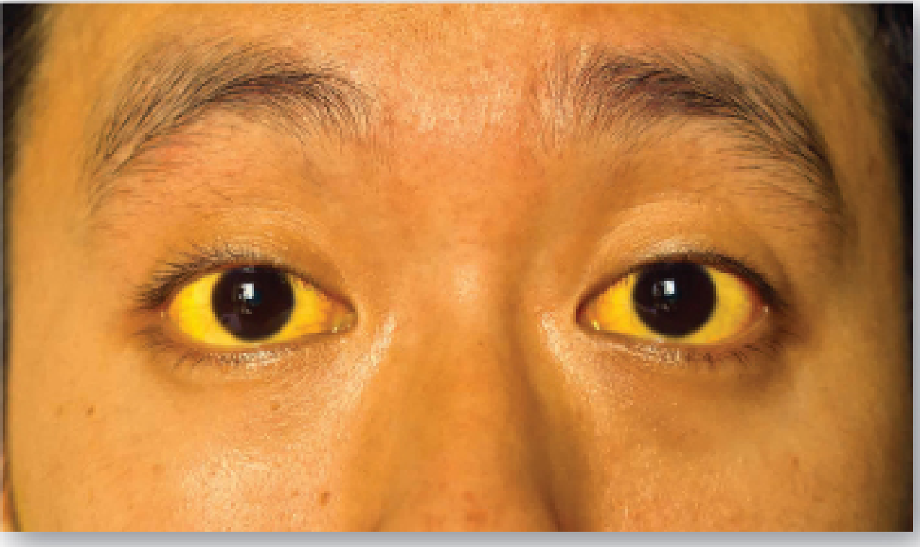
Jaundice (icterus) is a yellowish discoloration of the skin, nail beds, and sclera caused by deposition of bilirubin secondary to elevated blood bilirubin levels (hyperbilirubinemia).
- Normal blood bilirubin: ≤ 1 mg/dL
- Jaundice appears: at ≥ 2–3 mg/dL (approximately 3× normal)
- Can reach up to 40 mg/dL in severe conditions
Types of Jaundice
1. Hemolytic (Prehepatic) Jaundice
Mechanism: Excessive destruction of RBCs → bilirubin produced faster than liver can conjugate and excrete it → UCB accumulates in blood.
Features:
- Serum: ↑ Unconjugated (indirect) bilirubin
- Urine: No bilirubin (UCB not filtered by kidney); ↑ urobilinogen
- Stools: Normal or dark (↑ stercobilin)
- van den Bergh test: Indirect positive
Causes: Sickle cell anemia, hereditary spherocytosis, G6PD deficiency, ABO incompatibility, malaria, thalassemia
2. Hepatocellular (Hepatic) Jaundice
Mechanism: Liver cell damage impairs uptake, conjugation, and secretion of bilirubin → both UCB and CB accumulate.
Features:
- Serum: ↑ Both conjugated and unconjugated bilirubin
- Urine: Bilirubin present (dark urine); urobilinogen present
- Stools: Pale (reduced stercobilin)
- ↑ ALT, AST; ↑ prothrombin time
- van den Bergh test: Biphasic (both direct and indirect positive)
Causes: Viral hepatitis (A, B, C), alcoholic hepatitis, cirrhosis, toxic/drug-induced hepatitis, leptospirosis
3. Obstructive (Posthepatic/Cholestatic) Jaundice
Mechanism: Obstruction of the biliary system prevents CB from entering the intestine → CB regurgitates back into blood.
Features:
- Serum: ↑ Conjugated (direct) bilirubin
- Urine: Bilirubin present (dark/frothy urine, "Coca-Cola urine"); no urobilinogen
- Stools: Pale, clay-colored (no stercobilin)
- ↑ Alkaline phosphatase, ↑ GGT
- Pruritus (bile salts in skin)
- van den Bergh test: Direct positive
Causes: Gallstones (choledocholithiasis), carcinoma of pancreatic head, cholangiocarcinoma, biliary stricture, primary sclerosing cholangitis
4. Neonatal (Physiological) Jaundice
Mechanism: Immaturity of hepatic bilirubin UGT at birth → UCB accumulates; resolves in 4 weeks when UGT matures.
- Seen in 60% full-term and 80% preterm newborns
- Treated with phototherapy (blue light converts UCB to water-soluble photoisomers)
- Kernicterus: UCB crosses blood-brain barrier → toxic encephalopathy → damage to basal ganglia
Summary Comparison Table
| Feature | Hemolytic | Hepatocellular | Obstructive |
|---|
| Bilirubin (serum) | ↑ UCB | ↑ Both | ↑ CB |
| Urine bilirubin | Absent | Present | Present (dark) |
| Urinary urobilinogen | ↑↑ | ↑ | Absent |
| Stool color | Normal/dark | Pale | Clay/pale |
| van den Bergh | Indirect (+) | Both (+) | Direct (+) |
| ALT/AST | Normal | ↑↑ | Mild ↑ |
| Alk. Phosphatase | Normal | ↑ | ↑↑ |
SHORT Q5 — Normal Blood Urea Level & Conditions with Elevated Urea
Normal Blood Urea Level
- Blood urea: 15–40 mg/dL (or 2.5–6.7 mmol/L)
- Blood Urea Nitrogen (BUN): 7–20 mg/dL
- Normal plasma ammonia: 30–60 µM
Urea is synthesized in the liver from ammonia (urea cycle) and is filtered and excreted by the kidneys.
Conditions with Elevated Blood Urea (Uremia/Azotemia)
Two Major Causes:
1. Renal Failure (Most Common)
- Acute Kidney Injury (AKI) or Chronic Kidney Disease (CKD): Kidneys cannot filter and excrete urea → accumulates in blood
- Uremia: BUN > 100 mg/dL → nausea, vomiting, confusion, pericarditis, encephalopathy
- In CKD, elevated BUN promotes transfer of urea from blood into intestine → bacterial urease converts it to ammonia → absorbed back → contributes to hyperammonemia
2. Prerenal Azotemia (Dehydration, Hypovolemia)
- Conditions: Severe dehydration, hemorrhage, heart failure, burns, prolonged vomiting/diarrhea
- Reduced renal blood flow → decreased GFR → less urea excreted → elevated BUN
- BUN rises disproportionately to creatinine → BUN:Creatinine ratio > 20:1
Other causes of elevated blood urea:
- High protein intake (dietary or GI bleed — increased catabolism)
- Hypercatabolic states (sepsis, major surgery, severe burns)
- Liver disease (paradoxically, urea may be low; but hyperammonemia is the main concern)
- Urinary tract obstruction (postrenal azotemia)
*Sources: Biochemistry 8th ed. Lippincott Illustrated Reviews; Basic Medical Biochemistry – A Clinical Approach 6th ed.; Guyton & Hall Textbook of Medical Physiology; Harper's Illustrated Biochemistry 32nd Ed.*Your credits are exhausted. Your daily credits reset tomorrow, or you can purchase a top-up for more.