
Nervous system
Reading File
The query "Nervous system" is very broad. Let me clarify what you need before diving in.
Asking for Preferences
What aspect of the nervous system are you interested in?
Searching sources...
Finding Sources
Finding Sources
Reading File
Reading File
Reading File
Finding Sources
I now have excellent content from Gray's Anatomy for Students and Medical Physiology. Here is a thorough overview:
The Nervous System: Overview & Organization
What Is the Nervous System?
The nervous system is the body's primary communication network. It mediates everything from simple reflexes to complex behaviors like learning, memory, language, and consciousness. It integrates sensory input, processes information, and generates motor output to regulate the body and interact with the environment.
Major Divisions
The nervous system is traditionally divided into three overlapping subdivisions. The boundaries are somewhat arbitrary since all parts work closely together, but the framework is essential for understanding anatomy and clinical diagnosis.
1. Central Nervous System (CNS)
- Components: Brain (including cranial nerve II and the retina) and spinal cord
- Covered by three meninges: dura mater (outer), arachnoid (middle), and pia mater (inner, delicate)
- Within the CNS, neurons with similar functions cluster into nuclei
- Divided into gray matter (neuron cell bodies) and white matter (myelinated axons)
- In the brain: gray matter is on the cortical surface; white matter is deep
- In the spinal cord: the opposite - white matter is superficial, gray matter is central
- Myelination is provided by oligodendrocytes
- Axons in the CNS cannot regenerate after injury
2. Peripheral Nervous System (PNS)
- Components: Everything outside the dura mater - sensory receptors, peripheral portions of spinal and cranial nerves (except CN II), and peripheral portions of the autonomic nervous system
- Afferent nerves carry signals toward the CNS (sensory/input)
- Efferent nerves carry signals away from the CNS (motor/output)
- Peripheral ganglia are clusters of nerve cell bodies outside the CNS
- Myelination is provided by Schwann cells
- Axons in the PNS can regenerate after injury
3. Autonomic Nervous System (ANS)
- A functionally distinct division that regulates visceral functions: heart rate, blood pressure, digestion, temperature regulation, and reproductive function
- Anatomically composed of both CNS and PNS components
- Works via reflex arcs: visceral afferent neurons → CNS control centers → visceral motor output
- Further divided into sympathetic (fight-or-flight) and parasympathetic (rest-and-digest) subdivisions
Functional Organization
Functionally, the nervous system is split into two systems based on what they control:
| System | Function |
|---|---|
| Somatic nervous system | Carries conscious sensation from periphery to CNS; controls voluntary (skeletal) muscles |
| Visceral nervous system | Carries sensory info from viscera; autonomic motor output to regulate homeostatic functions (involuntary) |
The Brain: Structural Overview
The cerebral cortex is the outer surface of the brain - it consists of six layers of cell bodies (gray matter). Underneath lies the white matter, composed of myelinated axon bundles.
- The surface is folded into gyri (ridges) and sulci (grooves), dramatically increasing surface area
- Each hemisphere is divided into four lobes:
- Frontal - separated from parietal by the central sulcus (sulcus of Rolando)
- Parietal - separated from frontal anteriorly; from occipital by the parieto-occipital sulcus medially
- Temporal - separated from frontal by the lateral sulcus (fissure of Sylvius)
- Occipital - posterior visual processing lobe
- The two hemispheres are separated by the longitudinal (interhemispheric) fissure
- The insula is a small cortical region concealed by the operculum, visible only when the lateral sulcus is opened
White Matter Pathways
- Corona radiata: broad white matter sheet beneath the cortex
- Internal capsule: V-shaped condensation of white matter containing axons to/from cortex and deep structures
- Divided into anterior limb, genu, and posterior limb
Cells of the Nervous System
Two main cell types:
-
Neurons - the primary signaling units; vary greatly in shape but all conduct electrical signals
- The human brain contains approximately 100 billion (10¹¹) neurons
- Each neuron may connect with thousands of others
- The neuron doctrine (formalized by von Waldeyer, 1891; proven by Ramón y Cajal using Golgi's silver staining) established that neurons are discrete individual cells
-
Neuroglial cells (glia) - support cells; slightly outnumber neurons
- Oligodendrocytes (CNS) and Schwann cells (PNS): form myelin sheaths
- Astrocytes: structural and metabolic support
- Microglia: immune defense of the CNS
Regional Specialization and Clinical Significance
Different brain areas have neurons with unique shapes, physiological properties, and connections. One region cannot substitute for another that has failed. This is why:
- Damage to a specific brain area causes predictable, localizable symptoms
- Neurological diagnosis relies heavily on matching symptoms to anatomy (e.g., stroke localization)
- Any recovery reflects enhancement of existing circuits or recruitment of latent ones - not true regeneration of CNS tissue
Sources: Gray's Anatomy for Students | Medical Physiology (Boron & Boulpaep)
Reproductive system
Asking for Preferences
Which aspect of the reproductive system would you like to cover?
Searching sources...
Finding Sources
Finding Sources
Finding Sources
Finding Sources
Reading File
Reading File
Reading File
Reading File
Excellent content retrieved. Now let me fetch the key anatomy diagrams to include visually.
Loading Image
Loading Image
Here is a thorough overview of the reproductive system anatomy from authoritative medical textbooks:
The Reproductive System: Overview & Anatomy
MALE REPRODUCTIVE SYSTEM
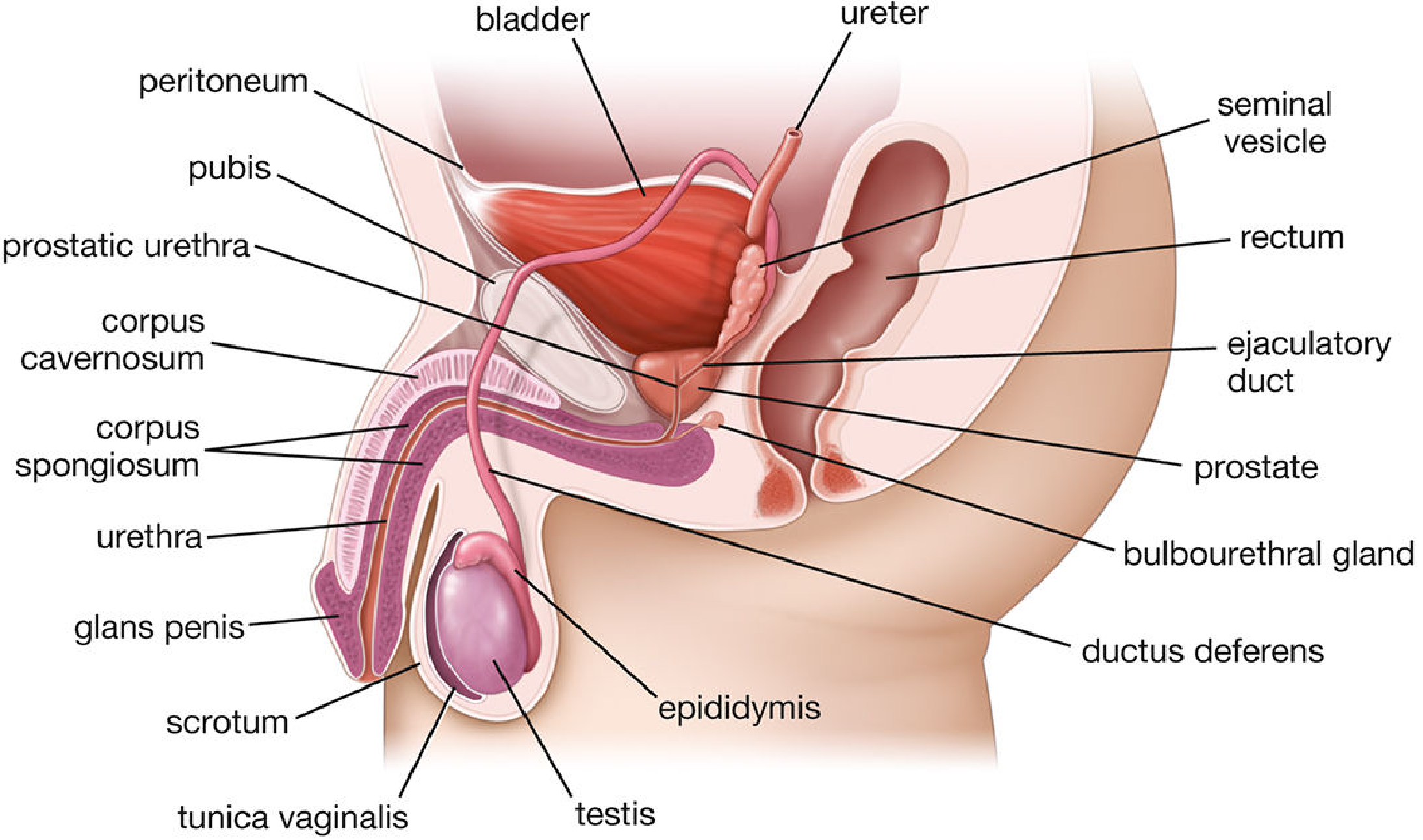
The male reproductive system consists of four major components: the testes, genital excurrent ducts, accessory sex glands, and external genitalia (penis and scrotum).
Testes
The testes are the primary male gonads. They serve two core functions:
- Spermatogenesis - production of sperm (male gametes)
- Steroidogenesis - synthesis of androgens, primarily testosterone
- Paired ovoid organs located in the scrotum - outside the body cavity
- The scrotal position is essential because spermatogenesis requires a temperature lower than core body temperature
- Each testis contains approximately 250 lobules, each holding 1-4 tightly coiled seminiferous tubules
- Development is triggered by the SRY gene on the Y chromosome, which produces testis-determining factor (TDF)
- Under testosterone and Müllerian-inhibiting factor (MIF), testes descend from the posterior abdominal wall into the scrotum during fetal development
- Dihydrotestosterone (DHT) drives development of external genitalia and accessory sex glands
Inside the Seminiferous Tubules
| Cell Type | Function |
|---|---|
| Sertoli cells | Support and nurture developing sperm; form the blood-testis barrier |
| Spermatogenic cells | Undergo mitosis/meiosis to produce sperm |
| Leydig (interstitial) cells | Located between tubules; produce testosterone and androgens |
| Myoid cells | Peritubular contractile cells in the tubule walls |
Spermatogenesis (Overview)
The process takes ~74 days and occurs in three phases:
- Spermatogonial phase - stem cells (spermatogonia) divide by mitosis, producing primary spermatocytes
- Spermatocyte phase - primary spermatocytes undergo meiosis I (22 days) → secondary spermatocytes → meiosis II (hours) → haploid spermatids
- Spermiogenesis - spermatids remodel into mature spermatozoa in association with Sertoli cells
Excurrent Duct System
Sperm travel from the seminiferous tubules through a series of ducts:
- Rete testis → Efferent ductules → Epididymis (site of sperm maturation and storage) → Ductus (vas) deferens → Ejaculatory duct → Urethra
Accessory Sex Glands
These glands produce components of seminal fluid:
| Gland | Location/Notes |
|---|---|
| Seminal vesicles | Paired glands posterior to bladder; contribute fructose-rich fluid (~60-70% of semen volume) |
| Prostate | Surrounds the urethra below the bladder; secretes fluid that activates sperm |
| Bulbourethral glands | Below the prostate; produce pre-ejaculatory lubricating fluid |
External Genitalia: Penis
The penis contains three cylindrical bodies of erectile tissue:
- Corpus cavernosum (x2) - dorsal erectile bodies
- Corpus spongiosum (x1) - ventral, surrounds the urethra, expands distally to form the glans penis
FEMALE REPRODUCTIVE SYSTEM
The female reproductive system is divided into internal and external structures.
Internal Organs
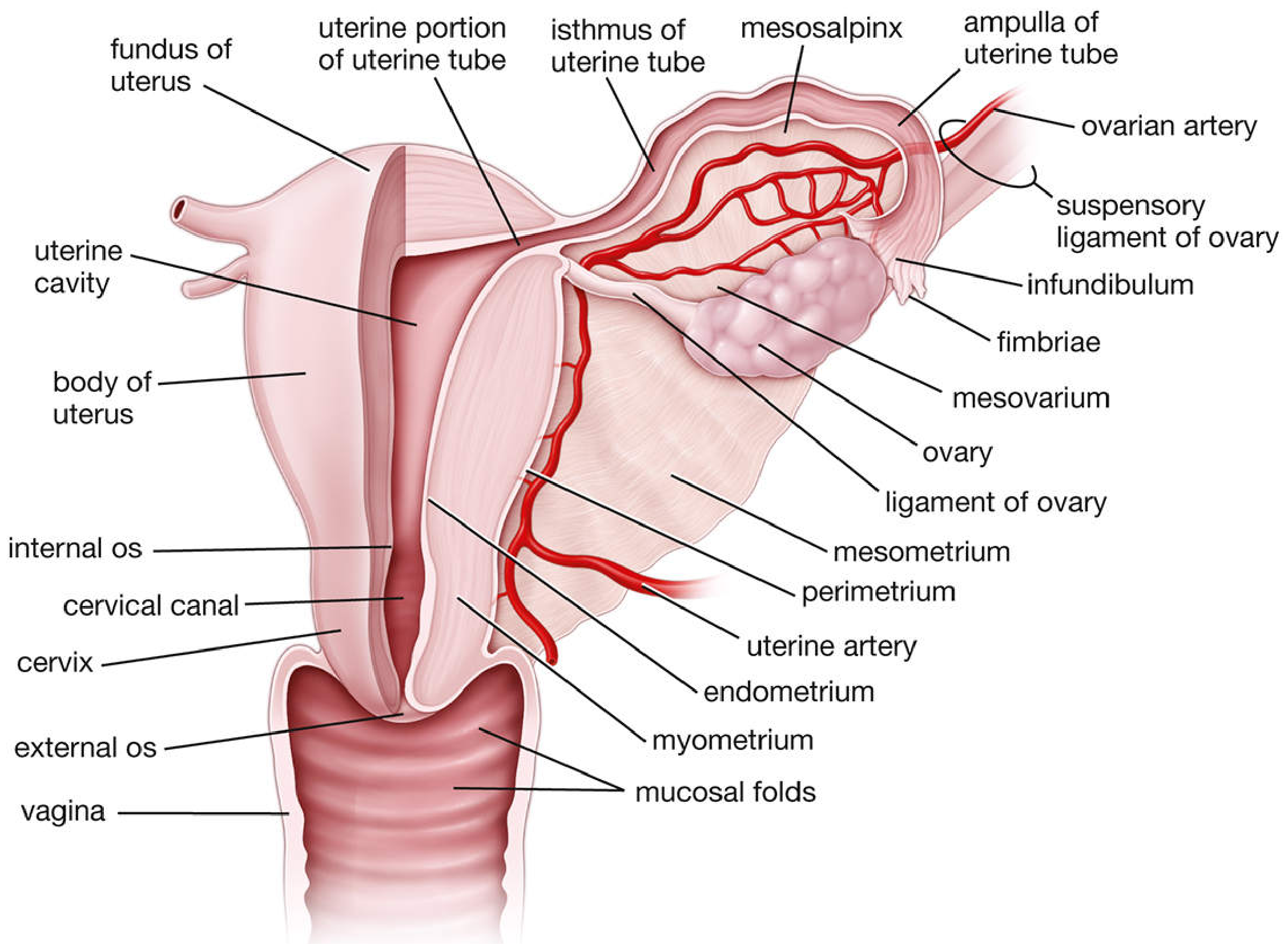
Located in the pelvis: ovaries, uterine (Fallopian) tubes, uterus, and vagina.
Ovaries
The female primary gonads, with two interrelated functions:
- Oogenesis - production of oocytes (female gametes)
- Steroidogenesis - secretion of estrogens and progestogens
Structure:
- Paired, almond-shaped structures (~3 cm × 1.5 cm × 1 cm in nulliparous females)
- Attached to the posterior surface of the broad ligament by the mesovarium
- Superior (tubal) pole attached to pelvic wall by the suspensory ligament of the ovary (carries ovarian vessels and nerves)
- Inferior (uterine) pole attached to uterus by the ovarian ligament (remnant of the gubernaculum)
- Surface becomes progressively scarred after each ovulation; shrinks to ~1/4 its reproductive-life size after menopause
- Covered by germinal epithelium (actually a mesothelium); underlain by a dense tunica albuginea
Internal structure:
- Cortex (peripheral): contains ovarian follicles embedded in cellular stroma
- Medulla (central): loose connective tissue with large contorted blood vessels, lymphatics, and nerves
Hormone actions:
- Estrogens - promote growth and maturation of sex organs; responsible for female secondary sex characteristics; stimulate mammary ductal and stromal growth
- Progestogens - prepare the uterus (especially the endometrium) for pregnancy; if implantation does not occur, the endometrium degenerates and menstruation follows
Uterine (Fallopian) Tubes
- Paired muscular tubes that transport the oocyte from the ovary to the uterus
- Segments from lateral to medial: infundibulum (funnel-shaped opening with fimbriae that sweep the ovum) → ampulla (widest part; usual site of fertilization) → isthmus → uterine portion (enters the uterine wall)
- Attached to broad ligament via the mesosalpinx
Uterus
A thick-walled muscular organ divided into:
- Fundus - dome-shaped top, above the entry of the uterine tubes
- Body - main cavity
- Cervix - inferior narrow portion connecting to the vagina via the cervical canal (with internal os above and external os below)
Wall layers (3):
| Layer | Description |
|---|---|
| Endometrium | Inner mucosal lining; undergoes cyclic changes with the menstrual cycle; site of implantation |
| Myometrium | Middle and thickest layer; smooth muscle; responsible for uterine contractions during labor |
| Perimetrium | Outer layer; peritoneal (serosal) covering |
Vagina
- Fibromuscular tube connecting the uterine cervix to the exterior
- Lined by mucosal folds
- Serves as the birth canal and receives the penis during intercourse
External Genitalia (Vulva)
Located in the anterior perineum:
- Mons pubis - fatty eminence over the pubic symphysis
- Labia majora - outer folds of skin
- Labia minora - inner mucosal folds
- Clitoris - erectile structure, homologous to the penis
- Vestibule - space between labia minora containing vaginal and urethral openings
- Hymen - thin mucosal fold partially covering the vaginal opening in virgins
Mammary Glands
Included as part of the female reproductive system because their development and function are directly tied to reproductive hormones. Estrogens stimulate ductal and stromal growth and fat accumulation; progestogens prepare glands for lactation during pregnancy.
Reproductive Lifespan (Female)
| Event | Age |
|---|---|
| Menarche (first menstruation) | 9-14 years (mean 12.4 years) |
| Menstrual cycle length | 21-34 days |
| Menopause (cessation of cycles) | 45-55 years (mean 51.4 years) |
At menopause, ovaries cease both oogenesis and hormone production, leading to changes in the vagina, mammary glands, and other estrogen-dependent tissues.
Sources: Histology: A Text and Atlas with Correlated Cell and Molecular Biology (Ross & Pawlina)
Lymphatic system
Asking for Preferences
Which aspect of the lymphatic system would you like to focus on?
Searching sources...
Finding Sources
Finding Sources
Finding Sources
Finding Sources
Reading File
Reading File
Reading File
Reading File
Loading Image
Finding Sources
Reading File
Finding Sources
Loading Image
Finding Sources
I now have rich content from three authoritative sources. Here is a comprehensive overview:
The Lymphatic System: Overview & Organization
What Is the Lymphatic System?
The lymphatic system is a network of specialized vessels, nodes, and organs that serves two fundamental roles:
- Fluid homeostasis - drains excess interstitial fluid from tissues and returns it to the bloodstream
- Immune defense - transports antigens and immune cells, initiating adaptive immune responses
Without a functioning lymphatic system, we would die within approximately 24 hours because proteins accumulating in the interstitial space cannot be removed any other way. - Guyton & Hall Medical Physiology
Components at a Glance
| Component | Type | Function |
|---|---|---|
| Lymphatic capillaries | Vessels | Absorb interstitial fluid and proteins |
| Collecting lymphatic vessels | Vessels | Transport lymph toward the heart |
| Thoracic duct & right lymphatic duct | Large ducts | Return lymph to venous circulation |
| Lymph nodes | Secondary lymphoid organs | Filter lymph; initiate immune responses |
| Spleen | Secondary lymphoid organ | Filters blood; immune surveillance |
| Thymus | Primary lymphoid organ | T lymphocyte maturation |
| Tonsils / MALT | Secondary lymphoid tissue | Mucosal immune defense |
1. Lymphatic Vessels
Lymphatic Capillaries
- Blind-ended channels found in the skin, epithelia, and parenchymal organs of almost all vascularized tissues
- Exceptions: superficial skin, muscle endomysium, bone (but these have prelymphatics that eventually drain into lymphatics)
- Lined by overlapping endothelial cells with no tight junctions and no continuous basement membrane - this allows free uptake of interstitial fluid, proteins, and even large particles like bacteria
- Attached to surrounding connective tissue by elastin anchoring filaments that pull them open when tissue swells
- One-way flap valves at the endothelial cell overlaps prevent backflow once fluid enters
How Lymph Flows
About 10% of capillary filtrate that exits blood vessels is not reabsorbed by venous capillaries. Instead it enters lymphatics and returns as lymph. Approximately 2-3 liters of lymph are returned to circulation per day.
Flow is driven by:
- Contraction of perilymphatic smooth muscle cells
- Pressure from musculoskeletal movement
- One-way intraluminal valves throughout all lymphatic vessels
Major Lymphatic Ducts
| Duct | Drains | Empties Into |
|---|---|---|
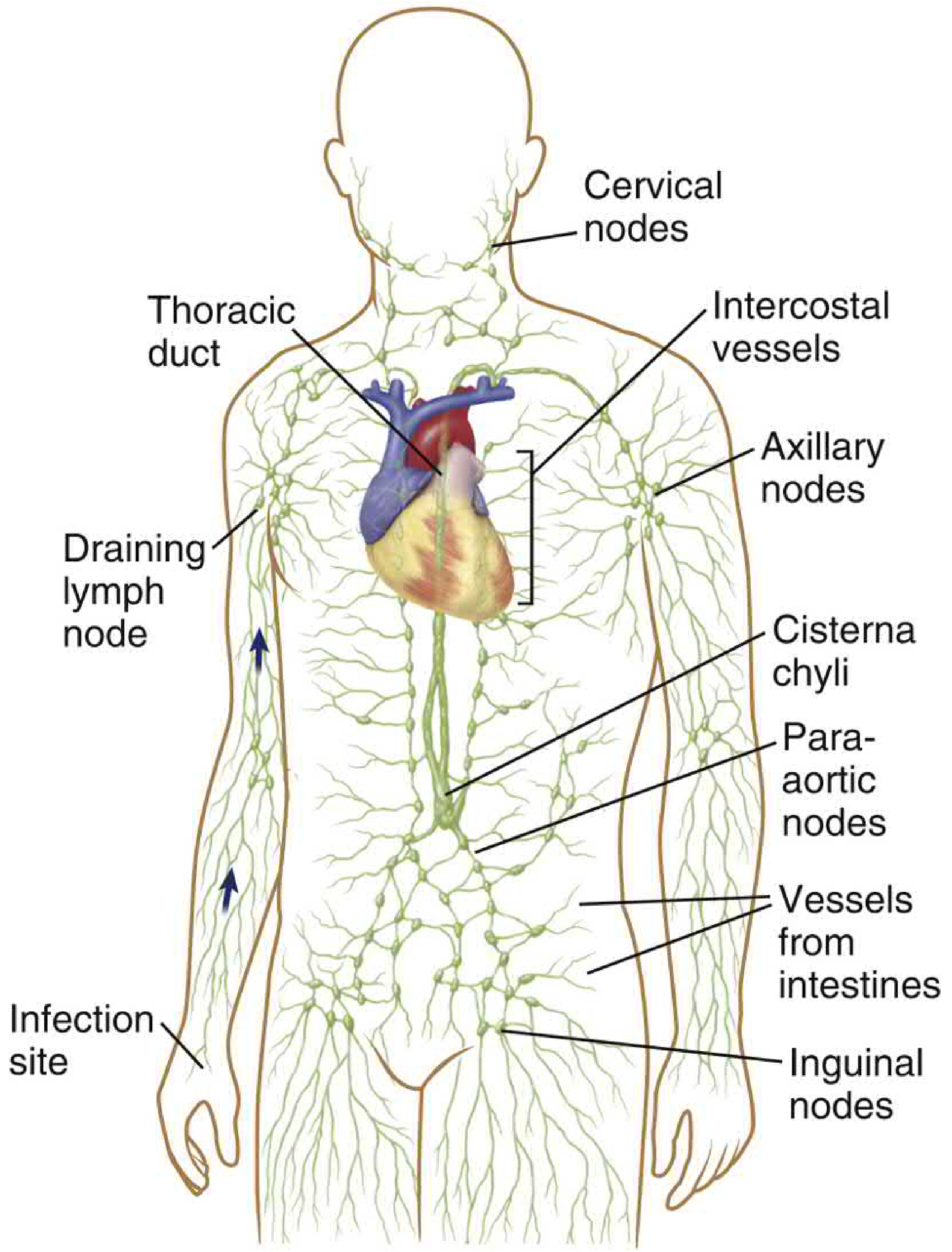
| Thoracic duct (largest) | Lower body, left upper trunk, left arm, left head/neck, intestines | Left internal jugular + left subclavian vein junction |
| Right lymphatic duct | Right head/neck, right arm, right thorax | Right subclavian + right internal jugular vein junction |
| Cisterna chyli | Intestines and lower limbs (collecting reservoir) | Into the thoracic duct |
2. Lymph Composition
Lymph begins as interstitial fluid - so its initial composition mirrors that fluid:
- Protein concentration: ~2 g/dL in most tissues
- Liver lymph: up to 6 g/dL (highest protein)
- Intestinal lymph: 3-4 g/dL
- Thoracic duct lymph (mixture of all): typically 3-5 g/dL
- After a fatty meal, thoracic duct lymph may contain up to 1-2% fat - the lymphatics are the primary route for dietary fat absorption (as chylomicrons via lacteals in the intestinal villi)
3. Lymph Nodes
There are approximately 500 lymph nodes in the human body, distributed along lymphatic vessels. They act as biological filters that sample lymph for antigens and pathogens.
Structure
- Surrounded by a fibrous capsule
- Afferent lymphatics deliver lymph → subcapsular sinus → medullary sinus → efferent lymphatics
- Macrophages in the subcapsular sinus phagocytose and destroy infectious organisms
Internal Zones
| Zone | Location | Cell Type |
|---|---|---|
| Outer cortex / follicles | Beneath capsule | B lymphocytes |
| Paracortex (T cell zone) | Deep cortex | T lymphocytes |
| Medulla | Center | Plasma cells, macrophages |
Follicles - Key Distinction
- Primary follicles (no germinal center): contain naive, resting B cells
- Secondary follicles (with germinal center): contain activated B cells; sites of B cell proliferation, affinity maturation, and generation of memory B cells and plasma cells
- Dark zone: proliferating B cells (centroblasts)
- Light zone: non-proliferating cells (centrocytes) being selected for high-affinity antibody production
Immune Role
- Dendritic cells (DCs) in tissues capture microbial antigens, enter lymphatics, and carry them to nodes
- T and B cells in the node then mount the adaptive immune response
- Each lymph node drains a specific body territory - this is why palpable lymphadenopathy in a region often indicates local infection or malignancy
4. The Spleen
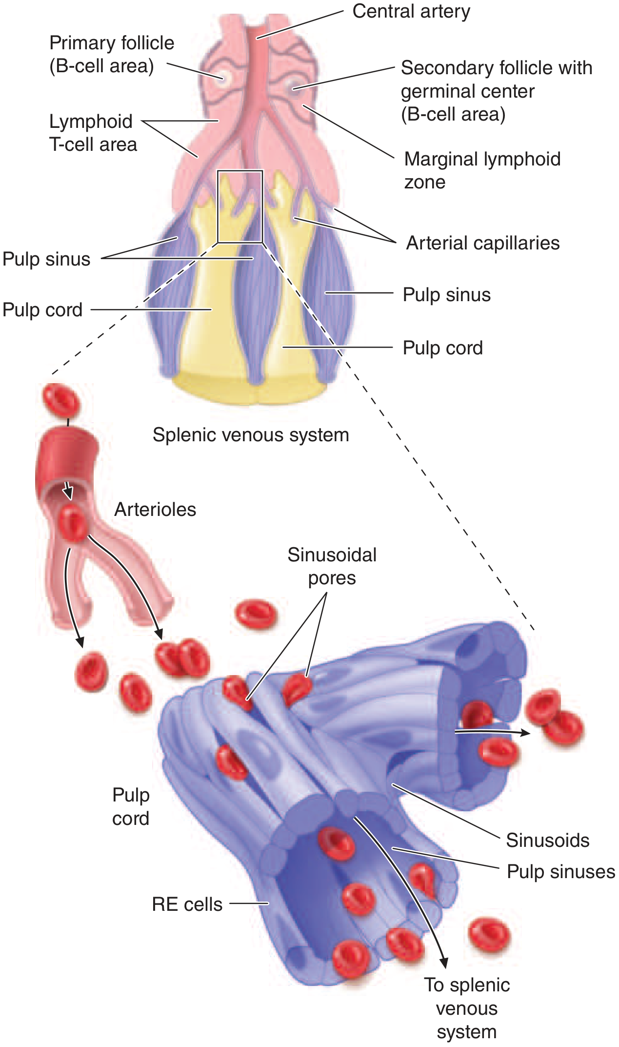
The spleen is the largest secondary lymphoid organ, located in the left upper quadrant. It sits in the portal circulation and receives ~150 mL/min of blood flow via the splenic artery.
Three Core Functions (Harrison's Principles)
- Quality control of erythrocytes (red pulp) - removes senescent and defective red blood cells
- Antibody synthesis (white pulp) - generates humoral immune responses to blood-borne antigens
- Clearance of opsonized microbes - removes antibody-coated bacteria and blood cells
Structure: Red vs. White Pulp
| Pulp | Composition | Function |
|---|---|---|
| Red pulp | Sinuses and macrophage-lined cords | RBC filtration; blood reservoir |
| White pulp | Lymphoid follicles (B cells), T cell sheaths around arterioles, marginal zone | Immune surveillance of blood |
How red cell filtration works: Blood from central arterioles flows into macrophage-lined cords. To re-enter circulation, red cells must squeeze through tiny sinusoidal slits. Old, stiff, or damaged RBCs cannot pass and are destroyed. This is also how the spleen removes red cell inclusions like Howell-Jolly bodies and Heinz bodies.
Accessory spleens occur in ~20% of people when developmental hillocks fail to fuse.
5. The Thymus (Primary Lymphoid Organ)
The thymus is a primary (central) lymphoid organ - meaning it is where lymphocytes mature and become immunocompetent, not where immune responses are initiated.
- Located in the anterior mediastinum
- Most active in childhood; involutes with age (replaced by fat)
- Site of T lymphocyte maturation (thymocytes → mature T cells)
- Divided into cortex (densely packed thymocytes, darker staining) and medulla (lighter, contains Hassall's corpuscles - concentric whorls of epithelial cells unique to the thymic medulla, function not fully understood)
- The blood-thymus barrier in the cortex shields developing T cells from self-antigens until they are fully tolerant
6. Other Lymphoid Tissues
Mucosa-Associated Lymphoid Tissue (MALT)
Lymphoid tissue distributed throughout mucosal surfaces, protecting the body's largest exposure points:
- GALT (gut-associated): Peyer's patches in the small intestine, appendix
- BALT (bronchus-associated): airways
- NALT (nasopharynx-associated): tonsils and adenoids (Waldeyer's ring)
The Brain's Lymphatic System - A Recent Discovery
True lymphatic vessels exist only in the meninges of the brain. However, a parallel system called the glymphatic system clears waste from deeper brain tissue:
- CSF flows into brain parenchyma along periarterial spaces (glial cell-lined)
- Exits via perivenous routes, carrying interstitial fluid and waste products (including amyloid-beta)
- Drains into meningeal lymphatics → blood
This system is most active during sleep and may be implicated in neurodegenerative diseases like Alzheimer's.
Clinical Relevance
| Disruption | Result |
|---|---|
| Lymphatic obstruction (tumor, filariasis) | Lymphedema - severe tissue swelling |
| Splenectomy | Impaired immunity to encapsulated bacteria (e.g. Streptococcus pneumoniae); Howell-Jolly bodies appear in blood |
| Lymph node metastasis | Indicates cancer spread; guides staging and treatment |
| Lymphoma | Malignancy of lymphoid tissue (Hodgkin vs. Non-Hodgkin) |
Sources: Cellular and Molecular Immunology (Abbas, Lichtman & Pillai) | Guyton and Hall Textbook of Medical Physiology | Harrison's Principles of Internal Medicine, 22e
This is a shared conversation. Sign in to Orris to start your own chat.