
Teach me elisa for my microbiology university exam
Reading File
Now let me search the medical library for ELISA content:
Finding Sources
Searching the Web
ELISA enzyme linked immunosorbent assay microbiology university exam types steps mechanism
Found content in Janeway's Immunobiology. Let me read the relevant sections:
Reading File
Finding Sources
Excellent - great content found. Let me fetch the ELISA diagram image and read more of the text:
Loading Image
Reading File
I now have excellent content from Janeway's Immunobiology. Let me also fetch the competitive ELISA diagram:
Loading Image
That was the affinity chromatography diagram (not ELISA). I now have all the key content I need. Let me compile a comprehensive exam-focused guide on ELISA.
ELISA - Enzyme-Linked Immunosorbent Assay
Source: Janeway's Immunobiology 10e
What is ELISA?
ELISA (Enzyme-Linked Immunosorbent Assay) is a solid-phase immunoassay used to detect and quantify either antigens or antibodies in a sample. It replaces the older radioimmunoassay (RIA) - same principle, but uses an enzyme label instead of radioactive label, making it safer, cheaper, and easier to read.
Key idea: Antibody-antigen binding is detected through an enzyme that converts a colorless substrate into a colored product. The color intensity is proportional to the amount of antigen or antibody present.
Core Components (Remember These!)
| Component | Role |
|---|---|
| Microtiter plate (96-well, plastic) | Solid support to immobilize reactants |
| Antigen or antibody | One is immobilized; the other is detected |
| Enzyme-linked antibody | Reporter - carries the signal |
| Substrate | Colorless molecule converted to colored product by the enzyme |
| Blocking agent (e.g., BSA, skim milk) | Blocks residual sticky sites on plastic to prevent false positives |
| Wash steps | Remove unbound reagents; critical for specificity |
Common enzymes used:
- Horseradish peroxidase (HRP) - most common
- Alkaline phosphatase (AP)
The 4 Types of ELISA
1. Direct ELISA
Purpose: Detect antigen in a sample.
Steps:
- Coat well with patient sample (antigen adsorbs to plastic)
- Block residual sites with irrelevant protein
- Add enzyme-linked antibody specific for the antigen
- Wash away unbound antibody
- Add substrate → color change if antigen is present
- Read absorbance
Result: Color = antigen present. No color = no antigen.
Pros: Simple, fast
Cons: Less sensitive; no signal amplification
2. Indirect ELISA
Purpose: Detect or quantify antibodies (e.g., HIV antibody testing in patient serum).
Steps:
- Coat well with known antigen
- Block
- Add patient serum (may contain specific antibody)
- Wash
- Add anti-human antibody linked to enzyme (secondary antibody)
- Wash
- Add substrate → color change if patient antibody is present
Key point: The secondary antibody amplifies the signal because multiple enzyme-linked secondaries can bind each primary antibody.
Example use: HIV serology, hepatitis B detection
3. Sandwich (Capture) ELISA
Purpose: Detect and quantify antigen with high sensitivity (used for cytokines, hormones, HIV p24 protein).
Steps:
- Coat well with capture antibody (specific for antigen)
- Block
- Add sample - antigen binds capture antibody
- Wash
- Add detection antibody (enzyme-linked, recognizes a different epitope on the same antigen)
- Wash
- Add substrate → color change
Why "sandwich"? The antigen is sandwiched between two antibodies.
Pros: Very sensitive; can detect very low concentrations; highly specific (two-antibody system)
Cons: Requires two antibodies that recognize different epitopes; antigen must be large enough to have two epitopes
Example use: Cytokine quantification (IL-6, TNF-α), HIV p24
4. Competitive (Inhibition) ELISA
Purpose: Quantify small antigens (haptens, drugs, hormones) that have only one epitope.
Principle: Unknown unlabeled antigen competes with a known amount of labeled antigen for binding to a fixed antibody.
Steps:
- Coat well with antibody specific for antigen
- Mix patient sample (unknown antigen) with a fixed amount of labeled antigen
- Add this mixture to the well
- Both labeled and unlabeled antigen compete for antibody binding
- Wash
- Add substrate → measure color
Interpretation (inverse relationship!):
- More antigen in sample → less labeled antigen binds → LESS color
- Less antigen in sample → more labeled antigen binds → MORE color
Uses: Drug level monitoring, hormone assays (estrogen, testosterone), detection of small molecules
ELISA vs. RIA - Comparison Table
| Feature | ELISA | RIA |
|---|---|---|
| Label | Enzyme | Radioactive isotope (¹²⁵I) |
| Detection | Color change (colorimetry) | Radioactivity counting |
| Safety | Safe | Radiation hazard |
| Equipment | Simple plate reader | Gamma counter required |
| Sensitivity | High | Very high |
| Preferred for | Viral diagnostics, antibody detection | Hormone measurement |
Two Critical Principles of ALL Serological Assays
(Directly from Janeway's - likely exam material)
- At least one reagent must be available in a pure, detectable form to obtain quantitative information.
- Bound fraction must be separated from unbound free fraction - achieved by immobilizing one partner on a solid support; unbound molecules are washed away.
Clinical Applications to Know
| Disease/Use | Type of ELISA | What is Detected |
|---|---|---|
| HIV diagnosis | Indirect ELISA (screening) | Anti-HIV antibodies in serum |
| HIV monitoring | Sandwich ELISA | p24 antigen |
| Hepatitis B | Indirect ELISA | Anti-HBs or anti-HBc antibodies |
| Cytokine quantification | Sandwich ELISA | IL-2, IL-6, TNF-α, etc. |
| Pregnancy test (hCG) | Sandwich ELISA | hCG hormone |
| Drug level monitoring | Competitive ELISA | Small drug molecules |
| Celiac disease | Indirect ELISA | Anti-tTG IgA antibodies |
Exam Memory Tips
"DISC" - the 4 types:
- Direct - simplest; enzyme antibody goes straight to coated antigen
- Indirect - detects antibodies in serum; two-antibody system
- Sandwich - antigen trapped between two antibodies; most sensitive
- Competitive - inverse result; for small molecules with one epitope
Blocking step - always done after coating to prevent nonspecific binding → false positives.
Washing steps - done between each addition to remove unbound reagents → reduce background noise.
Reading result: Measure absorbance (optical density) using a spectrophotometer/plate reader. Higher absorbance = more enzyme activity = more target bound (except in competitive ELISA, where it's the opposite).
Common Exam Questions
Q: Why is the blocking step needed?
A: Plastic wells bind proteins nonspecifically. After coating the antigen/antibody, remaining sticky sites must be blocked with an irrelevant protein (BSA or skim milk) to prevent the enzyme-linked antibody from binding nonspecifically - which would give a false positive.
Q: What is the advantage of sandwich ELISA over direct ELISA?
A: Sandwich ELISA uses two antibodies recognizing different epitopes, which greatly increases specificity and sensitivity. It can also concentrate dilute antigen from solution even at very low concentrations.
Q: Why does competitive ELISA give an inverse signal?
A: Unlabeled antigen in the patient's sample competes with labeled antigen for a limited number of antibody binding sites. More unlabeled antigen = less room for labeled antigen to bind = less enzyme activity = less color.
Q: Why is ELISA preferred over RIA?
A: ELISA avoids the hazards of radioactivity, uses simple colorimetric detection that can be read directly from the reaction tray with a plate reader, and is cheaper and easier to perform.
Q: What is the principle of signal amplification in indirect ELISA?
A: The secondary (anti-immunoglobulin) antibody amplifies the signal because at least two molecules of labeled secondary antibody can bind to each unlabeled primary antibody, increasing the detectable signal.
Janeway's Immunobiology 10e, Appendix A (pp. 822-823)
Tell me the principal of elisa
The principle of ELISA is based on the specific binding between an antigen and its complementary antibody, where one partner is immobilized on a solid surface and the other is labeled with an enzyme. The enzyme converts a colorless substrate into a colored product, allowing detection and quantification of the target molecule.
Searching sources...
Reading File
Here is the principle of ELISA explained clearly, straight from Janeway's Immunobiology:
Principle of ELISA
The Core Idea
ELISA is built on one fundamental immunological fact: an antibody binds its specific antigen with high affinity and specificity. ELISA exploits this binding to detect and quantify either an antigen or an antibody in a sample.
The word itself tells you the principle:
| Word | Meaning |
|---|---|
| Enzyme-Linked | An enzyme is chemically attached to an antibody as a reporter |
| Immunosorbent | Antigen or antibody is adsorbed (stuck) onto a solid surface |
| Assay | A test to measure something |
The 3-Part Principle
1. Immobilization on a solid support
One reaction partner (antigen or antibody) is physically attached to the surface of a plastic well in a microtiter plate. Plastic binds proteins nonspecifically - this is how they "stick." Residual sticky sites are then blocked with an irrelevant protein (like BSA or skim milk) to prevent false-positive binding later.
2. Specific binding
The other reaction partner, carrying an enzyme label, is added to the well. Under controlled conditions, it binds only to its specific target on the surface. Everything else that doesn't bind is removed by a wash step. This is the separation step - it physically separates bound reagent from unbound free reagent.
"There must be a means of separating the bound fraction of the labeled reagent from the unbound, free fraction." - Janeway's Immunobiology 10e, p.823
3. Enzymatic color development (detection)
A colorless substrate is added. The enzyme linked to the bound antibody converts this substrate into a colored product. The intensity of the color is directly proportional to how much antigen-antibody binding occurred, which is proportional to how much target was in the sample.
The color is read by a plate reader (spectrophotometer) measuring absorbance (optical density, OD).
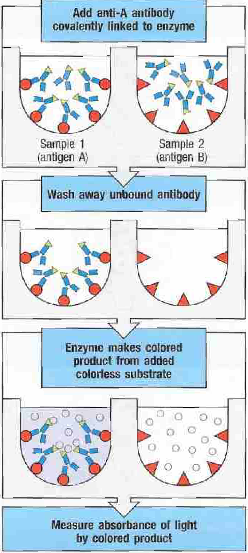
The Principle in One Diagram
From Janeway's Immunobiology 10e, Fig. A.5
- Sample 1 (contains antigen A): Enzyme-linked anti-A antibody binds → after wash, enzyme remains → substrate added → COLOR produced
- Sample 2 (contains antigen B, not A): Anti-A antibody does NOT bind → washed away → no enzyme remains → substrate added → NO COLOR
Two Non-Negotiable Requirements
Every ELISA must satisfy these two conditions (classic exam point):
- At least one reagent must be pure and detectable - you need a known, pure antibody or antigen to standardize the assay
- Bound and unbound fractions must be separated - achieved by the solid support + wash steps; without this, you cannot distinguish signal from background
Why Enzyme Instead of Radioactivity?
ELISA replaced RIA (radioimmunoassay) because:
- No radiation hazard
- Color can be read directly in the reaction tray with a simple plate reader
- Results are quantitative - the deeper the color, the more antigen/antibody
- Faster, cheaper, easier to run in high volumes
Summary Statement (Write This in Your Exam)
The principle of ELISA is based on the specific, high-affinity binding between an antigen and its complementary antibody, where one partner is immobilized on a solid surface (microtiter plate). The other partner, conjugated to an enzyme, binds only if the target is present. Unbound reagents are removed by washing. A colorless substrate is then added; the enzyme converts it to a colored product, whose absorbance is measured and is proportional to the amount of target in the sample.
- Janeway's Immunobiology 10e, Appendix A, pp. 822-823
Teach me agglutination reaction
Agglutination is the clumping of particles (bacteria, RBCs, latex beads) when antibodies cross-link antigens on their surfaces. It forms the basis of many diagnostic tests in microbiology.
Searching sources...
Finding Sources
Finding Sources
Searching the Web
agglutination reaction microbiology types direct indirect hemagglutination Widal Weil-Felix exam
Good - found relevant sections in multiple textbooks. Let me read them all in parallel:
Reading File
Reading File
Reading File
Excellent content from three textbooks. Let me also search for the Widal/Weil-Felix and prozone content:
Finding Sources
Finding Sources
Finding Sources
Finding Sources
I now have excellent content from multiple authoritative textbooks. Let me read the prozone section in full:
Reading File
I now have all the content I need from authoritative sources. Let me compile the full guide:
Agglutination Reaction - Complete Study Guide
What is Agglutination?
Agglutination is the visible clumping (aggregation) of particulate antigens when they react with their specific antibodies. The antibody acts as a bridge, cross-linking antigen-carrying particles into large visible clumps.
- Janeway's concept: antigen + antibody → large 3D lattice → precipitates or clumps
- The "particle" can be a bacterium, red blood cell (RBC), latex bead, or charcoal particle
- Agglutination requires multivalent antigen and bivalent antibody to form cross-links
"The amount of antigen or antibody necessary to produce a visible immunologic reaction can be reduced if either is on the surface of a relatively large particle." - Sherris & Ryan's Medical Microbiology 8e
The Principle
- Antigen is present on the surface of a particle (naturally, like bacteria, or artificially coated)
- Specific antibody is added
- Antibody has two binding sites (bivalent) - it bridges two antigen-carrying particles
- Multiple antibodies cross-link multiple particles → large visible clump (agglutinate)
Key requirement: Lattice formation needs both antigen and antibody in roughly equal proportions. Too much of either one breaks down the lattice (see Prozone Effect below).
IgM vs. IgG in Agglutination
| Antibody | Agglutinating ability | Why |
|---|---|---|
| IgM | Excellent - produces complete agglutination | Pentameric; 10 binding sites; large size overcomes particle repulsion |
| IgG | Poor alone - may cause incomplete agglutination | Only 2 binding sites; smaller; cannot always overcome zeta potential |
Zeta potential - Particles like bacteria and RBCs carry a net negative charge in solution, causing mutual repulsion. IgM can overcome this; IgG often cannot → needs enhancement.
Enhancement of IgG-based agglutination:
- Lower ionic strength of medium
- Add polymeric molecules: albumin, dextran, PEG, Polybrene
- Use the Coombs test (antiglobulin method)
Types of Agglutination
1. Direct (Active) Agglutination
The antigen is naturally present on the particle surface. No coating needed.
Examples:
| Test | Antigen | Antibody detected | Disease |
|---|---|---|---|
| Widal test | Salmonella typhi O & H antigens (whole bacteria) | Anti-Salmonella antibodies | Typhoid fever |
| Weil-Felix test | Proteus OX2, OX19, OXK antigens | Heterophile antibodies in Rickettsial disease | Typhus, Rocky Mountain spotted fever |
| Blood grouping | ABO/Rh antigens on RBCs | Anti-A, Anti-B antibodies | Blood typing |
| Brucella agglutination | Brucella organisms | Anti-Brucella antibodies | Brucellosis |
Note on Weil-Felix: A heterophile agglutination test - the patient does NOT have Proteus infection. It works because Rickettsia and Proteus share cross-reacting antigens. Antibodies made against Rickettsia happen to agglutinate Proteus bacteria.
2. Passive (Indirect) Agglutination
A soluble antigen is artificially coated onto a carrier particle (RBC, latex, gelatin). The coated particle is then agglutinated by specific antibody in the patient's serum.
Common particles used:
- Sheep or human RBCs (→ passive hemagglutination)
- Latex beads (→ latex agglutination)
- Gelatin particles
- Charcoal particles
Examples:
| Test | Particle | Antigen coated | Detects |
|---|---|---|---|
| TPHA (Treponema Pallidum Hemagglutination) | Fixed RBCs | T. pallidum antigen | Syphilis antibody |
| RA latex test | Latex | IgG (acts as antigen) | Rheumatoid factor (IgM anti-IgG) |
| ASO (anti-streptolysin O) | RBCs | Streptolysin O | Antibodies post-Streptococcal infection |
| RPR (Rapid Plasma Reagin) | Charcoal/cardiolipin | Lipid antigen | Syphilis (screening) |
3. Reverse Passive Agglutination
Here antibody is coated onto the particle, not antigen. Used to detect soluble antigen in the specimen.
- Patient's sample contains antigen
- Antibody-coated particle binds the antigen
- Cross-linking → agglutination = antigen present
Example: Detecting HBsAg (Hepatitis B surface antigen), hCG pregnancy test
4. Hemagglutination
Specifically uses red blood cells (RBCs) as the carrier particle.
a) Direct hemagglutination - antigen naturally on RBC surface
- ABO blood grouping, Rh typing
b) Passive (indirect) hemagglutination - foreign antigen coated onto RBCs
- Widely used; RBCs are good passive carriers, easily obtained and stored
- Example: TPHA for syphilis
c) Hemagglutination Inhibition (HAI)
- Principle: a specific antigen in the patient's sample blocks (inhibits) hemagglutination by competing with the particle-bound antigen for the antibody
- If inhibition occurs → antigen is present in sample
- Used for: influenza typing, rubella serology, pregnancy tests (historically)
5. Agglutination Inhibition
Based on the competitive principle - a hapten or soluble antigen in the patient's sample blocks agglutination.
Steps:
- Patient sample (unknown antigen) is mixed with a fixed, known amount of antibody
- The sample antigen binds and neutralizes some antibody
- The remaining unoccupied antibody is added to the coated particles
- Less free antibody → less agglutination
Result:
- Inhibition of agglutination = antigen IS present in the sample
- Agglutination occurs = antigen is NOT present (antibody is free)
Used for: small molecules (haptens), drugs, hormones (estrogen, hCG)
6. Coagglutination
Antibodies are adsorbed onto Staphylococcus aureus Protein A, which has high affinity for the Fc region of IgG. The antibody Fab arms point outward and react with specific antigen → agglutination of the Staph.
Uses: Rapid identification of bacterial pathogens (Streptococcus, N. meningitidis)
7. Coombs Test (Antiglobulin Test)
Specifically designed for incomplete agglutination - detects IgG antibodies that have coated RBCs but cannot cause visible agglutination alone (due to electrostatic repulsion).
Direct Coombs Test (DCT)
- Detects antibodies already coating the patient's RBCs in vivo
- Add anti-human immunoglobulin (Coombs reagent) → bridges IgG-coated RBCs → agglutination
- Uses: Autoimmune hemolytic anemia, hemolytic disease of the newborn (HDN)
Indirect Coombs Test (ICT)
- Detects free antibodies in patient's serum
- Mix patient serum + normal RBCs → incubate → then add Coombs reagent
- Uses: Cross-matching blood transfusions, antenatal antibody screening
The Prozone Effect (Hook Effect)
This is a critical exam topic that explains false-negative agglutination.
What happens: When antibody is present in very large excess, every antigen-binding site on every particle is saturated by individual antibody molecules. No free antigen sites remain to be bridged → no cross-linking → no agglutination despite antibodies being present.
"A vast excess of antigen can completely saturate all antibody-binding sites, leading to a negative signal (i.e., no agglutination). This phenomenon is sometimes referred to as the prozone effect." - Henry's Clinical Diagnosis and Management by Laboratory Methods
Also occurs with antigen excess: If antigen greatly exceeds antibody, each antibody molecule is blocked on both binding sites by individual antigen particles → no bridge → no agglutination.
Real-world example:
- Secondary syphilis - antibody titers are very high → prozone at low dilutions
- Solution: serial dilution of the patient's serum → as dilution increases, paradoxically the agglutination appears positive
Zones explained:
| Zone | Condition | Result |
|---|---|---|
| Prozone | Antibody excess | No agglutination (false negative) |
| Zone of equivalence | Antigen ≈ Antibody | Maximum agglutination |
| Postzone | Antigen excess | No agglutination (false negative) |
Agglutination vs. Precipitation - Quick Comparison
| Feature | Agglutination | Precipitation |
|---|---|---|
| Antigen form | Particulate (on a cell or bead) | Soluble |
| Reaction product | Visible clumps | Precipitin line / cloudy precipitate |
| Sensitivity | High | Moderate |
| Equipment needed | None (slide) or simple reader | Gel plates, capillary tubes |
| Example | Widal test, Coombs test | Ouchterlony, immunoelectrophoresis |
Named Agglutination Tests - Exam Quick Reference
| Test | Organism/Antigen | Disease | Type |
|---|---|---|---|
| Widal | Salmonella typhi O, H antigens | Typhoid | Direct |
| Weil-Felix | Proteus OX strains | Typhus/Rickettsia | Heterophile direct |
| Paul-Bunnell | Sheep RBCs | Infectious mononucleosis | Heterophile hemagglutination |
| TPHA | T. pallidum on RBCs | Syphilis | Passive hemagglutination |
| RPR | Charcoal-cardiolipin | Syphilis (screening) | Flocculation/agglutination |
| RA latex | IgG on latex | Rheumatoid arthritis | Passive latex |
| Coombs (DCT) | Anti-IgG reagent | AIHA, HDN | Antiglobulin |
| Blood grouping | RBC surface antigens | Transfusion medicine | Direct hemagglutination |
Exam Memory Framework
"DPRHCAG" - types of agglutination:
- Direct - antigen naturally on particle
- Passive - antigen artificially coated on particle
- Reverse passive - antibody on particle, detects antigen
- Hemagglutination - RBC as carrier
- Coagglutination - Protein A of Staph aureus
- Agglutination inhibition - competitive, inverse result
- G (Coombs) - antiglobulin for incomplete reactions
Prozone = Antibody excess → False Negative → Fix with serial dilution
Sources: Tietz Textbook of Laboratory Medicine 7e; Henry's Clinical Diagnosis and Management by Laboratory Methods; Sherris & Ryan's Medical Microbiology 8e; Jawetz Melnick & Adelbergs Medical Microbiology 28e
Teach me leprosy
Your credits are exhausted. You can purchase a top-up or subscribe to a plan to continue using Orris. If you're already subscribed, your daily credits will reset tomorrow.
This is a shared conversation. Sign in to Orris to start your own chat.