
Anatomy of female reproductive system
anatomy of female reproductive system uterus ovary fallopian tube
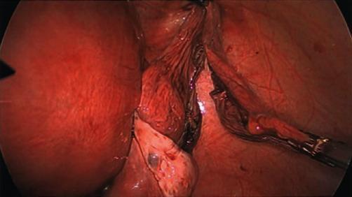
This laparoscopic clinical photograph depicts an intra-abdominal view during a surgical procedure involving the female reproductive system. On the left, a large, smooth, erythematous, and distended gravid uterus is clearly visible, occupying a significant portion of the pelvic field. Adjacent to the uterus, the right adnexal region is shown following the excision of a large cyst. A surgical grasper is seen on the right, retracting or manipulating the adnexal tissue near the pelvic sidewall. Central to the image are the dissected remains of the right adnexa, including the right ovary and the elongated right fallopian tube. The tissues show areas of dark red discoloration consistent with surgical dissection and cauterization (haemostasis). The image demonstrates the surgical anatomy of a second-trimester pregnancy and the spatial constraints encountered during a laparoscopic adnexal cystectomy in a gravid patient. It serves as an educational example of obstetric surgery and gynaecological laparoscopy.
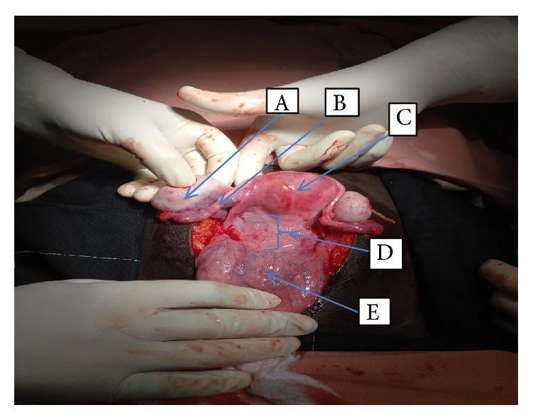
This clinical photograph captures an intraoperative view during a laparotomy, demonstrating female pelvic anatomy and a large occupying mass. The image provides clear visualization of the reproductive system with labeled landmarks: (A) the right ovary, which is smooth-surfaced and ovoid; (B) the right fallopian tube; and (C) the uterus, which appears pink and normally shaped but is displaced superiorly. Centrally, a large vaginal wall mass (D) is observed, located between the lower uterine segment and the urinary bladder. This mass is firm and appears to be pushing the surrounding structures, particularly the uterus, in a cephalad direction. The urinary bladder (E) is visible inferiorly, reflected away from the vaginal wall and mass. The photo illustrates the distorted anatomical relationships caused by a pelvic tumor, likely a cervical or vaginal myoma or angiofibroma, and is significant for teaching surgical approaches to pelvic masses and identification of retroperitoneal and pelvic landmarks.
"Genitalia, Female"[MeSH Terms] AND "anatomy"
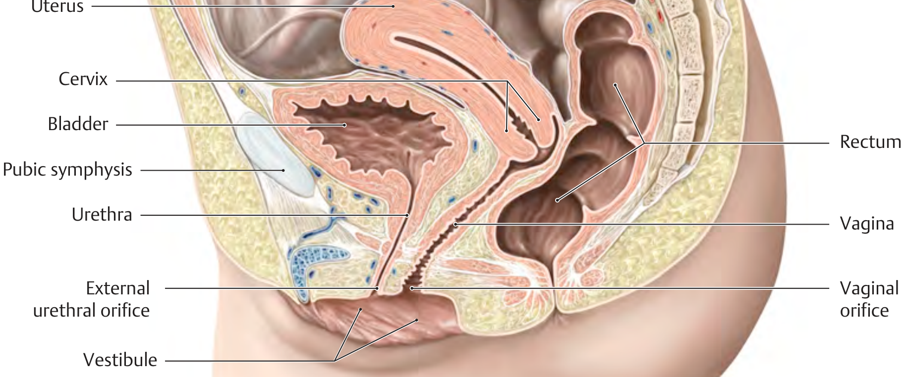
female pelvis sagittal section uterus vagina bladder rectum peritoneal pouches
Anatomy of the Female Reproductive System
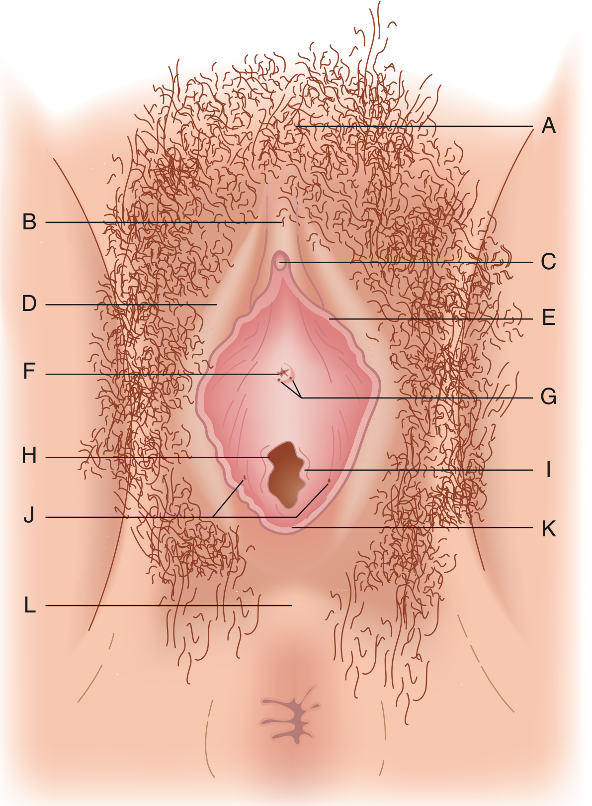
1. External genitalia (vulva)
- Mons pubis is the fatty tissue over the pubic symphysis. The pudendal cleft divides its lower part into the labia majora.
- Labia majora are pigmented skin folds containing smooth muscle, sebaceous, sweat and scent glands. They join at the anterior and posterior commissures.
- Labia minora fuse anteriorly to form the clitoral hood (prepuce) and posteriorly to form the vestibule.
- Clitoris has a glans, with the vestibular bulbs lying beneath the superficial perineal structures.
- Vestibule contains the urethral meatus and the vaginal introitus, which is outlined by the hymenal ring. Hart's line marks its outer border. Its anterior boundary is the clitoral frenulum and its posterior boundary is the posterior fourchette.
- Bartholin glands open at the fossa navicularis, posterolateral to the introitus. They are homologues of the male bulbourethral glands.
- Skene glands lie beside the urethral meatus. They are homologues of the prostate.
- Both glands provide lubrication.
- Perineum: a line between the ischial tuberosities divides it into an anterior urogenital triangle and a posterior anal triangle.
2. Internal genitalia
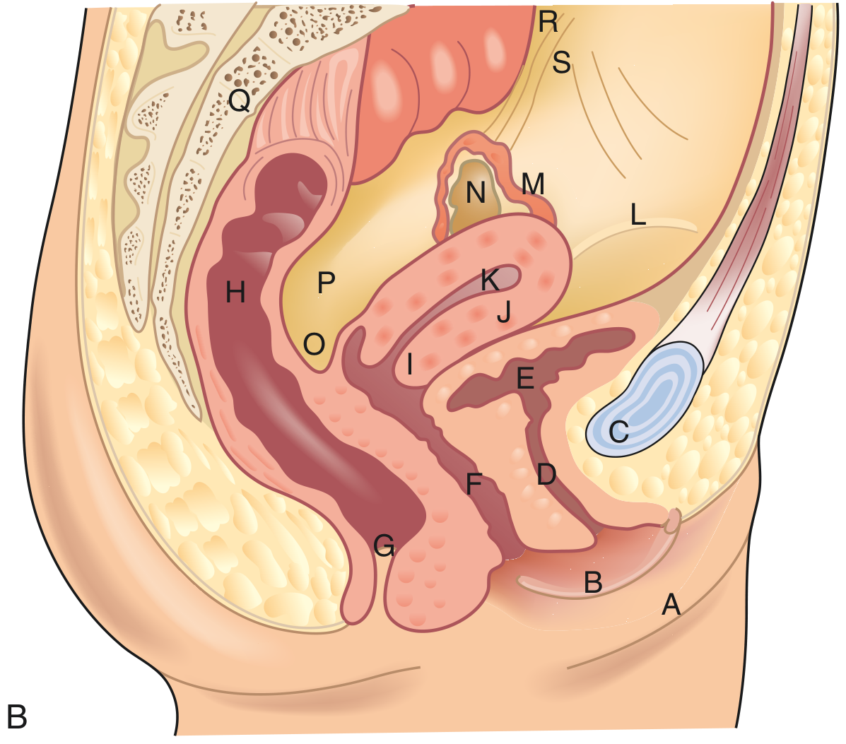
Vagina
- It is a flattened fibromuscular tube.
- Its layers, from inside out, are stratified squamous mucosa, lamina propria, muscularis and areolar connective tissue.
- Development: the lower part comes from the urogenital sinus. The upper part comes from the Müllerian ducts.
- Blood supply: the vaginal artery (from the internal iliac), with anastomoses from the uterine, internal pudendal, inferior vesical and middle hemorrhoidal arteries.
- Nerve supply: S2-S4, ending in the pudendal nerve. Sensation is concentrated in the distal vagina and is sparse in the upper part.
Cervix
- It is about 3 cm long, with a central canal (the os) connecting the vagina to the uterine cavity.
- The ectocervix has squamous epithelium and the endocervix has mucus-secreting columnar epithelium. The junction is the transition zone, where cervical intraepithelial neoplasia (CIN) arises.
- Blood supply: descending branches of the uterine artery run at the 3 and 9 o'clock positions. Azygos arteries run anteriorly and posteriorly in the midline.
- Innervation: parasympathetic (S2-S4), mostly in the endocervix.
Uterus
- It is an intraperitoneal muscular organ lying behind the bladder and in front of the rectum. The nonpregnant uterus weighs 40-80 g.
- Parts: fundus, corpus (body), isthmus and cervix.
- Wall layers: serosa (perimetrium), myometrium (smooth muscle in three layers) and endometrium.
- Supports and ligaments:
- The broad ligament is a peritoneal fold.
- The round ligament runs from the cornu through the inguinal canal to the labia majora.
- The cardinal (transverse cervical) ligaments support the cervix and carry the uterine arteries.
- The uterosacral ligaments run from the posterior cervix to S3.
- Blood supply: uterine arteries (from the internal iliac) and ovarian arteries (from the aorta).
- Innervation: sympathetic via the hypogastric and ovarian plexuses, and parasympathetic S2-S4. Visceral afferents travel at T11-T12.
Fallopian tubes (uterine tubes)
| Part | Features |
|---|---|
| Interstitial (intramural) | 1-2 cm, within the myometrium |
| Isthmus | About 4 cm, narrow, most muscular |
| Ampulla | 4-6 cm, wider, the usual site of fertilization |
| Infundibulum | Funnel-shaped, with fimbriae that surround the ostium |
- The mucosa has ciliated columnar, secretory and peg cells.
- Blood supply: ovarian and uterine arteries via the mesosalpinx.
Ovaries
- They are paired, oval and about 2-3 cm in diameter. They lie intraperitoneally in the ovarian fossa, between the internal and external iliac arteries. The ureter lies close behind.
- Attachments:
- The mesovarium attaches to the posterior broad ligament.
- The suspensory (infundibulopelvic) ligament carries the ovarian vessels.
- The ovarian (uteroovarian) ligament runs to the uterine tubal angle.
- Microscopic structure: the tunica albuginea is covered by a cuboidal surface epithelium. The cortex holds follicles at various stages and the corpus luteum. The medulla holds vessels, nerves and hilar cells.
- A newborn has about 500,000 to 1-2 million primordial follicles, many of which are lost by puberty. The oocytes are arrested in prophase of meiosis.
- Blood supply: the ovarian artery from the aorta, which anastomoses with the uterine artery.
- Clinical note: 80-90% of ovarian cancers arise from the surface epithelium.
3. Key clinical points
- Uterine artery and ureter: the ureter passes beneath the uterine artery in the cardinal ligament, so it is at risk in hysterectomy.
- The ovarian vessels in the infundibulopelvic ligament must be ligated in oophorectomy, and the ureter lies close by.
- Müllerian fusion failure causes uterine and tubal anomalies. Transverse vaginal septae can present at menarche.
- The ampulla is the commonest site of tubal ectopic pregnancy. This comes from standard teaching, not the pages I read.