
A 65 years old male on antipsychotic medications came with the C/o difficulty in walking, resting tremors and difficulty in swallowing a) What is the diagnosis. b)How will you treat this patient. c)Describe the mechanism of action, importance of combination therapy and adverse effects of Levodopa.
a) Diagnosis: Drug-Induced Parkinsonism (DIP)
- Resting tremors (pill-rolling type)
- Difficulty walking (bradykinesia, shuffling gait, rigidity)
- Dysphagia (difficulty swallowing - involvement of bulbar musculature)
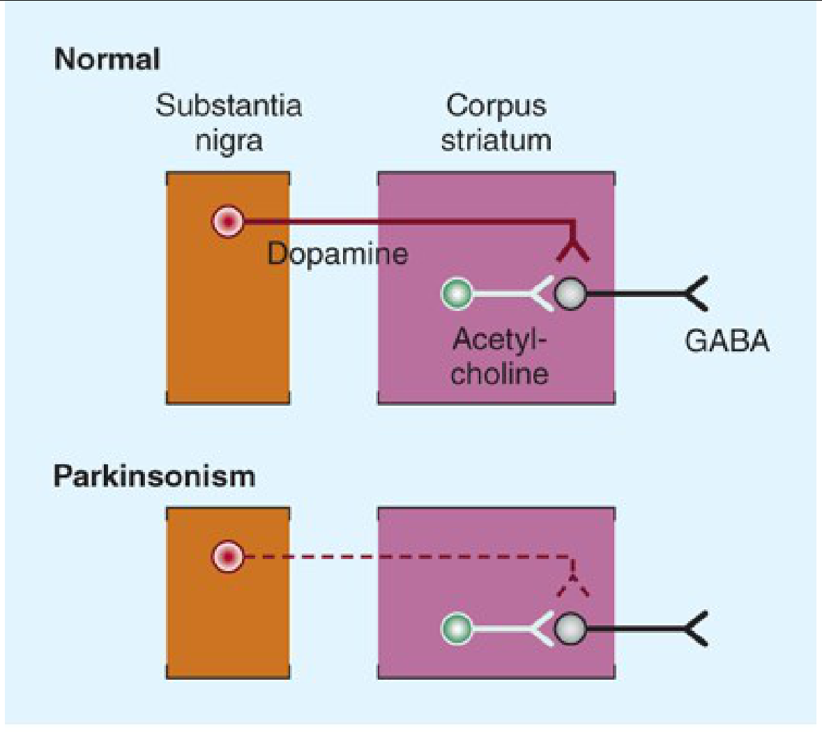
b) Treatment
Step 1 - Reassess the antipsychotic
- If clinically possible, reduce the dose or switch to an atypical antipsychotic with lower D2-blocking potency (e.g., quetiapine, clozapine). These cause far less DIP.
- Note: Levodopa is NOT helpful if the neuroleptic drug is continued - it cannot overcome the D2 blockade and may aggravate the psychiatric disorder for which the antipsychotic was originally prescribed.
- If the antipsychotic is withdrawn, parkinsonian symptoms clear over several weeks to months. In elderly patients, this can take up to 3 months.
Step 2 - Symptomatic treatment for parkinsonism
| Drug | Mechanism | Notes |
|---|---|---|
| Anticholinergics (Benztropine/Cogentin, Trihexyphenidyl) | Restore dopamine-ACh balance | First-line for DIP; preferred treatment |
| Amantadine | Increases dopamine release, anticholinergic | Alternative option |
| Diphenhydramine (Benadryl) | Anticholinergic antihistamine | Useful, especially if IV/IM needed |
- Anticholinergics should be withdrawn after 4-6 weeks to assess whether tolerance to the parkinsonian effects has developed. About half of patients require continued treatment.
- Even after stopping the antipsychotic, continue anticholinergic until symptoms fully resolve.
c) Levodopa - Mechanism of Action, Combination Therapy, and Adverse Effects
Mechanism of Action
- Benefits primarily depend on stimulation of D2 receptors (postsynaptic striatal neurons), though D1 stimulation may also contribute to maximal effect
- Levodopa is particularly effective at relieving bradykinesia and related disabilities
Importance of Combination Therapy (Levodopa + Carbidopa)
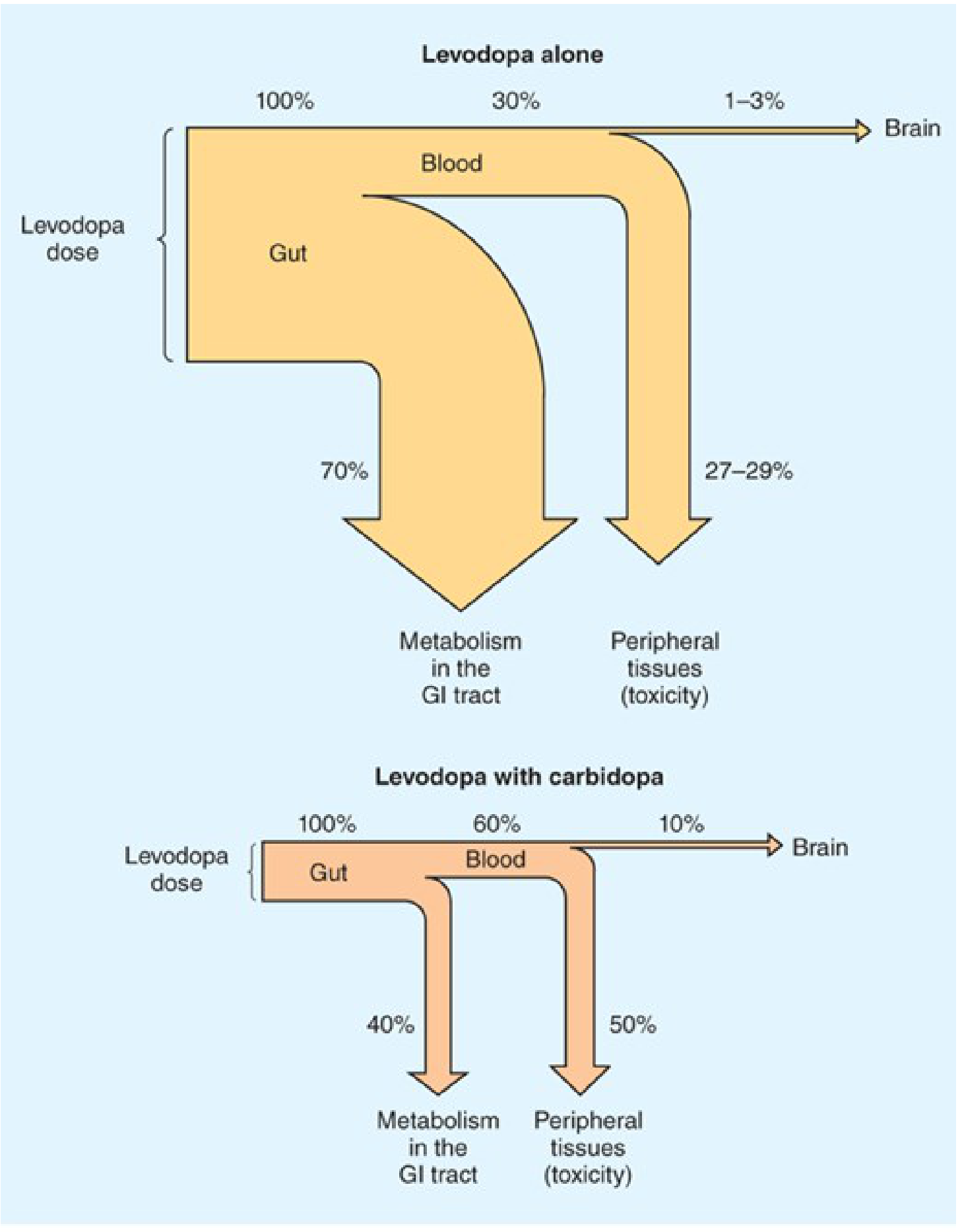
- Reduces daily levodopa requirement by ~75% - less drug needed for the same effect
- Plasma levels are higher and half-life longer - more drug available to reach the brain
- Brain delivery increases from ~1-3% to ~10% of the administered dose
- Dramatically reduces peripheral side effects - nausea/vomiting drops from ~80% to <20%; cardiac arrhythmias also reduced
- Allows much faster titration to therapeutic doses
Adverse Effects of Levodopa
- Anorexia, nausea, vomiting in ~80% when used alone (reduced to <20% with carbidopa)
- Due to stimulation of the chemoreceptor trigger zone (CTZ) - located outside the BBB
- Management: take with food, add extra carbidopa (Lodosyn 25 mg), use domperidone or ondansetron
- Avoid phenothiazine antiemetics - they worsen parkinsonism
- Cardiac arrhythmias (tachycardia, ventricular extrasystoles, rarely atrial fibrillation) - from peripheral catecholamine formation; reduced significantly with carbidopa co-administration
- Postural hypotension - common but often asymptomatic; tends to improve with continued treatment
- Hypertension - if combined with non-selective MAO inhibitors or sympathomimetics (dangerous combination)
- Depression, anxiety, agitation, insomnia, somnolence, confusion
- Delusions, hallucinations, nightmares, euphoria
- More common when levodopa is combined with carbidopa (more drug reaches the brain)
- Managed with atypical antipsychotics (quetiapine, clozapine) or pimavanserin (5-HT2A inverse agonist) for Parkinson psychosis
- Choreiform, athetoid, or dystonic movements - most common long-term complication
- Particularly affects the face, mouth, tongue, and limbs
- Related to peak plasma levodopa levels; managed by dose reduction or adding amantadine
- "Wearing-off" effect - symptom recurrence before next dose as plasma levodopa falls
- "On-off" phenomenon - unpredictable sudden shifts between mobility (on) and immobility (off), unrelated to dosing schedule
- Managed with: extended-release carbidopa/levodopa (Rytary), COMT inhibitors (entacapone, opicapone), MAO-B inhibitors (rasagiline), or deep brain stimulation
- Darkening of urine (levodopa metabolites - not harmful)
- Rarely, neuroleptic malignant-like syndrome on abrupt withdrawal
Summary Table
| Feature | Detail |
|---|---|
| Diagnosis | Drug-induced Parkinsonism (D2 blockade by antipsychotic) |
| First-line treatment | Reduce/switch antipsychotic + Anticholinergics (benztropine) |
| Why NOT levodopa for DIP? | Cannot overcome D2 blockade; may worsen psychiatric condition |
| Levodopa MOA | Precursor crosses BBB → converted to dopamine → D2 stimulation |
| Why add carbidopa? | Blocks peripheral decarboxylation → reduces dose by 75%, cuts GI/CV side effects |
| Key adverse effects | Nausea, arrhythmias, hypotension, hallucinations, dyskinesias, on-off fluctuations |
Neurohumoral transmission
Neurohumoral Transmission
1. Historical Background and Evidence
- The physiologically active compound and its biosynthetic enzymes are present at appropriate anatomical sites
- The compound can be recovered from perfusate of an innervated structure during nerve stimulation but not at rest
- The compound produces responses identical to nerve stimulation when administered exogenously
- Responses to nerve stimulation and the administered compound are modified identically by pharmacological antagonists
Note: Nitric oxide (NO) is an exception - it is a neurotransmitter in some postganglionic parasympathetic nerves but is synthesized on demand rather than stored and released by exocytosis.
2. Steps in Neurohumoral Transmission
Step 1 - Axonal Conduction
- At rest: interior is ~70 mV negative relative to exterior (resting membrane potential)
- Depolarization to threshold opens voltage-sensitive Na+ channels → rapid Na+ influx → depolarization with positive overshoot
- Followed by K+ efflux → repolarization
- Local circuit currents propagate the AP along the axon in a forward direction
- The depolarized region enters a brief refractory state to ensure unidirectional propagation
Step 2 - Junctional (Synaptic) Transmission
a) Storage and Release of Transmitter
- Non-peptide neurotransmitters (biogenic amines) are synthesized in axonal terminals and stored in synaptic vesicles
- Vesicular storage is driven by a vesicular proton pump (vesicular ATPase)
- Synaptic vesicle proteins include: synapsin, synaptophysin, synaptogyrin
- The exocytosis machinery involves SNARE proteins: synaptobrevin (vesicular membrane), SNAP-25 and syntaxin (plasma membrane), and synaptotagmin (Ca²+ sensor)
- Docking - Munc18 binds syntaxin, stabilizing the SNARE complex
- Priming I - Syntaxin assembles with SNAP-25, allowing synaptobrevin to bind
- Priming II - Complexin binds the SNARE complex; synaptotagmin binds Ca²+
- Fusion - Ca²+ entry triggers full membrane fusion and exocytosis
- Reset - NSF ATPase and SNAP adapters dissociate the SNARE complex
Botulinum toxin cleaves SNARE proteins (synaptobrevin) and blocks ACh release at the NMJ. Tetanus toxin acts similarly on inhibitory neurons in the CNS.
b) Combination with Postjunctional Receptor
- Released transmitter binds specific receptors on the postsynaptic membrane
- Ionotropic receptors (ligand-gated ion channels): rapid - opens within milliseconds
- Excitatory: nicotinic, glutamate, 5-HT3 → Na+ influx → EPSP (depolarization)
- Inhibitory: GABA, glycine → Cl- influx → IPSP (hyperpolarization)
- Metabotropic receptors (G protein-coupled): slower, via second messengers
- E.g., muscarinic receptors, α and β adrenergic receptors
- Signal through cAMP (adenylyl cyclase), IP3/DAG (phospholipase C), or modulation of K+/Ca²+ channels
c) Initiation of Response
- Summation of microscopic channel-opening events generates the excitatory postsynaptic potential (EPSP)
- Sufficient depolarization triggers a new action potential in the postjunctional cell
d) Termination of Transmitter Action
- Enzymatic hydrolysis (e.g., ACh by acetylcholinesterase)
- Reuptake into the nerve terminal (primary for catecholamines)
- Diffusion away from the synapse
e) Non-electrogenic (Trophic) Functions
- Neurotransmitters also control enzyme turnover, receptor density, and synaptic plasticity through trophic actions
3. Cholinergic Transmission
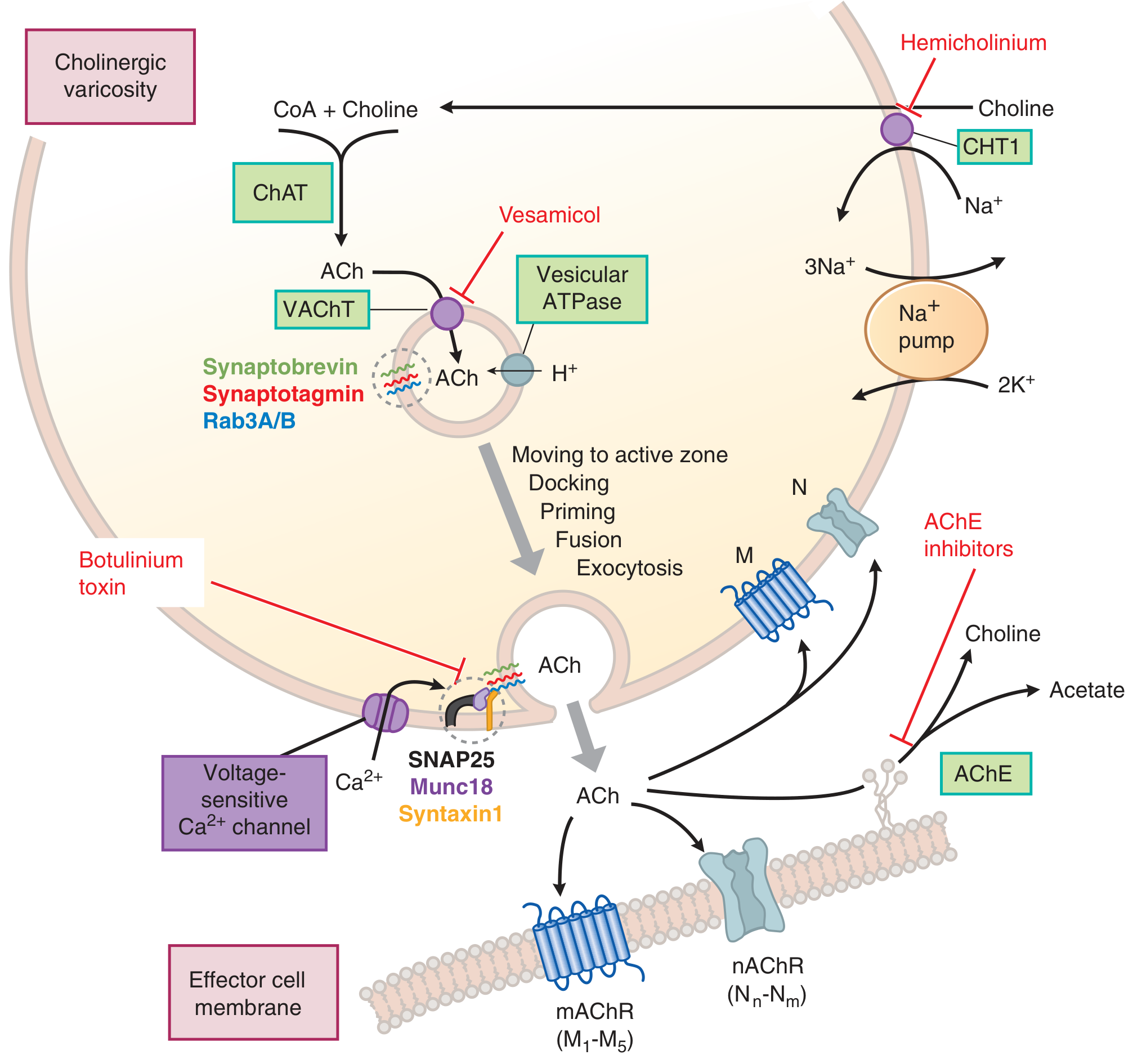
Synthesis of Acetylcholine (ACh)
- Choline acetyltransferase (ChAT) catalyzes: Choline + Acetyl CoA → ACh
- Rate-limiting step = uptake of choline via high-affinity choline transporter (CHT1) from the extracellular fluid (Na+-dependent)
- Blocked by: hemicholinium
- Choline is recycled after ACh hydrolysis
- ACh is packaged into vesicles by vesicular ACh transporter (VAChT)
- Blocked by: vesamicol
Release of ACh
- Action potential → Ca²+ entry via voltage-gated Ca²+ channels → SNARE-mediated exocytosis
- Two pools: readily releasable pool (near membrane, newly synthesized) and reserve pool (replenishes the first)
Degradation of ACh
- Acetylcholinesterase (AChE) hydrolyzes ACh → choline + acetate (within <1 millisecond at the NMJ)
- Choline is recycled back into the terminal
- Butyrylcholinesterase (pseudocholinesterase) - found in liver/plasma; physiologically hydrolyzes ingested plant esters
Cholinergic Receptors
| Receptor | Type | Location | Mechanism |
|---|---|---|---|
| Nicotinic (nAChR) | Ionotropic (ligand-gated Na+/K+ channel) | NMJ (Nm), Autonomic ganglia (Nn), CNS | EPSP/depolarization |
| Muscarinic M1,3,5 | Metabotropic (Gq) | Glands, smooth muscle, CNS | ↑ IP3/DAG → ↑ Ca²+ |
| Muscarinic M2,4 | Metabotropic (Gi) | Heart, presynaptic terminals | ↓ cAMP, ↑ K+ conductance |
4. Adrenergic (Catecholamine) Transmission
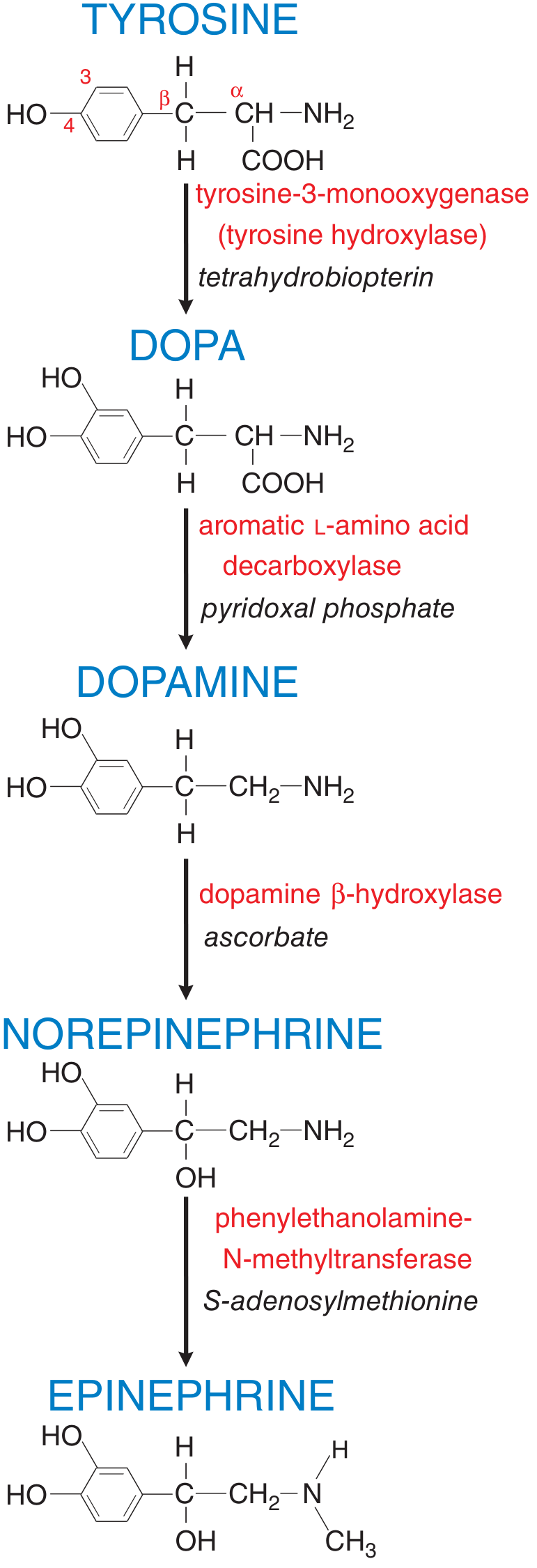
Synthesis of Norepinephrine (NE)
- Tyrosine → DOPA by tyrosine hydroxylase (TH) [rate-limiting step; cofactor: tetrahydrobiopterin]
- DOPA → Dopamine by aromatic L-amino acid decarboxylase (dopa decarboxylase) [cofactor: pyridoxal phosphate]
- Dopamine → Norepinephrine by dopamine β-hydroxylase (DβH) [cofactor: ascorbate] - inside storage vesicles
- Norepinephrine → Epinephrine by phenylethanolamine-N-methyltransferase (PNMT) [cofactor: S-adenosylmethionine] - only in adrenal medulla and a few brainstem neurons
Release of NE
- Action potential → Ca²+ entry → exocytosis of vesicular contents (NE, ATP, NPY, chromogranins, DβH)
- SNARE proteins (SNAP-25, syntaxin, synaptobrevin) mediate fusion
Termination of NE Action
| Transporter | Also called | Location | Affinity | Inhibitors |
|---|---|---|---|---|
| NET (SLC6A2) | Uptake 1 | Sympathetic nerves, adrenal medulla | High affinity for NE > EPI | Cocaine, TCAs (desipramine, imipramine) |
| OCT3/ENT (SLC22A3) | Uptake 2 | Nonneuronal cells (heart, liver) | Low affinity, prefers EPI > NE | Corticosterone, normetanephrine |
- MAO (monoamine oxidase) - intraneuronally; MAO-B selective for DA and phenylethylamines; MAO-A selective for 5-HT and NE
- COMT (catechol-O-methyltransferase) - extraneuronally
Adrenergic Receptors
| Receptor | G protein | Key Location | Effect |
|---|---|---|---|
| α1 | Gq | Smooth muscle, liver | Vasoconstriction, glycogenolysis |
| α2 | Gi | Presynaptic terminals, platelets | ↓ NE release (autoreceptor), platelet aggregation |
| β1 | Gs | Heart | ↑ Heart rate, ↑ contractility |
| β2 | Gs | Bronchi, blood vessels | Bronchodilation, vasodilation |
| β3 | Gs | Adipose tissue | Lipolysis |
5. Summary of Key Neurotransmitter Systems
| Feature | Cholinergic | Adrenergic |
|---|---|---|
| Neurotransmitter | Acetylcholine | Norepinephrine / Epinephrine |
| Precursor | Choline + Acetyl CoA | Tyrosine |
| Synthesis enzyme | Choline acetyltransferase (ChAT) | Tyrosine hydroxylase (rate-limiting) |
| Vesicular transporter | VAChT (blocked by vesamicol) | VMAT2 (blocked by reserpine) |
| Release trigger | Ca²+ entry via depolarization | Ca²+ entry via depolarization |
| Termination | Hydrolysis by AChE | Reuptake by NET; MAO/COMT metabolism |
| Receptors | Nicotinic (ionotropic), Muscarinic (metabotropic) | α1, α2, β1, β2, β3 (all metabotropic/GPCR) |
| Blocked by (release) | Botulinum toxin | - |
| Blocked by (synthesis) | Hemicholinium (choline uptake) | α-methyltyrosine (TH inhibitor) |
25 years male RTA victim presented with rapid pulse, cold clammy cyanotic skin, tachypnea and landed in emergency ward in unconscious state what is the diagnosis? Classify the disease write in detail about the pathogenesis, morphology evolution of the disease.
Diagnosis: Hypovolemic Shock (Hemorrhagic Shock)
- Rapid (tachycardic) pulse - compensatory response to falling cardiac output
- Cold, clammy, cyanotic skin - cutaneous vasoconstriction shunting blood to vital organs
- Tachypnea - compensatory hyperventilation to improve oxygenation and blow off CO2
- Unconsciousness - end-organ (brain) hypoperfusion
Classification of Shock
| Type | Clinical Example | Principal Mechanism |
|---|---|---|
| Hypovolemic | Hemorrhage, burns, vomiting, diarrhea, trauma | Inadequate blood or plasma volume → low cardiac output |
| Cardiogenic | Myocardial infarction, arrhythmia, cardiac tamponade, pulmonary embolism | Myocardial pump failure → low cardiac output |
| Septic | Gram-positive/negative sepsis, fungi | Peripheral vasodilation, vascular leak, DIC, cytokine cascade |
| Neurogenic | Spinal cord injury, general anesthesia | Loss of vascular tone → acute vasodilation → hypotension |
| Anaphylactic | IgE-mediated hypersensitivity | Systemic vasodilation + increased vascular permeability |
Pathogenesis of Shock
Core Mechanism
Massive hemorrhage → ↓ circulating blood volume → ↓ venous return → ↓ cardiac output → ↓ tissue perfusion → cellular hypoxia → organ dysfunction → death (if uncorrected)
Compensatory (Neurohumoral) Responses Activated
- Baroreceptor reflexes - carotid/aortic baroreceptors detect falling BP → reflex sympathetic activation
- Catecholamine release (epinephrine from adrenal medulla, NE from sympathetics) → tachycardia, peripheral vasoconstriction
- Renin-Angiotensin-Aldosterone System (RAAS) activation → angiotensin II causes vasoconstriction; aldosterone promotes renal Na+ and water retention
- ADH (vasopressin) release from posterior pituitary → water reabsorption by kidney
- Generalized sympathetic stimulation → redistributes blood away from skin, gut, and kidneys toward heart and brain
- Tachycardia
- Peripheral vasoconstriction (cold, clammy, pale/cyanotic skin)
- Oliguria (urine output falls)
- Relative preservation of cardiac and cerebral perfusion initially
Evolution (Stages) of Shock
Stage 1 - Nonprogressive (Compensated) Stage
- Reflex compensatory mechanisms are fully activated
- Vital organ (heart, brain) perfusion is maintained
- Clinically: tachycardia, tachypnea, cool clammy skin, mild anxiety
- Cellular injury is reversible at this stage
- Blood is shunted away from skin/gut to heart and brain
- Cutaneous vasoconstriction accounts for the cold, clammy, cyanotic appearance
Stage 2 - Progressive Stage
- Underlying cause is not corrected → widespread tissue hypoxia develops
- Aerobic respiration fails → anaerobic glycolysis → lactic acid production → metabolic lactic acidosis
- Lactic acidosis lowers tissue pH → vasomotor response is blunted
- Arterioles dilate → blood pools in the microcirculation
- Peripheral pooling worsens cardiac output (vicious cycle)
- Endothelial anoxic injury → risk of Disseminated Intravascular Coagulation (DIC)
- Vital organs (heart, brain, kidney) begin to fail
- Clinically: worsening hypotension, deepening coma, oliguria/anuria, worsening lactic acidosis
Stage 3 - Irreversible Stage
- Severe, sustained tissue injury → widespread cell death
- Lysosomal enzyme leakage from necrotic cells further worsens the shock state (autolytic injury)
- Myocardial contractile function deteriorates (partly from excess NO synthesis reducing cardiac muscle tone)
- Ischemic bowel allows gut bacteria to enter the circulation → superimposed bacteremic shock (septic shock on top of hemorrhagic shock)
- Acute tubular necrosis (ATN) of kidneys from sustained ischemia → acute renal failure
- Despite heroic therapeutic efforts, death is inevitable once this stage is reached
- The terminal pathway is multi-organ failure (MOF)
Morphology of Shock
1. Brain
- Ischemic encephalopathy - neuronal necrosis in the most vulnerable areas: hippocampus (Sommer's sector), cerebellar Purkinje cells, and neocortical neurons
- Watershed (border-zone) infarcts in areas between the end-territories of major cerebral arteries (ACA-MCA and MCA-PCA junction zones)
- Neurons are the most sensitive cells to hypoxia - irreversible damage after just 3-5 minutes of complete anoxia
2. Heart
- Subendocardial hemorrhage and necrosis - the subendocardial myocardium is the zone most vulnerable to ischemia (farthest from coronary supply, highest oxygen demand)
- Focal areas of myocardial necrosis (contraction band necrosis) - seen in hemorrhagic shock
- Reduced myocardial contractility with worsening shock (partly mediated by TNF, IL-1, and NO in later stages)
3. Kidneys
- Acute Tubular Necrosis (ATN) - the most characteristic morphological finding
- Patchy ischemic necrosis of tubular epithelium, especially the proximal tubule and thick ascending limb of Henle
- Tubular casts (pigmented) in the distal tubules and collecting ducts
- Interstitial edema
- Intact tubular basement membrane (important - allows regeneration if patient survives)
- Clinically: oliguria, rising BUN and creatinine, electrolyte disturbances
- Prognosis: tubular cells can regenerate - reversible if perfusion is restored
4. Lungs
- Lungs are relatively resistant to hypoxic injury in pure hemorrhagic shock
- However, when shock is complicated by trauma or sepsis: Diffuse Alveolar Damage (DAD) develops
- Interstitial and alveolar edema
- Hyaline membrane formation (protein-rich exudate)
- Type II pneumocyte hyperplasia
- This is the pathological substrate of "Shock Lung" = Acute Respiratory Distress Syndrome (ARDS)
- Clinically: hypoxia, bilateral infiltrates on CXR, respiratory failure
5. Adrenal Glands
- Cortical cell lipid depletion - a non-specific stress response reflecting increased use of stored cholesterol/lipids for cortisol synthesis
- In severe septic shock complicated by DIC: adrenal hemorrhage → Waterhouse-Friderichsen syndrome → adrenal insufficiency
- Loss of cortisol in late stages contributes to hemodynamic collapse
6. Gastrointestinal Tract
- Mucosal ischemia → stress ulcers (Curling's ulcers in the stomach/duodenum)
- Mucosal barrier breakdown → bacterial translocation (gut bacteria enter the bloodstream)
- This is a key mechanism for converting hypovolemic shock into septic shock in the later stages
- Can also develop hemorrhagic gastroenteritis and ischemic colitis
7. Liver
- Centrilobular necrosis (zone 3 of hepatic acinus - most distal from hepatic artery supply, most sensitive to hypoxia)
- "Nutmeg liver" appearance grossly (congested, red-yellow mottled)
- Rising liver enzymes (transaminases) → jaundice in severe/prolonged shock
Summary Flowchart
RTA → Hemorrhage
↓
↓ Circulating Blood Volume
↓
↓ Cardiac Output
↓
↓ Tissue Perfusion
↓
┌──────────────────────────────────────────┐
│ COMPENSATORY STAGE │
│ ↑ HR, ↑ Catecholamines, ↑ RAAS, ↑ ADH │
│ Peripheral vasoconstriction │
│ Cold clammy cyanotic skin │
└──────────────────────────────────────────┘
↓ (if not corrected)
┌──────────────────────────────────────────┐
│ PROGRESSIVE STAGE │
│ Anaerobic glycolysis → Lactic acidosis │
│ Arteriolar dilation → Microvascular │
│ pooling → DIC risk │
│ Vital organ dysfunction begins │
└──────────────────────────────────────────┘
↓ (if not corrected)
┌──────────────────────────────────────────┐
│ IRREVERSIBLE STAGE │
│ Lysosomal rupture, ↓ myocardial │
│ contractility, gut translocation, │
│ ATN, ARDS, MOF → DEATH │
└──────────────────────────────────────────┘
Clinical Note
Prognosis: More than 90% of young, otherwise healthy patients with hypovolemic shock survive with appropriate management (aggressive IV fluid resuscitation, blood transfusion, surgical hemorrhage control). This compares favorably with cardiogenic or septic shock, which carry substantially worse outcomes even with state-of-the-art care. The key is rapid intervention before the irreversible stage is reached.
20 years male following antibiotic intake had total leukocyte count of 1200 cells / mm3 and absolute neutrophil count of 120 cells. What is your diagnosis? What is the pathogenesis of this condition?
Diagnosis
Primary Diagnosis: Drug-Induced Agranulocytosis
| Parameter | Patient's Value | Normal Range | Interpretation |
|---|---|---|---|
| Total Leukocyte Count (TLC) | 1200/mm³ | 4000-11,000/mm³ | Severe leukopenia |
| Absolute Neutrophil Count (ANC) | 120/mm³ | 1500-8000/mm³ | Agranulocytosis (<500/mm³) |
- Neutropenia = ANC < 1500/mm³
- Severe neutropenia = ANC < 500/mm³
- Agranulocytosis = ANC < 100-500/mm³ (marked reduction, near absence of neutrophils)
Classification of Neutropenia/Agranulocytosis
Broad Mechanism-Based Classification
AGRANULOCYTOSIS
├── A. Decreased Production (Inadequate/Ineffective Granulopoiesis)
│ ├── 1. Suppression of Hematopoietic Stem Cells (HSCs)
│ │ ├── Aplastic anemia
│ │ ├── Marrow infiltration (leukemia, granulomas, tumors)
│ │ └── Myelotoxic chemotherapy (affects all cell lines)
│ ├── 2. Suppression of Committed Granulocytic Precursors
│ │ └── Drug toxicity (selective - other lineages spared)
│ ├── 3. Ineffective Hematopoiesis
│ │ ├── Megaloblastic anemia (B12/folate deficiency)
│ │ └── Myelodysplastic neoplasms
│ └── 4. Congenital
│ └── Kostmann syndrome (severe congenital neutropenia)
│
└── B. Increased Destruction/Sequestration
├── 1. Immune-mediated neutrophil destruction (drug-induced or idiopathic)
├── 2. Splenomegaly (splenic sequestration)
├── 3. Overwhelming infection (bacterial, fungal, rickettsial)
└── 4. LGL leukemia (CD8+ cytotoxic T cell suppression of myelopoiesis)
Drug-Based Classification of Causes
| Mechanism | Drug Class | Examples |
|---|---|---|
| Predictable, dose-related myelosuppression | Antineoplastics | Alkylating agents, antimetabolites |
| Idiosyncratic - direct toxic to precursors | Antipsychotics | Chlorpromazine, clozapine, phenothiazines |
| Idiosyncratic - immune-mediated | Antibiotics | Penicillins, sulfonamides, chloramphenicol |
| - | Antithyroidal | Methimazole, propylthiouracil, carbimazole |
| - | Anticonvulsants | Valproate, carbamazepine |
| - | Anti-inflammatory | Sulfasalazine |
| - | Antiarrhythmic | Procainamide |
Pathogenesis
Mechanism 1: Direct Toxic/Myelosuppressive Effect
Drug enters bone marrow
↓
Direct toxic effect on granulocytic precursors
(myeloblasts, promyelocytes, myelocytes)
↓
Selective destruction of granulocyte precursors
(erythroid and megakaryocytic lineages spared)
↓
Maturation arrest at promyelocyte/myelocyte stage
↓
↓↓ Release of mature neutrophils into blood
↓
Agranulocytosis
Mechanism 2: Immune-Mediated (Hapten) Mechanism
STEP 1: SENSITIZATION PHASE
Drug (hapten) binds to neutrophil surface proteins
→ Forms drug-protein complex (neoantigen)
→ Presented to immune system as foreign antigen
→ Antibody production against drug-neutrophil complex
(IgG/IgM anti-neutrophil antibodies generated)
STEP 2: SUBSEQUENT EXPOSURE
Re-exposure to the same drug
↓
Drug binds to neutrophil surface again
↓
Pre-formed antibodies (IgG) attach to drug-neutrophil complex
↓
Two pathways of destruction:
├─ COMPLEMENT ACTIVATION (IgM/IgG):
│ Complement fixed on neutrophil surface
│ → Membrane Attack Complex (MAC)
│ → Direct neutrophil lysis in circulation
│
└─ OPSONIZATION + PHAGOCYTOSIS (IgG):
Fc receptors on macrophages/monocytes
recognize IgG-coated neutrophils
→ Phagocytosis in spleen and liver
→ Peripheral neutrophil destruction
↓
Rapid fall in circulating neutrophils
↓
AGRANULOCYTOSIS
Additional Immune Mechanism: Autoimmune Neutropenia
- Antibodies directed against neutrophil surface antigens
- Idiopathic or associated with SLE, rheumatoid arthritis (Felty syndrome)
- Neutrophils opsonized → destroyed in spleen
Morphology
Bone Marrow Changes
| Type of Mechanism | Bone Marrow Appearance |
|---|---|
| Immune-mediated peripheral destruction | Hypercellular - compensatory increase in granulocytic precursors (the marrow tries to compensate for peripheral loss) |
| Direct toxic suppression of precursors | Hypocellular (specifically reduced granulocytic series; erythroid and megakaryocytes preserved) |
| Myelotoxic chemotherapy | Hypocellular all lineages reduced |
Peripheral Blood
- Near-absent neutrophils (ANC <500)
- Other cell lines (RBCs, platelets, lymphocytes) relatively preserved in pure drug-induced cases
Sites of Infection (Consequences of Agranulocytosis)
- Oral cavity most characteristic: Necrotizing ulcerative lesions of the gingiva, floor of mouth, buccal mucosa, and pharynx
- Deep, undermined ulcers covered by gray-to-green-black necrotic membranes
- Masses of bacteria/fungi visible histologically with minimal leukocytic inflammatory response (no neutrophils available)
- Lungs, urinary tract, kidneys: Severe invasive bacterial or fungal infections
- Deep fungal infections: Candida and Aspergillus (characteristically opportunistic)
- All sites share the hallmark: massive microbial growth with absent neutrophilic infiltrate
Clinical Features and Danger Threshold
| ANC | Risk Level | Clinical Implication |
|---|---|---|
| 1000-1500/mm³ | Mild | Increased susceptibility to infection |
| 500-1000/mm³ | Moderate | Significant infection risk |
| <500/mm³ | Severe | Risk rises sharply - infections become life-threatening |
| <100/mm³ | Agranulocytosis | Overwhelming infections; can cause death within hours to days |
- Malaise, chills, high-grade fever (often the first sign)
- Oropharyngeal ulcers/mucositis
- Weakness and fatigue
- If untreated: rapidly progressive sepsis → death
Management Principles
- Stop the offending drug immediately (causative antibiotic)
- Broad-spectrum antibiotics at first sign of infection (do not wait for culture results - infections are fulminant)
- G-CSF (Granulocyte Colony-Stimulating Factor / Filgrastim) - stimulates granulocyte production from marrow precursors, accelerates recovery
- Reverse isolation (protective environment to minimize infection exposure)
- Bone marrow biopsy to assess cellularity and mechanism
- Antifungal prophylaxis (fluconazole/voriconazole) given risk of Candida/Aspergillus
Prognosis: With early diagnosis, cessation of the offending drug, and G-CSF therapy, most patients recover. Mortality is high if diagnosis is delayed and fulminant sepsis develops.
A 25 year old male was admitted in the hospital four days after Road Traffic Accident (RTA). On examination, crush injury right foot with soiled gauze contaminated with soil was observed. There was edema, crushed muscles and pain at the site. On palpation crepitus was felt. a. What is the clinical condition? b. Mention the etiological agent responsible for this condition? c. Describe the pathogenesis and clinical manifestations of this condition? d. Describe in detail the laboratory diagnosis and treatment of this disease?
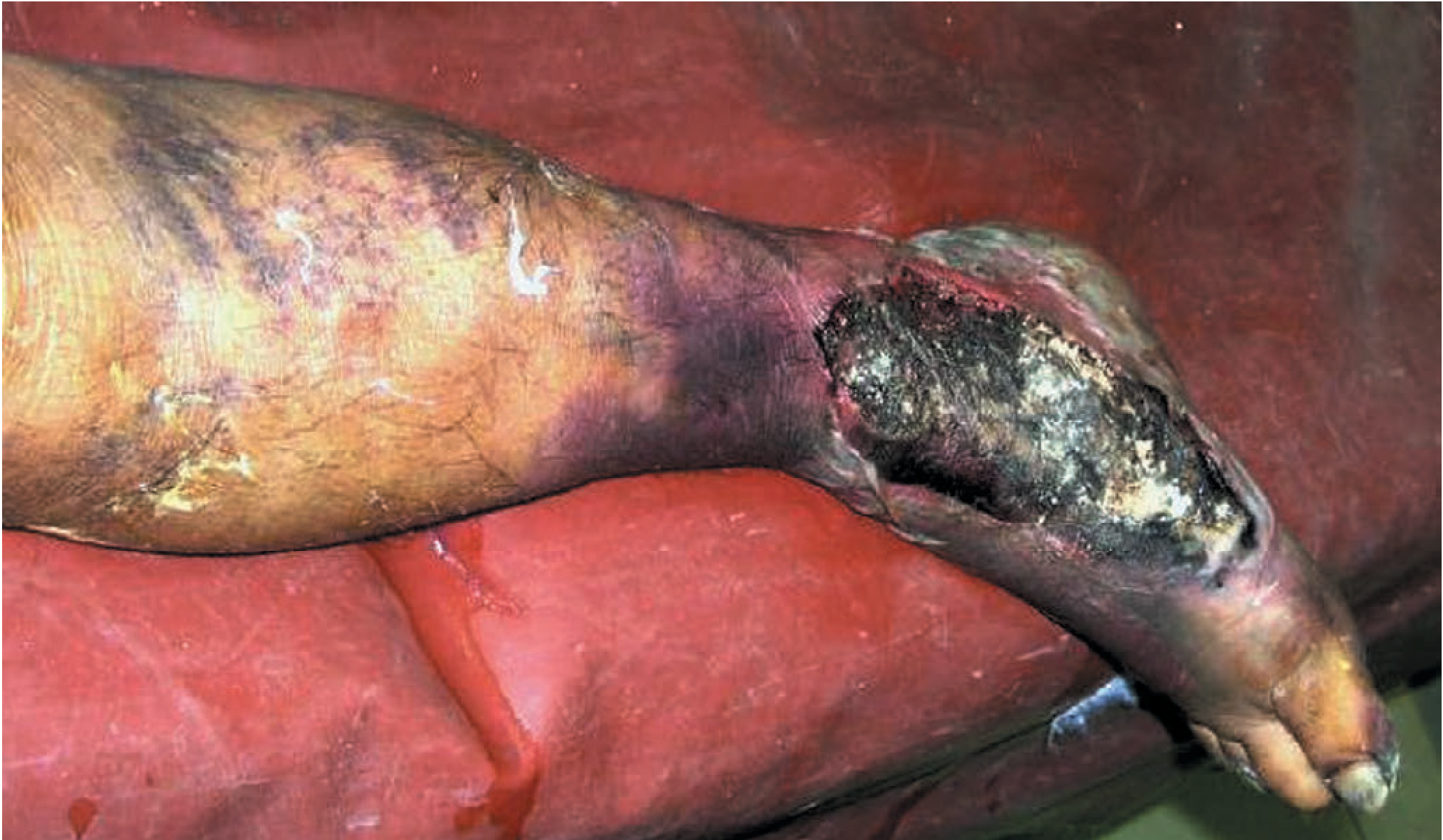
a) Clinical Condition: Gas Gangrene (Clostridial Myonecrosis)
| Feature | Significance |
|---|---|
| RTA with crush injury to right foot | Devitalized tissue with disrupted blood supply - creates anaerobic microenvironment |
| Soil-contaminated wound | Soil is the primary reservoir of Clostridium spores |
| 4-day delay before presentation | Sufficient incubation time (6 hours to 4 days) for spore germination and toxin production |
| Edema + crushed muscles + severe pain | Myonecrosis and spreading infection |
| Crepitus on palpation | Pathognomonic - gas (CO₂ and H₂S) produced by clostridial fermentation in tissues |
b) Etiological Agent
Primary Agent: Clostridium perfringens (also called C. welchii)
| Property | Detail |
|---|---|
| Gram stain | Large Gram-positive bacilli with blunt ends |
| Morphology | Large rectangular rods, "boxcar-shaped" |
| Oxygen requirement | Obligate anaerobe |
| Spore formation | Yes - forms spores (allows survival in soil for years) |
| Spore position | Subterminal, oval spores |
| Motility | Non-motile |
| Capsule | Encapsulated |
| Habitat | Soil, human/animal intestine, feces, dust |
Other Clostridial Species Causing Gas Gangrene:
- Clostridium septicum
- Clostridium novyi (Type A)
- Clostridium sordellii
- Clostridium bifermentans
- Clostridium histolyticum
- Clostridium sporogenes
Note: Non-clostridial gas-producing organisms (coliforms, mixed aerobic/anaerobic bacteria) are found in 60-85% of gas gangrene cases as co-infectors.
Types of Clostridial Wound Infection:
| Type | Characteristic |
|---|---|
| Type 1 | Clostridial contamination - positive culture, NO clinical signs of infection |
| Type 2 | Clostridial cellulitis - gas in tissue, foul smell, NO systemic involvement |
| Type 3 | Gas gangrene (Clostridial myonecrosis) - systemic signs of severe infection |
c) Pathogenesis and Clinical Manifestations
Pathogenesis
Step 1 - Setting the Stage (Prerequisite Conditions)
Crush injury + Soil contamination
↓
Clostridial spores introduced into wound
↓
Crushed/devitalized tissue → disrupted blood supply
↓
Reduced tissue oxygen tension (low redox potential)
↓
Anaerobic microenvironment created
↓
Spores GERMINATE into vegetative (actively dividing) form
- Devitalized (dead/ischemic) muscle tissue
- Soil contamination
- Retained foreign bodies
- Delayed wound treatment / wound closure without debridement
- Immunosuppression, diabetes, peripheral vascular disease
Step 2 - Toxin Production (Core Mechanism)
| Toxin | Biochemical Nature | Action |
|---|---|---|
| Alpha (α) toxin | Lecithinase (phospholipase C) | Most important - destroys phospholipids in RBC, WBC, platelet, muscle cell, and fibroblast membranes → hemolysis, myonecrosis, leukocyte destruction, capillary damage |
| Theta (θ) / Perfringolysin O | Cholesterol-dependent cytolysin | Hemolysis + myocardial suppression (cardiotoxicity) |
| Kappa (κ) toxin | Collagenase | Destruction of connective tissue and blood vessel walls |
| Mu (μ) toxin | Hyaluronidase ("spreading factor") | Breaks down hyaluronic acid → allows infection to spread through tissue planes |
| Nu (ν) toxin | DNase (deoxyribonuclease) | Cell necrosis |
| Lambda (λ) toxin | Protease | Tissue destruction |
- It is a phospholipase C that hydrolyzes lecithin (phosphatidylcholine) - a major component of all cell membranes
- This directly disrupts membrane integrity of red blood cells (hemolysis), platelets (thrombocytopenia), neutrophils (immune evasion), and muscle cells (myonecrosis)
- Also causes widespread capillary damage and microvascular thrombosis
Step 3 - Vicious Cycle of Tissue Destruction
Alpha-toxin → destroys muscle cell membranes
↓
Muscle necrosis + capillary thrombosis
↓
Further tissue ischemia → deeper anaerobic conditions
↓
More bacterial growth + more toxin production
↓
Spreading necrosis along muscle planes
↓
Gas production (H₂S + CO₂) from fermentation
→ spreads along fascial planes → CREPITUS
↓
Systemic absorption of toxins
↓
Hemolysis → hemolytic anemia + hemoglobinuria
Thrombocytopenia → coagulopathy
Myocardial suppression (theta toxin) → cardiogenic shock
↓
MULTI-ORGAN FAILURE → DEATH (if untreated)
Clinical Manifestations
| Stage | Local Signs |
|---|---|
| Early (first hours) | Sudden onset of severe, rapidly worsening pain - often out of proportion to apparent injury (most important early symptom) |
| Tense edema of the wound | |
| Established (24-48 hours) | Serosanguineous (thin, brownish, watery) discharge from wound |
| Characteristic sickly-sweet or "mousy" foul odor | |
| Soft-tissue crepitus (gas tracking along fascial planes) - pathognomonic | |
| Gas visible on X-ray as streaks in soft tissue | |
| Advanced | Skin darkens from pale → bronze → blue-brown → black |
| Hemorrhagic bullae (blood-filled blisters) over overlying skin | |
| Skin becomes gangrenous | |
| Muscles appear pale/gray, brick-red → black on exposure; do NOT bleed or contract |
| System | Manifestation |
|---|---|
| General | High fever, chills; disproportionate tachycardia |
| Extreme anxiety and terror (characteristic psychological feature - patient senses doom) | |
| Profound weakness and prostration | |
| Hematological | Hemolytic anemia (from alpha-toxin lysing RBCs) |
| Jaundice (from hemolysis) | |
| Hemoglobinuria (dark tea-colored urine) | |
| Cardiovascular | Tachycardia → hypotension → septic shock |
| Myocardial suppression (theta toxin) | |
| Renal | Acute tubular necrosis → acute kidney injury |
| Hemoglobinuric nephropathy | |
| CNS | Altered mental status → delirium → coma |
| Respiratory | Tachypnea, potential ARDS |
Without treatment: 100% fatal. Death usually occurs from septic shock, renal failure, and cardiovascular collapse within 48-72 hours of established infection.
d) Laboratory Diagnosis and Treatment
Laboratory Diagnosis
I. Microscopy (Most Rapid and Clinically Useful)
- Large, rectangular Gram-positive bacilli ("boxcar" shaped)
- Paucity or complete absence of polymorphonuclear leukocytes (neutrophils)
- This is a hallmark finding - the toxins destroy neutrophils, so no inflammatory response is seen
- May see Gram-negative bacilli as co-infectors
II. Culture (Confirmatory)
| Medium/Method | Findings |
|---|---|
| Anaerobic blood agar | Specimen collected in anaerobic transport medium (sodium thioglycolate) |
| Double zone of hemolysis around colonies (inner zone of complete hemolysis + outer zone of incomplete hemolysis) | |
| Robertson's cooked meat broth | "Stormy fermentation" - rapid gas production shatters the meat particles; characteristic of C. perfringens |
| Blackening of meat particles (H₂S gas) | |
| Rancid odor | |
| Egg yolk agar (Nagler's plate) | Nagler's reaction - inoculated with antitoxin on one half and plain on other half |
| Alpha-toxin (lecithinase) produces opalescent haze on egg-yolk agar (lecithin breakdown) | |
| Haze is inhibited by antitoxin on the protected half - confirms C. perfringens | |
| Litmus milk | Stormy clot/stormy fermentation - coagulation of milk followed by violent gas production that tears the clot apart |
| Blood cultures | Occasionally positive in septicemic cases - considered diagnostic when positive |
- Grows rapidly (generation time ~8 minutes - fastest of all pathogens)
- Non-motile (unlike other clostridia)
- Produces lecithinase (alpha-toxin) - detected by Nagler reaction
- Ferments lactose, glucose, maltose, sucrose with acid and gas
- Does NOT ferment mannitol
III. Imaging Studies
- Plain X-ray of the limb: Gas shadows (feathery pattern) tracking along muscle planes between fascial layers - highly characteristic
- CT scan / MRI: More sensitive for detecting early gas in deep tissue planes, especially in chest and abdominal involvement
- Gas in soft tissue is non-specific but very suggestive in the clinical context
IV. Blood Investigations
| Test | Finding |
|---|---|
| Complete blood count | Anemia (hemolytic), ↓ WBC (leukocyte destruction by toxins), ↓ platelets |
| Peripheral blood smear | Fragmented RBCs (schistocytes), evidence of hemolysis |
| Serum bilirubin | Elevated (unconjugated - hemolytic jaundice) |
| LDH, haptoglobin | ↑ LDH, ↓ haptoglobin (markers of hemolysis) |
| Urine | Hemoglobinuria (dark-colored urine) |
| Renal function | Elevated creatinine, urea (acute kidney injury) |
| Coagulation profile | May show DIC (PT/aPTT prolonged, ↓ fibrinogen, ↑ D-dimers) |
| MALDI-TOF | FDA-approved for identifying C. perfringens from cultures |
V. Histopathology (if tissue biopsied)
- Extensive myonecrosis with coagulative necrosis of muscle fibers
- Ghost-like muscle cells with no nuclei
- Massive bacterial colonies
- Absent or minimal neutrophilic infiltrate (toxins destroy neutrophils)
- Gas bubbles within tissue
Treatment
1. Resuscitation and ICU Monitoring
- IV access + aggressive IV fluid resuscitation
- Close monitoring: hemolysis, renal function, cardiac function
- Correct anemia (blood transfusion), coagulopathy (FFP, platelets)
- Endotracheal intubation for respiratory compromise
- Vasopressors if septic shock
2. Surgical Treatment (PRIMARY and Most Important)
| Procedure | Indication |
|---|---|
| Immediate wound opening | Release tissue tension, improve tissue oxygenation |
| Wide surgical debridement | Remove ALL necrotic skin, subcutaneous tissue, fascia, and muscle |
| Tissue with questionable viability left and re-inspected at 24-48 hour re-exploration | |
| Repeat debridements every 24-48 hours | Until clear demarcation of healthy vs. dead tissue |
| Fasciotomy | Relieve compartment pressure |
| Amputation | Life-saving and should NOT be delayed in established gas gangrene with systemic toxicity |
| Amputation stump is left OPEN (never closed primarily) | |
| Wound lightly packed with saline-soaked gauze | |
| Vacuum-assisted closure (VAC) | After multiple debridements to aid wound closure |
3. Antibiotic Therapy
| Drug | Details |
|---|---|
| High-dose Penicillin G (First-line) | 10-24 million units/day IV in divided doses; excellent activity against clostridia |
| + Clindamycin (Combination preferred) | Inhibits protein synthesis (ribosomal 50S subunit) → directly inhibits toxin production - this benefit is in addition to bactericidal effect; combination is superior to penicillin alone |
| Metronidazole | Excellent anti-anaerobic activity; good alternative |
| Meropenem / Ertapenem | Carbapenem for polymicrobial/severe cases |
| Chloramphenicol | Alternative in penicillin allergy |
| Cephalosporin + Aminoglycoside | For large dirt/grease-contaminated wounds with mixed infection |
The combination of penicillin G + clindamycin is the treatment of choice. Clindamycin's ability to inhibit toxin synthesis is its critical advantage in this setting.
4. Hyperbaric Oxygen Therapy (HBO)
| Aspect | Detail |
|---|---|
| Mechanism | Increases tissue pO₂ to 250-300 mmHg at 3.0 ATA → directly halts alpha-toxin production and inhibits clostridial growth (obligate anaerobes cannot survive high O₂) |
| Allows host defenses to detoxify patient rapidly | |
| Protocol | 3.0 ATA for 90 minutes, 3 times/day for first 24 hours, then twice daily for 2-5 days |
| Benefit | Survival rates 72-81%; limb salvage rates 80-82% (vs. 40-50% with surgery alone) |
| Important note | Should NOT delay surgical debridement; HBO supplements surgery |
| Results | No mortality reported after third HBO session in several series |
5. Antitoxin
- Polyvalent gas gangrene antitoxin is available but its role is limited - passive immunity from antitoxin may help neutralize circulating toxin
- Not routinely recommended in current guidelines; surgical removal of source is paramount
Summary of Treatment Approach
EMERGENCY MEASURES
│
├─ ICU admission + monitoring
├─ IV fluids + hemodynamic support
│
├─ SURGERY (IMMEDIATE - do not delay)
│ ├─ Wound opening and exploration
│ ├─ Radical debridement of all necrotic tissue
│ ├─ Fasciotomy if needed
│ └─ Amputation if life-threatening systemic toxicity
│
├─ ANTIBIOTICS (START IMMEDIATELY)
│ └─ Penicillin G + Clindamycin IV
│
└─ HYPERBARIC OXYGEN
└─ 3.0 ATA × 90 min, 3x/day × day 1, then 2x/day × 2-5 days
Prognosis
| Situation | Mortality |
|---|---|
| Without any treatment | 100% fatal |
| With optimal treatment (surgery + antibiotics + HBO) | 5-30% (extremity infections) |
| Delay in treatment | Mortality increases rapidly with each hour |
Anti tuberculosis drugs
Anti-Tuberculosis Drugs
Classification
First-Line Drugs (RIPE mnemonic)
| Abbreviation | Drug |
|---|---|
| R | Rifampicin (Rifampin) |
| I | Isoniazid (INH) |
| P | Pyrazinamide (PZA) |
| E | Ethambutol (EMB) |
| + | Streptomycin (S) - older regimens |
| + | Rifapentine (RPT), Moxifloxacin - newer 4-month regimen |
Second-Line Drugs
| Group | Drugs |
|---|---|
| Injectable aminoglycosides | Amikacin, Capreomycin, Kanamycin |
| Fluoroquinolones | Moxifloxacin, Levofloxacin, Gatifloxacin |
| Oral bacteriostatic agents | Ethionamide, Cycloserine, Para-aminosalicylic acid (PAS), Thioacetazone |
| Newer agents | Bedaquiline, Delamanid, Pretomanid |
| Repurposed drugs | Linezolid, Clofazimine, Imipenem-cilastatin, Amoxicillin-clavulanate |
| Rifamycins | Rifabutin |
Standard Treatment Regimens
Traditional 6-Month Regimen (Drug-susceptible TB)
| Phase | Duration | Drugs | Purpose |
|---|---|---|---|
| Intensive phase | 2 months | RIPE (Rifampin + INH + PZA + EMB) | Rapid bacterial kill, prevent resistance |
| Continuation phase | 4 months | RI (Rifampin + INH) | Sterilize residual organisms; prevent relapse |
Mnemonic: "2 RIPE → 4 RI"
Newer 4-Month Regimen
- Intensive phase (8 weeks): Rifapentine + Moxifloxacin + INH + PZA
- Continuation phase (9 weeks): Rifapentine + Moxifloxacin + INH
- Non-inferior to 6-month regimen for drug-susceptible pulmonary TB in persons ≥12 years weighing ≥40 kg
First-Line Drugs: Detailed Profile
1. Isoniazid (INH)
- INH is a prodrug - activated by KatG (mycobacterial catalase-peroxidase enzyme)
- The activated form forms a covalent complex with AcpM (acyl carrier protein) and KasA (β-ketoacyl carrier protein synthetase)
- This blocks mycolic acid synthesis - mycolic acids are unique, long-chain fatty acids essential for the mycobacterial cell wall
- Result: disruption of cell wall integrity → bactericidal effect
- Active against both extracellular and intracellular organisms (penetrates macrophages)
- Bactericidal for actively growing tubercle bacilli; bacteriostatic for dormant organisms
- Mutation/deletion of katG gene → cannot activate INH → high-level resistance
- Overexpression of inhA (NADH-dependent acyl carrier protein reductase) → low-level resistance + cross-resistance to ethionamide
- Mutations in ksaA gene
- Well absorbed orally (best on empty stomach; fatty meals reduce peak by 50%)
- Widely distributed in all tissues and body fluids including CSF (20-100% of serum levels)
- Metabolized in liver by N-acetyltransferase (NAT2) - acetylation
- Genetic polymorphism in acetylation:
- Slow acetylators - higher plasma levels, longer half-life (~3 hours), more prone to toxicity (neuropathy, hepatitis)
- Rapid acetylators - lower plasma levels, shorter half-life (<1 hour), may need higher doses with intermittent therapy
- Excreted in urine; no dose adjustment needed in renal failure
- Inhibits CYP450 enzymes → ↑ phenytoin, carbamazepine, benzodiazepine levels
| Effect | Details |
|---|---|
| Peripheral neuropathy | Most common (10-20% at doses >5 mg/kg); due to pyridoxine (Vitamin B6) deficiency (INH competes with B6); prevented/treated with pyridoxine 10-25 mg/day |
| Hepatotoxicity | Most serious - hepatitis in ~1% of recipients; fatal if not stopped promptly. Risk increases with age: <1% (20-35 yr), 1.2% (36-50 yr), 2.3% (>50 yr). Alcohol use disorder increases risk. Asymptomatic transaminase elevation (up to 3-4x) in 10-20% - does not require stopping |
| CNS effects | Memory loss, psychosis, ataxia, seizures (rare; respond to pyridoxine) |
| Hematological | Hemolytic anemia, sideroblastic anemia |
| Drug interactions | ↑ Phenytoin toxicity (slurred speech, ataxia) |
| SLE-like syndrome | Drug-induced lupus (rare) |
2. Rifampicin (Rifampin)
- Inhibits bacterial DNA-dependent RNA polymerase by binding to its β-subunit (rpoB gene product)
- Blocks the initiation step of RNA transcription → prevents mRNA synthesis
- Bactericidal for mycobacteria
- Human RNA polymerase does NOT bind rifampin - basis for selective toxicity
- Kills organisms in all locations: extracellular, intracellular (within macrophages), in abscess walls, in lung cavities - unique advantage
- Point mutations in rpoB gene → reduced binding of rifampin to RNA polymerase
- Resistance emerges rapidly if used as a single agent (1 in 10⁶ organisms are naturally resistant)
- Cross-resistance to all rifamycins (rifabutin, rifapentine)
- Well absorbed orally; take on empty stomach
- Excreted mainly via bile (enterohepatic circulation); small amount in urine
- Excreted as deacetylated metabolite in feces
- Penetrates widely into tissues including CSF, abscesses, and cells
| Effect | Details |
|---|---|
| Hepatotoxicity | Cholestatic jaundice, hepatitis (especially combined with INH) |
| Flu-like syndrome | Fever, chills, myalgia - more common with intermittent dosing |
| Orange discoloration | Urine, tears, sweat, saliva, sputum - harmless but alarming; stains contact lenses |
| Thrombocytopenia | Immune-mediated |
| Hemolytic anemia | With high-dose intermittent therapy |
| Acute renal failure | Rare; with intermittent therapy |
| Drug interactions | Potent CYP450 inducer (CYP3A4, 2C9, 2C19) → dramatically reduces levels of: oral contraceptives (use backup), antiretrovirals (especially protease inhibitors), warfarin, corticosteroids, methadone, azole antifungals |
Rifampin + INH/PZA together have additive hepatotoxicity risk.
3. Pyrazinamide (PZA)
- A nicotinamide analog (prodrug)
- Taken up by macrophages; converted inside the acidic lysosomal environment by mycobacterial pyrazinamidase (pncA gene product) into pyrazinoic acid - the active form
- Pyrazinoic acid disrupts mycobacterial cell membrane metabolism and transport functions
- Unique activity: kills organisms residing within the ACIDIC environment of macrophage lysosomes (pH 5.5) - other drugs are inactive here
- This is why PZA is the critical "sterilizing agent" in short-course regimens - eliminates the last reservoir of dormant intracellular bacilli that can cause relapse
- Inactive at neutral pH
- Impaired uptake of PZA
- Mutations in pncA gene → impaired conversion to pyrazinoic acid
- No cross-resistance with INH or other drugs
- Well absorbed orally; widely distributed including inflamed meninges
- Half-life: 8-11 hours
- Metabolized by liver; metabolites renally cleared
- Reduce frequency (not dose) in renal failure (CrCl <30 mL/min: thrice weekly dosing)
| Effect | Details |
|---|---|
| Hyperuricemia | Occurs in virtually ALL patients (pyrazinoic acid inhibits renal urate excretion); usually asymptomatic; not a reason to stop therapy unless gout develops |
| Hepatotoxicity | 1-5% of patients; dose-related; most serious |
| Nausea, vomiting | Common GI effects |
| Photosensitivity | Skin becomes sensitive to sunlight |
| Drug fever | Non-specific |
| Arthralgia | Related to hyperuricemia; non-gouty polyarthralgia common |
4. Ethambutol (EMB)
- Inhibits arabinosyl transferase (embB gene product)
- This enzyme is responsible for the synthesis of arabinogalactan - a key cell wall polysaccharide that links the peptidoglycan core to the mycolic acid outer layer
- Disrupts mycobacterial cell wall synthesis
- Primarily bacteriostatic at standard doses; bactericidal at high doses
- Active against M. tuberculosis and M. kansasii
- Mutations in embB gene (codon 306 mutation most common)
- Well absorbed orally
- Widely distributed; penetrates CSF only when meninges are inflamed
- Excreted mainly in urine (dose adjustment needed in renal failure)
| Effect | Details |
|---|---|
| Retrobulbar (Optic) Neuritis | Most important and dose-related toxicity; presents as: decreased visual acuity, loss of red-green color discrimination, visual field defects; usually reversible if drug stopped early; regular ophthalmologic monitoring mandatory |
| GI disturbances | Nausea, vomiting, abdominal pain |
| Hyperuricemia | Reduces uric acid excretion |
| Peripheral neuropathy | Uncommon |
| Hepatotoxicity | Rare |
Contraindicated in young children and infants who cannot be tested for visual changes.
5. Streptomycin (S)
- Aminoglycoside antibiotic
- Binds irreversibly to 30S ribosomal subunit (16S rRNA)
- Inhibits protein synthesis by:
- Blocking initiation of translation
- Causing misreading of mRNA codons → incorporation of wrong amino acids
- Bactericidal
- Active only against extracellular bacilli (does NOT penetrate cells well)
- Does NOT penetrate CSF well (even with inflamed meninges)
- Not absorbed orally - given by IM or IV injection
- Renally excreted; dose adjustment essential in renal failure
- Ototoxic - auditory and vestibular damage (monitor audiometry)
| Effect | Details |
|---|---|
| Ototoxicity | Auditory toxicity (high-frequency hearing loss → cochlear nerve damage); Vestibular toxicity (vertigo, ataxia, nystagmus) - cumulative and often irreversible |
| Nephrotoxicity | Acute tubular necrosis; monitor creatinine |
| Neuromuscular blockade | Rare |
| Hypersensitivity | Skin rash, fever, eosinophilia |
Contraindicated in pregnancy (causes fetal sensorineural deafness - vestibulotoxic to fetus)
Second-Line Drugs: Key Agents
Fluoroquinolones (Moxifloxacin, Levofloxacin)
- Inhibit DNA gyrase (topoisomerase II) and topoisomerase IV
- Moxifloxacin is the most active against M. tuberculosis
- Now incorporated into first-line 4-month regimen
- Used in MDR-TB regimens
- Adverse effects: QT prolongation (moxifloxacin), tendinopathy, CNS effects
Bedaquiline
- Novel mechanism - first new TB drug approved since 1971 (FDA 2012)
- Inhibits mycobacterial ATP synthase (F₀ subunit) → blocks energy production
- Active against both replicating and non-replicating bacilli
- Key drug in MDR-TB and XDR-TB regimens
- Used with linezolid + pretomanid for XDR-TB (BPaL regimen)
- Adverse effects: QT prolongation (ECG monitoring required), hepatotoxicity, nausea
Linezolid
- Oxazolidinone antibiotic
- Inhibits 50S ribosomal subunit (23S rRNA) → blocks protein synthesis
- Used in MDR-TB and XDR-TB (BPaL regimen: Bedaquiline + Pretomanid + Linezolid)
- Adverse effects: bone marrow suppression (anemia, thrombocytopenia), irreversible peripheral and optic neuropathy with prolonged use; serotonin syndrome risk
Ethionamide
- Structural analog of INH; also inhibits mycolic acid synthesis (via inhA)
- Cross-resistance with low-level INH resistance (inhA mutations)
- Adverse effects: severe GI intolerance (nausea, vomiting - dose-limiting), hepatotoxicity, neurotoxicity, hypothyroidism (with prolonged use)
Cycloserine
- Inhibits D-alanine racemase and D-Ala-D-Ala ligase → blocks peptidoglycan cell wall synthesis
- Adverse effects: CNS toxicity - anxiety, depression, psychosis, seizures (major limiting factor; use pyridoxine supplementation)
Para-Aminosalicylic Acid (PAS)
- Competitive inhibitor of folate synthesis (similar to sulfonamides) in mycobacteria
- Bacteriostatic
- Adverse effects: GI intolerance (severe nausea/vomiting), hypersensitivity syndrome, hypothyroidism (long-term)
Rifabutin
- Rifamycin derivative; similar mechanism to rifampin (rpoB binding)
- Less potent CYP450 inducer than rifampin → preferred over rifampin in HIV patients on antiretroviral therapy (protease inhibitors, NNRTIs)
- Adverse effects: similar to rifampin + leukopenia, thrombocytopenia, optic neuritis, orange body fluid discoloration
Comparative Summary Table
| Drug | Target | Activity | Key Toxicity | Dose |
|---|---|---|---|---|
| Isoniazid | Mycolic acid synthesis (KatG activation → AcpM/KasA inhibition) | Bactericidal (active growers) | Hepatitis, peripheral neuropathy (B6 deficiency) | 300 mg/day |
| Rifampicin | RNA polymerase β-subunit | Bactericidal (all locations) | Hepatitis, orange discoloration, CYP inducer | 600 mg/day |
| Pyrazinamide | Cell membrane metabolism (pyrazinoic acid) | Bactericidal (intracellular, acidic) | Hepatitis, hyperuricemia, photosensitivity | 25 mg/kg/day |
| Ethambutol | Arabinosyl transferase → arabinogalactan | Bacteriostatic | Optic neuritis (red-green color blindness) | 15-25 mg/kg/day |
| Streptomycin | 30S ribosome | Bactericidal (extracellular) | Ototoxicity, nephrotoxicity | 15 mg/kg/day IM |
| Moxifloxacin | DNA gyrase/Topo IV | Bactericidal | QT prolongation | 400 mg/day |
| Bedaquiline | ATP synthase | Bactericidal + sterilizing | QT prolongation, hepatotoxicity | 400 mg/day |
| Linezolid | 50S ribosome | Bacteriostatic/cidal | Bone marrow suppression, neuropathy | 600-1200 mg/day |
MDR-TB and XDR-TB
| Type | Definition | Treatment |
|---|---|---|
| MDR-TB | Resistant to both INH and Rifampin | Fluoroquinolone + injectable + 3-4 second-line drugs for 18-24 months |
| XDR-TB | MDR-TB + resistance to fluoroquinolones + injectable | BPaL regimen: Bedaquiline + Pretomanid + Linezolid for 6-9 months |
| Pre-XDR-TB | MDR-TB + resistance to any fluoroquinolone | As for XDR-TB |
Key Principle: Why Combination Therapy?
Treatment of hypertension pharmacology
Treatment of Hypertension - Pharmacology
Classification of Antihypertensive Drugs
I. DIURETICS
A. Thiazides - Hydrochlorothiazide, Chlorthalidone, Indapamide
B. Loop diuretics - Furosemide, Bumetanide, Torsemide
C. Potassium-sparing - Spironolactone, Eplerenone, Amiloride, Triamterene
II. RENIN-ANGIOTENSIN-ALDOSTERONE SYSTEM (RAAS) BLOCKERS
A. ACE Inhibitors (ACEIs) - Captopril, Enalapril, Lisinopril, Ramipril
B. Angiotensin Receptor Blockers (ARBs) - Losartan, Valsartan, Candesartan
C. Direct Renin Inhibitor - Aliskiren
D. Aldosterone Antagonists - Spironolactone, Eplerenone (MRAs)
III. CALCIUM CHANNEL BLOCKERS (CCBs)
A. Dihydropyridines - Amlodipine, Nifedipine, Felodipine, Nicardipine
B. Non-dihydropyridines - Verapamil (phenylalkylamine), Diltiazem (benzothiazepine)
IV. DRUGS ACTING ON SYMPATHETIC NERVOUS SYSTEM
A. Beta-blockers
1. Non-selective - Propranolol, Nadolol
2. Cardioselective (β1) - Metoprolol, Atenolol, Bisoprolol
3. With α-blocking - Labetalol, Carvedilol
B. Alpha-1 blockers - Prazosin, Doxazosin, Terazosin
C. Central sympatholytics - Clonidine, Methyldopa
D. Adrenergic neuron blockers - Guanethidine (historical)
E. Ganglion blockers - Mecamylamine (historical)
V. VASODILATORS
A. Arteriolar - Hydralazine, Minoxidil, Diazoxide
B. Arteriovenous - Sodium Nitroprusside (parenteral)
VI. NEWER/OTHER AGENTS
- Sacubitril/Valsartan (ARNi), Azilsartan, Olmesartan
I. Diuretics
A. Thiazide Diuretics
- Inhibit the Na⁺-Cl⁻ co-transporter (NCC) in the distal convoluted tubule → ↑ Na⁺ and water excretion
- Acutely: ↓ blood volume → ↓ cardiac output → ↓ BP
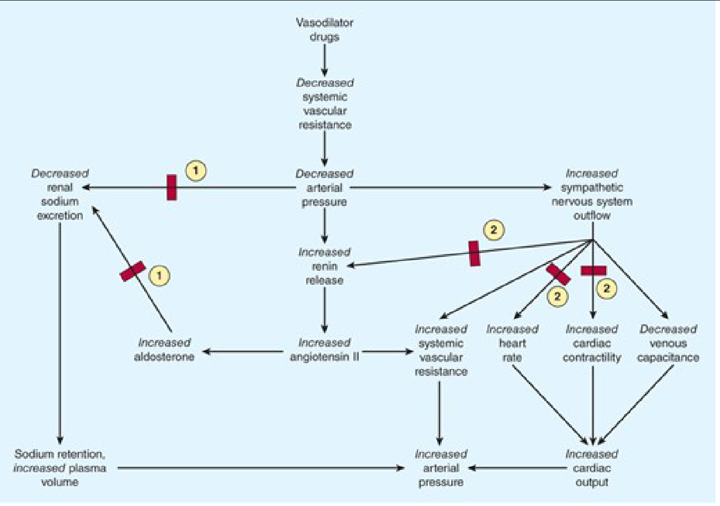
- Chronically (6-8 weeks): cardiac output normalizes, but peripheral vascular resistance decreases (primary sustained mechanism - related to reduced intracellular Ca²⁺ in vessel walls from altered Na⁺-Ca²⁺ exchange)
- Effective in lowering BP by 10-15 mmHg as monotherapy
| Effect | Details |
|---|---|
| Hypokalemia | Most common; increases arrhythmia risk (serious in elderly) |
| Hyperuricemia | Precipitates gout |
| Hyperglycemia | Worsens type 2 diabetes |
| Hyperlipidemia | ↑ LDL, triglycerides |
| Hyponatremia | Especially in elderly |
| Erectile dysfunction | |
| Hypercalcemia | (unlike loop diuretics which cause Ca²⁺ loss) |
B. Loop Diuretics
C. Potassium-Sparing Diuretics / Aldosterone Antagonists
- Spironolactone/Eplerenone: block mineralocorticoid (aldosterone) receptors → prevent Na⁺ retention and K⁺ loss
- Amiloride/Triamterene: block epithelial Na⁺ channels (ENaC) in collecting duct
II. Renin-Angiotensin-Aldosterone System (RAAS) Blockers
A. ACE Inhibitors (ACEIs)
- Block Angiotensin-Converting Enzyme (ACE/kininase II) which normally:
- Converts Angiotensin I → Angiotensin II (a potent vasoconstrictor + aldosterone stimulator)
- Degrades bradykinin (a vasodilator)
- Net effects: ↓ Angiotensin II → ↓ peripheral vascular resistance + ↓ aldosterone → ↓ Na⁺ retention
- Accumulation of bradykinin → additional vasodilation (but also causes cough and angioedema)
- No reflex tachycardia (unique advantage vs. direct vasodilators) - due to baroreceptor resetting and enhanced parasympathetic tone
- Most are prodrugs (enalapril → enalaprilat; lisinopril is active as given)
- Most excreted renally (reduce dose in renal failure except fosinopril, moexipril)
- Captopril: short-acting, requires 2-3x/day dosing; rest can be given once daily
- Diabetic nephropathy: Reduce glomerular efferent arteriolar resistance → ↓ intraglomerular pressure → reduce proteinuria and slow CKD progression (first-line even without hypertension in diabetics)
- Post-MI: Reduce mortality and prevent cardiac remodeling
- Heart failure: Cornerstone of management (↓ preload and afterload)
- Left ventricular hypertrophy: Regression of LVH
| Effect | Mechanism | Notes |
|---|---|---|
| Dry cough | Bradykinin accumulation | In ~15%; class effect; switch to ARB |
| Angioedema | Bradykinin accumulation | Rare but life-threatening; switch to ARB |
| Hyperkalemia | ↓ aldosterone | Especially in CKD, diabetes |
| Acute renal failure | Efferent arteriole dilation | In bilateral renal artery stenosis (contraindicated) |
| First-dose hypotension | Especially in hypovolemic patients (diuretic users) | |
| Teratogenicity | Fetal renal toxicity | Contraindicated in 2nd and 3rd trimester of pregnancy |
B. Angiotensin Receptor Blockers (ARBs)
- Block AT₁ receptors (Angiotensin II type 1) → prevent all actions of Angiotensin II
- Do NOT affect bradykinin → no cough or angioedema (major advantage over ACEIs)
- Same cardiovascular and renal protective benefits as ACEIs
Note: ACEIs and ARBs should NOT be combined - dual RAAS blockade increases adverse effects without additive benefit.
C. Direct Renin Inhibitor
III. Calcium Channel Blockers (CCBs)
- Block voltage-gated L-type Ca²⁺ channels in vascular smooth muscle and cardiac cells
- In vascular smooth muscle: ↓ Ca²⁺ entry → vasodilation → ↓ peripheral vascular resistance → ↓ BP
- Different drugs have different selectivity:
| Subclass | Drug | Selectivity | Effect on Heart | Notes |
|---|---|---|---|---|
| Dihydropyridines (DHP) | Amlodipine, Nifedipine, Felodipine | Predominantly vascular | Minimal cardiac depression; may cause reflex tachycardia | Preferred for hypertension, angina |
| Phenylalkylamines | Verapamil | Cardiac > vascular | Strong cardiac depression: ↓ HR, ↓ AV conduction, ↓ cardiac output | Used for rate control in AF; angina |
| Benzothiazepines | Diltiazem | Intermediate | Intermediate cardiac depression | Used for angina, rate control |
- Elderly patients (safe and effective)
- Isolated systolic hypertension
- Angina pectoris (dihydropyridines + non-DHPs)
- Vasospastic (Prinzmetal's) angina
- Atherosclerosis (CCBs preferred)
- Raynaud's phenomenon
| Drug | Adverse Effects |
|---|---|
| All DHPs | Peripheral edema (ankle), flushing, headache, reflex tachycardia (more with short-acting) |
| Verapamil | Constipation (most common), bradycardia, heart block, negative inotropy (avoid in heart failure), hypotension |
| Diltiazem | Bradycardia, heart block (less than verapamil), constipation |
| Nifedipine (short-acting) | Excessive reflex tachycardia, increased MI risk |
IV. Drugs Acting on the Sympathetic Nervous System
A. Beta-Adrenoceptor Blockers (β-Blockers)
- Non-selective (β1+β2): Propranolol, Nadolol, Timolol
- Cardioselective (β1 > β2): Metoprolol, Atenolol, Bisoprolol, Nebivolol
- Mixed α+β: Labetalol, Carvedilol
- ↓ Cardiac output via β1 blockade (↓ HR and contractility)
- ↓ Renin release from juxtaglomerular cells (β1 mediated) → ↓ RAAS activation → ↓ Angiotensin II
- Central sympatholytic effect (reduced CNS sympathetic outflow)
- Peripheral presynaptic β blockade → ↓ NE release
- Post-MI (reduce mortality)
- Heart failure (metoprolol, bisoprolol, carvedilol - reduce mortality)
- Angina pectoris
- Aortic dissection (labetalol)
- Hyperthyroidism-induced hypertension
- Hypertension during pregnancy (labetalol is drug of choice)
| Effect | Mechanism |
|---|---|
| Bronchospasm | β2 blockade in airways; contraindicated in asthma/COPD |
| Bradycardia / Heart block | β1 blockade |
| Worsening heart failure (acute) | Negative inotropy (but useful long-term) |
| Masking hypoglycemia symptoms | β2 blockade (tachycardia masked); caution in diabetics |
| Peripheral vasoconstriction | β2 blockade; worsens Raynaud's |
| Lipid abnormalities | ↑ Triglycerides, ↓ HDL |
| CNS effects | Fatigue, depression, nightmares (lipid-soluble propranolol) |
| Withdrawal syndrome | Rebound tachycardia, angina, hypertension on abrupt discontinuation - taper gradually |
B. Alpha-1 Adrenoceptor Blockers
- First-dose phenomenon: Severe orthostatic hypotension and syncope after first dose (especially prazosin) - give at bedtime with low initial dose
- Reflex tachycardia
- Fluid retention (use with diuretic)
- Nasal stuffiness
C. Central Sympatholytics
- Mechanism: Stimulates α2 receptors (and imidazoline receptors) in the brainstem vasomotor centers → ↓ sympathetic outflow → ↓ HR and BP; retains baroreceptor sensitivity
- Uses: Hypertension (oral); hypertensive urgency (oral/TTS patch); opioid/alcohol withdrawal; ADHD
- Adverse Effects: Sedation, dry mouth, bradycardia, depression
- Critical: Abrupt withdrawal causes rebound hypertension (sometimes severe - hypertensive crisis) - taper slowly
- Mechanism: Prodrug → α-methyldopamine → α-methylnorepinephrine (false transmitter) → stimulates central α2 receptors → ↓ sympathetic outflow
- Uses: Drug of choice for hypertension in pregnancy (proven safety record); also used in elderly
- Adverse Effects: Sedation, dry mouth, positive Coombs test (hemolytic anemia in ~1%), hepatotoxicity, drug fever, retroperitoneal fibrosis (rare)
V. Direct Vasodilators
Hydralazine
- Mechanism: Direct arteriolar smooth muscle relaxation (exact mechanism unclear; may involve NO release and inhibition of inositol trisphosphate-induced Ca²⁺ release)
- Dilates arterioles >> venules → ↓ peripheral resistance
- Uses: Moderate-severe hypertension (usually with β-blocker + diuretic); hypertension in pregnancy (IV for hypertensive emergencies); heart failure (hydralazine + nitrate combination)
- Adverse Effects:
- Reflex tachycardia (must combine with β-blocker)
- Fluid retention (combine with diuretic)
- Drug-induced SLE (lupus-like syndrome) - dose-related; more in slow acetylators; presents with joint pains, rash, pleuritis
- Headache, flushing, palpitations
Minoxidil
- Mechanism: Opens K⁺-ATP channels in vascular smooth muscle → hyperpolarization → Ca²⁺ channel closure → relaxation → vasodilation
- Most powerful oral vasodilator available
- Uses: Severe, refractory hypertension unresponsive to other agents
- Adverse Effects:
- Hypertrichosis (hirsutism) - abnormal hair growth (basis for topical use as Rogaine® for baldness)
- Severe fluid retention and edema (requires loop diuretic)
- Reflex tachycardia (requires β-blocker)
- Pericardial effusion (with prolonged use)
Sodium Nitroprusside (IV only)
- Mechanism: Releases NO (nitric oxide) → activates guanylyl cyclase → ↑ cGMP → vascular smooth muscle relaxation → dilates both arteries and veins
- Uses: Hypertensive emergencies (ICU only); aortic dissection; acute heart failure
- Adverse Effects: Cyanide toxicity (nitroprusside metabolized to cyanide) - limit duration; give thiosulfate to prevent; tachycardia, hypotension
Diazoxide (IV)
- Opens K⁺-ATP channels (similar to minoxidil) → arteriolar dilation
- Also inhibits insulin secretion → hyperglycemia
- Used in hypertensive emergencies
VI. Comparative Summary Table
| Drug Class | Mechanism | ↓ CO | ↓ PVR | Key ADR | Compelling Indications |
|---|---|---|---|---|---|
| Thiazide diuretics | ↓ Na⁺/volume | Initial; then↓PVR | Yes | Hypokalemia, gout, hyperglycemia | Elderly, isolated systolic HTN, Black patients |
| ACE Inhibitors | ↓ Ang II, ↑ bradykinin | No | Yes | Cough, angioedema, hyperK⁺ | Diabetes, CKD, heart failure, post-MI |
| ARBs | Block AT₁ receptor | No | Yes | HyperK⁺; NO cough | Same as ACEI; ACEI intolerance |
| Calcium channel blockers (DHP) | ↓ Ca²⁺ in VSM | No | Yes | Edema, flushing, reflex tachycardia | Elderly, angina, atherosclerosis |
| Beta-blockers | ↓ HR/contractility, ↓ renin | Yes | No | Bronchospasm, fatigue, masking hypoglycemia | Post-MI, heart failure, angina |
| Alpha-1 blockers | Block α1 VSM | No | Yes | First-dose hypotension, fluid retention | BPH + HTN |
| Clonidine | Central α2 agonist | Yes | Yes | Sedation, rebound HTN on withdrawal | Opioid withdrawal, pregnancy |
| Methyldopa | False transmitter - central α2 | Yes | Yes | Sedation, positive Coombs, hepatitis | Drug of choice in pregnancy |
| Hydralazine | Direct arteriolar dilation | No | Yes | Drug-induced SLE, reflex tachycardia | Pregnancy hypertension (IV) |
| Minoxidil | Opens K⁺-ATP channels | No | Yes | Hirsutism, fluid retention | Resistant hypertension |
| Sodium nitroprusside | NO → ↑ cGMP | No | Yes | Cyanide toxicity | Hypertensive emergencies |
VII. Drug Choice in Special Situations
| Clinical Condition | Preferred Drug(s) | Drugs to Avoid |
|---|---|---|
| Diabetes mellitus | ACEI or ARB (renoprotective) | Thiazides (hyperglycemia), β-blockers (mask hypoglycemia) |
| CKD / Proteinuria | ACEI or ARB | NSAIDs |
| Heart failure | ACEI/ARB + β-blocker + diuretic + MRA | CCBs (verapamil/diltiazem - negative inotropy) |
| Post-MI | β-blocker + ACEI/ARB | - |
| Angina pectoris | β-blocker or CCB | Hydralazine (reflex tachycardia worsens angina) |
| Atrial fibrillation (rate control) | β-blocker or non-DHP CCB (verapamil/diltiazem) | - |
| BPH + Hypertension | α1-blocker (doxazosin) | - |
| Pregnancy | Methyldopa (1st line), Labetalol, Hydralazine, Nifedipine | ACEIs/ARBs (teratogenic - CONTRAINDICATED) |
| Black patients | Thiazide or CCB | ACEIs/ARBs (less effective as monotherapy) |
| Elderly | Thiazide, CCB, ACEI/ARB | High-dose β-blockers (falls risk) |
| Hypertensive emergency | IV Sodium nitroprusside, IV Labetalol, IV Nicardipine | - |
| Pheochromocytoma | Phenoxybenzamine (α-blocker first), then β-blocker | β-blocker ALONE (hypertensive crisis from unopposed α stimulation) |
| Resistant HTN | Add spironolactone (MRA) | - |
| Aortic dissection | IV Labetalol or β-blocker + nitroprusside | Vasodilators alone (reflex tachycardia increases aortic shear stress) |
VIII. Treatment Principles
- BP target: <130/80 mmHg (AHA/ACC 2017); <140/90 mmHg (many other guidelines)
- Lifestyle modifications first: sodium restriction, weight loss, exercise, alcohol reduction, DASH diet
- First-line drug classes: ACEIs/ARBs, CCBs, thiazide diuretics (all equivalent for most patients without compelling indications)
- Combination therapy: Most patients with stage 2 HTN (>160/100) require 2 or more drugs from different classes
- Do NOT combine: ACEI + ARB (dual RAAS blockade increases harm)
- Once-daily dosing preferred for compliance (trough-peak ratio >50%)
- Blood pressure lowering itself is the most important therapeutic goal - no one class is clearly superior when BP is equivalently reduced
Megaloblastic anemia pathology
Megaloblastic Anemia - Pathology
Definition
Etiology and Classification
| Cause | Mechanism | Examples |
|---|---|---|
| Vitamin B12 (cobalamin) deficiency | Decreased intake | Strict vegetarian/vegan diet (B12 only in animal products) |
| Impaired absorption (intrinsic factor deficiency) | Pernicious anemia (autoimmune), gastrectomy | |
| Malabsorption states | Diffuse intestinal disease, ileal resection/ileitis | |
| Competitive uptake | Fish tapeworm (Diphyllobothrium latum), bacterial overgrowth | |
| Inactivation | Nitrous oxide (recreational use) | |
| Folic acid deficiency | Decreased intake | Alcoholism, malnutrition, infancy |
| Impaired absorption | Malabsorption syndromes, anticonvulsants, oral contraceptives | |
| Increased loss | Hemodialysis | |
| Increased requirement | Pregnancy, infancy, disseminated cancer, hemolytic anemia | |
| Impaired utilization | Methotrexate, trimethoprim (folate antagonists) | |
| Unresponsive to B12/folate | Metabolic inhibitors | Hydroxyurea, 5-fluorouracil, cytosine arabinoside (inhibit DNA synthesis directly) |
Biochemical Pathogenesis
The Core Problem: DNA Synthesis Failure

Normal Pathway
FOOD (B12-containing)
↓ [Pepsin releases B12]
B12 + Salivary haptocorrin
↓ [Pancreatic proteases in duodenum release B12 from haptocorrin]
B12 + INTRINSIC FACTOR (from gastric parietal cells)
↓ [B12-IF complex binds CUBILIN receptor in terminal ileum]
B12 absorbed → binds TRANSCOBALAMIN II in ileal cells
↓ [Delivered to liver and bone marrow]
METHYLCOBALAMIN (active B12)
↓ [cofactor for METHIONINE SYNTHASE]
N5-methyl FH4 → FH4 (tetrahydrofolate - active folate)
↓
N5,10-methylene FH4 (via FH4 derivatives)
↓ [Thymidylate synthetase]
dUMP → dTMP (deoxythymidine monophosphate)
↓
DNA SYNTHESIS
In B12 Deficiency: "Folate Trap"
↓ Methylcobalamin → Methionine synthase BLOCKED
↓
N5-methyl FH4 CANNOT be converted to FH4
↓
Folate is TRAPPED as N5-methyl FH4
↓
↓ FH4 → ↓ N5,10-methylene FH4
↓
Thymidylate synthetase STARVED of substrate
↓
↓ dTMP → ↓ DNA synthesis → MEGALOBLASTOSIS
"Folate trap hypothesis" - in B12 deficiency, folate accumulates in a form the cell cannot use, functionally mimicking folate deficiency. This explains why folate administration partially corrects the anemia of B12 deficiency but NOT the neurological complications.
Two Reactions Requiring B12
| Reaction | Enzyme | Cofactor | Clinical Consequence of Deficiency |
|---|---|---|---|
| Homocysteine → Methionine | Methionine synthase | Methylcobalamin | ↓ FH4 → impaired DNA synthesis → megaloblastosis; ↑ serum homocysteine |
| Methylmalonyl-CoA → Succinyl-CoA | Methylmalonyl-CoA mutase | Adenosylcobalamin | ↑ methylmalonic acid and propionate → abnormal fatty acid synthesis → myelin breakdown → subacute combined degeneration of spinal cord |
Critical distinction: The neurological damage from B12 deficiency is NOT corrected by folate (and may be worsened). Folate supplementation without B12 can mask hematological findings while neuropathy progresses unchecked.
In Folate Deficiency
↓ Dietary folate (or impaired absorption/increased demand)
↓
↓ FH4 → ↓ N5,10-methylene FH4
↓
↓ dTMP → ↓ DNA synthesis → MEGALOBLASTOSIS
↓
No neurological complications (folate not needed for myelin)
Morphology
1. Peripheral Blood Smear
| Finding | Description | Significance |
|---|---|---|
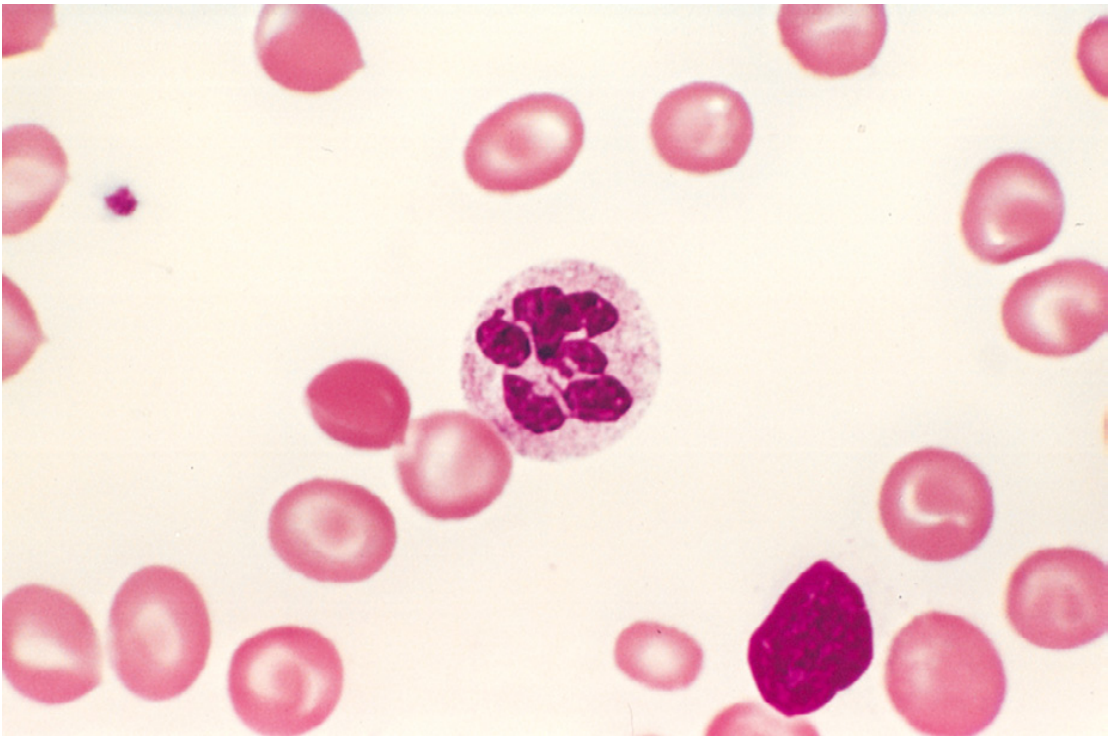
| Macro-ovalocytes (macro-ovalocytes) | Large, oval red cells (MCV >100 fL); lack central pallor; "hyperchromic" appearance (though MCHC normal) | Most characteristic RBC finding |
| Hypersegmented neutrophils | Neutrophils with ≥5 nuclear lobes (normally 3-4); a neutrophil with ≥1 six-lobed nucleus is virtually diagnostic | Most specific and early finding; precedes anemia |
| Anisocytosis | Marked variation in red cell size | Reflects ineffective erythropoiesis |
| Poikilocytosis | Variation in red cell shape | |
| Low reticulocyte count | Reflects ineffective hematopoiesis | |
| Pancytopenia | ↓ RBCs, ↓ WBCs, ↓ platelets | All rapidly dividing cell lines affected |
| Nucleated RBC precursors | Occasionally seen in severe anemia |
2. Bone Marrow (Aspirate)
| Finding | Description |
|---|---|
| Hypercellularity | Compensatory increase in hematopoietic precursors (driven by ↑ erythropoietin) - paradoxically increased cellularity despite anemia |
| Megaloblasts | Abnormally large erythroid precursors at all stages of development: promegaloblasts, basophilic megaloblasts, polychromatophilic megaloblasts, orthochromatic megaloblasts |
| Nuclear-cytoplasmic asynchrony (dissociation) | Key diagnostic feature: cytoplasm matures normally (accumulates hemoglobin) while nucleus remains immature with fine, open ("lacy") chromatin - does NOT condense to the expected pyknotic clump |
| Giant metamyelocytes and band forms | Dysmaturation of granulocytic precursors due to impaired DNA synthesis |
| Abnormal megakaryocytes | Large, hyperlobated megakaryocyte nuclei |
| Ineffective hematopoiesis | Despite hypercellularity, most precursors undergo apoptosis in the marrow before maturation → paradoxical pancytopenia |
The key paradox: Bone marrow is hyperactive (hypercellular) yet peripheral blood counts are low (pancytopenia). This is the hallmark of ineffective hematopoiesis.
3. Hemolysis
- A mild degree of intramedullary and peripheral hemolysis compounds the anemia
- Mechanism: the fragile, abnormal megaloblasts are destroyed by macrophages in the marrow and spleen before they can mature
- Results in ↑ unconjugated bilirubin, ↑ LDH (very high), ↓ haptoglobin
Pernicious Anemia (Specific B12 Deficiency Subtype)
Autoimmune Pathogenesis
Autoreactive T cells attack gastric parietal cells
↓
Chronic atrophic gastritis (fundic gland atrophy)
↓
Loss of parietal cells → loss of intrinsic factor + HCl
↓
Three types of autoantibodies:
Type I (75%) - blocks B12 binding to IF
Type II (large proportion) - blocks IF-B12 complex from binding cubilin
Type III (85-90%) - against α/β subunits of gastric proton pump (H+/K+ ATPase)
↓
↓ IF → B12 not absorbed from terminal ileum
↓
Gradual depletion of hepatic B12 stores (takes years - stores last 3-5 years)
↓
Megaloblastic anemia + Subacute combined degeneration
Morphology of Gastric Lesion
- Diffuse chronic atrophic gastritis of the fundus/body
- Loss of parietal cells (which secrete IF and HCl) and chief cells
- Replacement by mucus-secreting goblet cells: intestinal metaplasia (intestinalization)
- Megaloblastic changes in mucosal epithelial cells
- Dense lymphocyte and plasma cell infiltrate in lamina propria
- Important: Gastric changes persist after B12 replacement; marrow changes reverse rapidly
Neurological Lesion: Subacute Combined Degeneration (SCD)
- Seen in ~75% of florid pernicious anemia cases
- Mechanism: ↑ methylmalonyl-CoA → abnormal fatty acid synthesis → incorporated into neuronal lipids → myelin breakdown
- Pathology: Demyelination of posterior (dorsal) and lateral columns of spinal cord
- Clinical:
- Posterior column: sensory ataxia, loss of vibration and proprioception, positive Romberg sign
- Lateral column (corticospinal tracts): spastic paraparesis, extensor plantars
- Peripheral neuropathy: symmetric distal paresthesias ("glove and stocking")
- Cognitive changes, dementia, optic neuropathy
- Critical: Neurological damage may be IRREVERSIBLE if untreated; duration and severity before treatment inversely correlates with recovery
Clinical Features
Hematological
- Insidious onset; often severe anemia before diagnosis
- Pallor, fatigue, weakness, dyspnea
- Mild jaundice (from intramedullary hemolysis)
- Palpitations, heart failure in severe cases
Gastrointestinal
- Atrophic glossitis: tongue becomes smooth, shiny, "beefy red" with loss of papillae (Hunter's glossitis)
- Anorexia, nausea, weight loss
- Diarrhea from megaloblastic changes in intestinal epithelium
Neurological (B12 deficiency only, NOT folate)
- Bilateral symmetric distal paresthesias (tingling, numbness - earliest symptom)
- Loss of vibration sense and proprioception (posterior column dysfunction)
- Unsteady gait, sensory ataxia
- Spastic paraparesis, extensor plantars (lateral column)
- Lhermitte's sign (electric shock sensation down spine on neck flexion)
- Cognitive impairment, dementia, behavioral changes
- Neuropsychiatric manifestations can precede anemia
Laboratory Diagnosis
| Test | Finding |
|---|---|
| CBC | ↓ Hemoglobin; ↑ MCV (macrocytosis, MCV >100 fL, often >115); ↓ WBC, ↓ platelets (pancytopenia) |
| Peripheral blood smear | Macro-ovalocytes, hypersegmented neutrophils (≥5 lobes), anisocytosis, poikilocytosis |
| Reticulocyte count | Low (inappropriate for degree of anemia) |
| Serum B12 | ↓ (<200 pg/mL) in B12 deficiency |
| Serum folate | ↓ in folate deficiency; RBC folate is more reliable (reflects tissue stores) |
| Serum LDH | Markedly elevated (intramedullary destruction of megaloblasts - one of highest LDH levels in medicine) |
| Serum bilirubin | ↑ Unconjugated (mild - hemolysis) |
| Serum homocysteine | ↑ in BOTH B12 AND folate deficiency |
| Serum methylmalonic acid (MMA) | ↑ in B12 deficiency ONLY (not folate) - most specific |
| Schilling test (historical) | Distinguishes pernicious anemia from other B12 deficiency causes by testing B12 absorption with and without IF |
| Anti-intrinsic factor antibodies (Type I) | Specific for pernicious anemia (75% sensitivity) |
| Anti-parietal cell antibodies (Type III) | Sensitive (85-90%) but not specific (also in idiopathic gastritis) |
| Bone marrow aspirate | Hypercellular; megaloblasts at all stages; giant metamyelocytes; nuclear-cytoplasmic asynchrony |
| Serum gastrin | ↑ (from loss of acid feedback inhibition - achlorhydria) in pernicious anemia |
Distinguishing B12 from Folate Deficiency
| Feature | B12 Deficiency | Folate Deficiency |
|---|---|---|
| Neurological signs | YES - subacute combined degeneration | NO |
| ↑ Methylmalonic acid | YES | NO |
| ↑ Homocysteine | YES | YES |
| Low serum B12 | YES | Normal |
| Low serum/RBC folate | Normal | YES |
| Response to folate alone | Anemia corrects, neuro worsens | Full correction |
Treatment
| Condition | Treatment |
|---|---|
| Pernicious anemia | IM Cyanocobalamin 1000 μg/day × 7 days, then weekly × 4, then monthly for life (oral B12 1000-2000 μg/day also effective via passive diffusion - avoids injections) |
| Dietary B12 deficiency (vegetarians) | Oral B12 supplementation |
| Folate deficiency | Oral folic acid 1-5 mg/day × 4 months (or lifetime if cause persists) |
| Response to treatment | Reticulocytosis within 5-7 days; hemoglobin normalizes in 1-2 months; thrombocytopenia and leukopenia correct within 2 weeks; neurological improvement variable |
| Important: | Never give folate alone in undifferentiated macrocytic anemia without first ruling out B12 deficiency - folate will correct anemia but allow neurological damage to progress undetected |
1 year male child was brought with loss of vision and mass lesion of the eyeball. There was a family H/o sarcoma of the long bone in the sibling. What is your diagnosis? What is the molecular mechanism involved in this family. Describe the normal cell cycle and how this gene affects cell cycle, Name 3 other genes involved in familial syndromes.