
Veins histology description with pictures
vein histology tunica intima media adventitia histology reference
histology micrograph vein wall cross section tunica intima media adventitia valve
veins histology
Veins: histological description
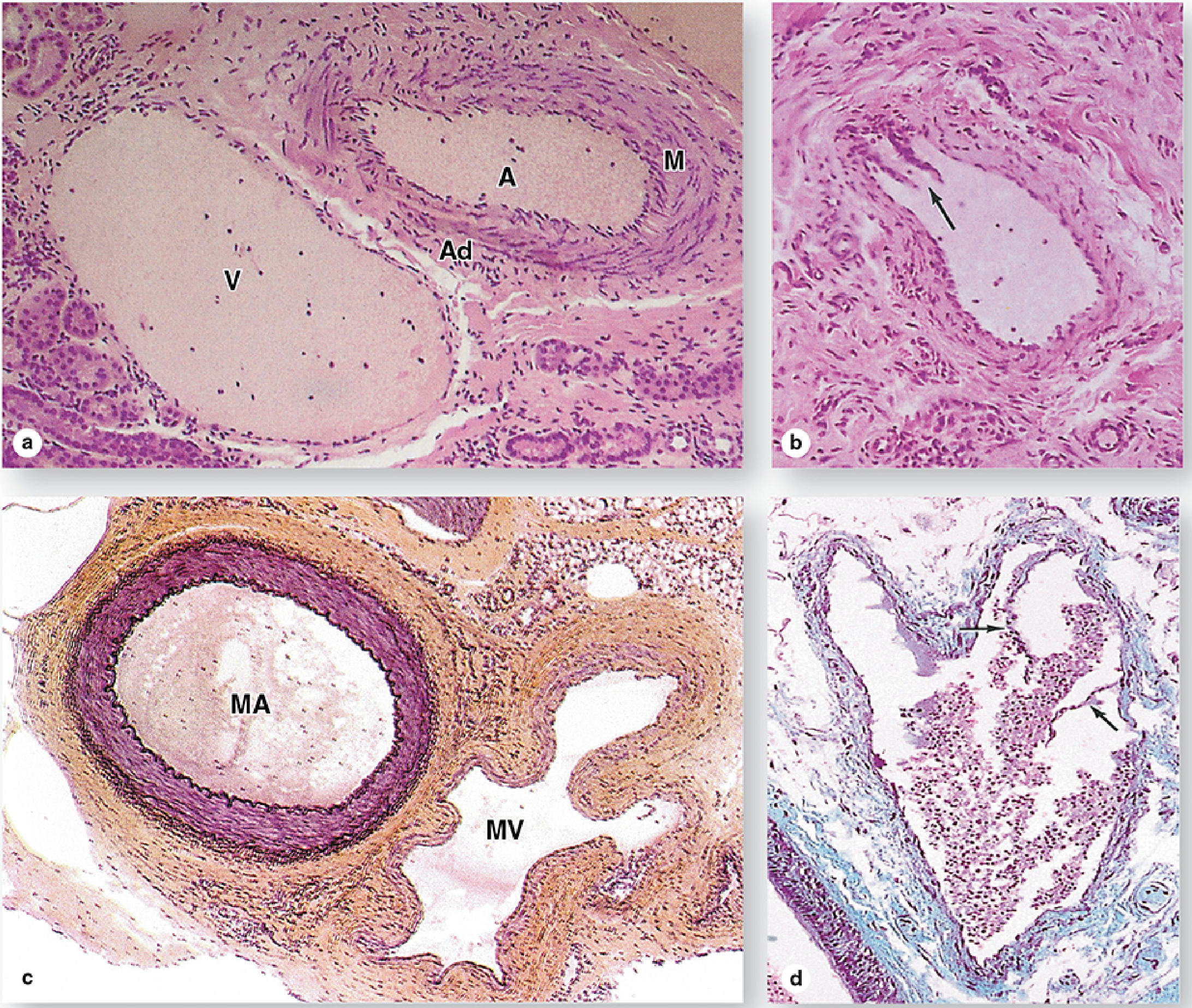
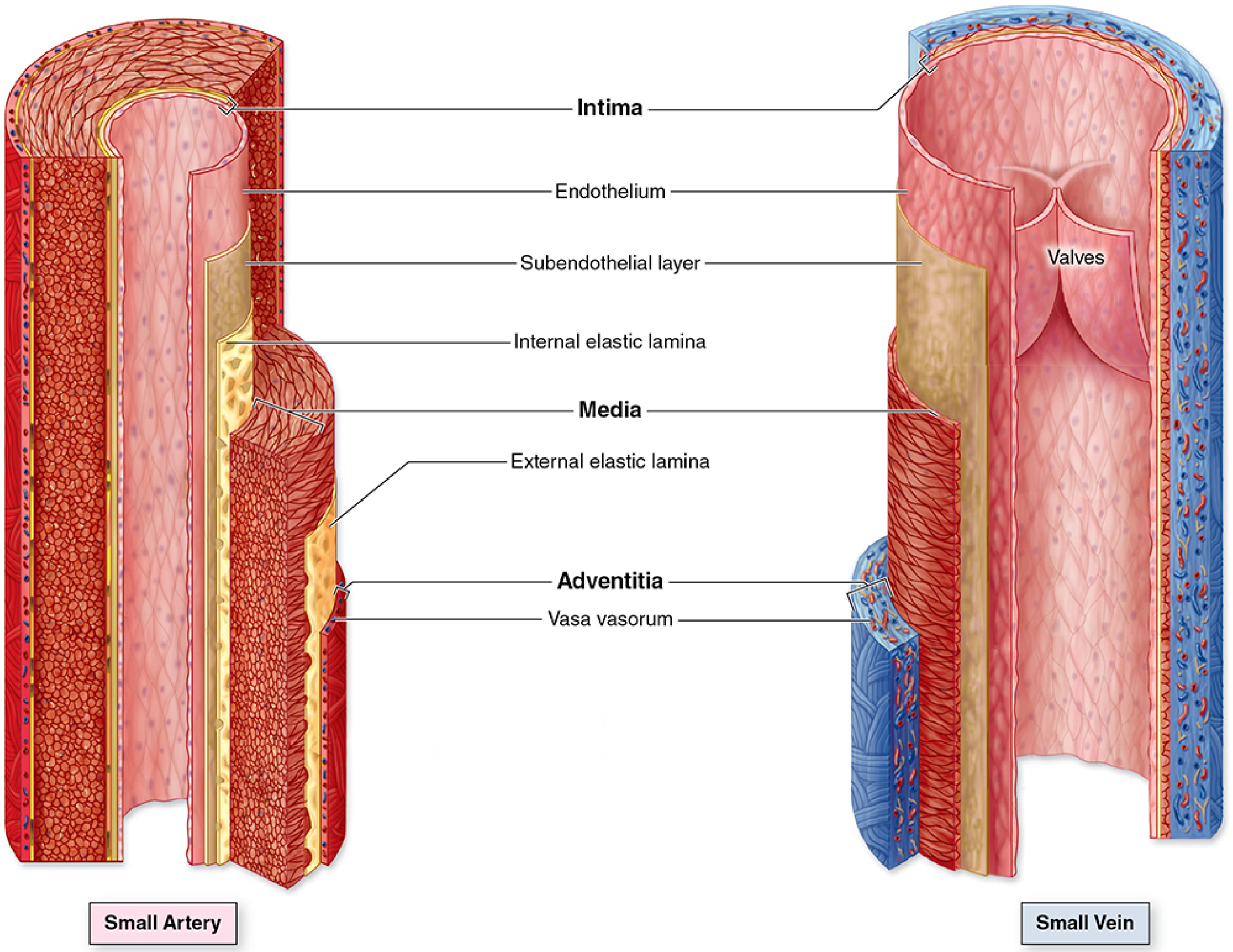
Layers of the venous wall
| Layer | Histological features |
|---|---|
| Tunica intima | Very thin. It consists of a simple squamous endothelial lining, its basal lamina, and scant subendothelial connective tissue. The internal elastic lamina is usually inconspicuous or absent. |
| Tunica media | Much thinner than in an artery. It contains relatively few circular smooth muscle cells, with collagen, reticular fibers, and delicate elastic fibers. |
| Tunica adventitia | Usually the thickest coat, especially in large veins. It is collagen-rich connective tissue with elastic fibers, fibroblasts, nerves, and vasa vasorum. In large veins, it often has prominent longitudinal smooth-muscle bundles. |
Venous valves
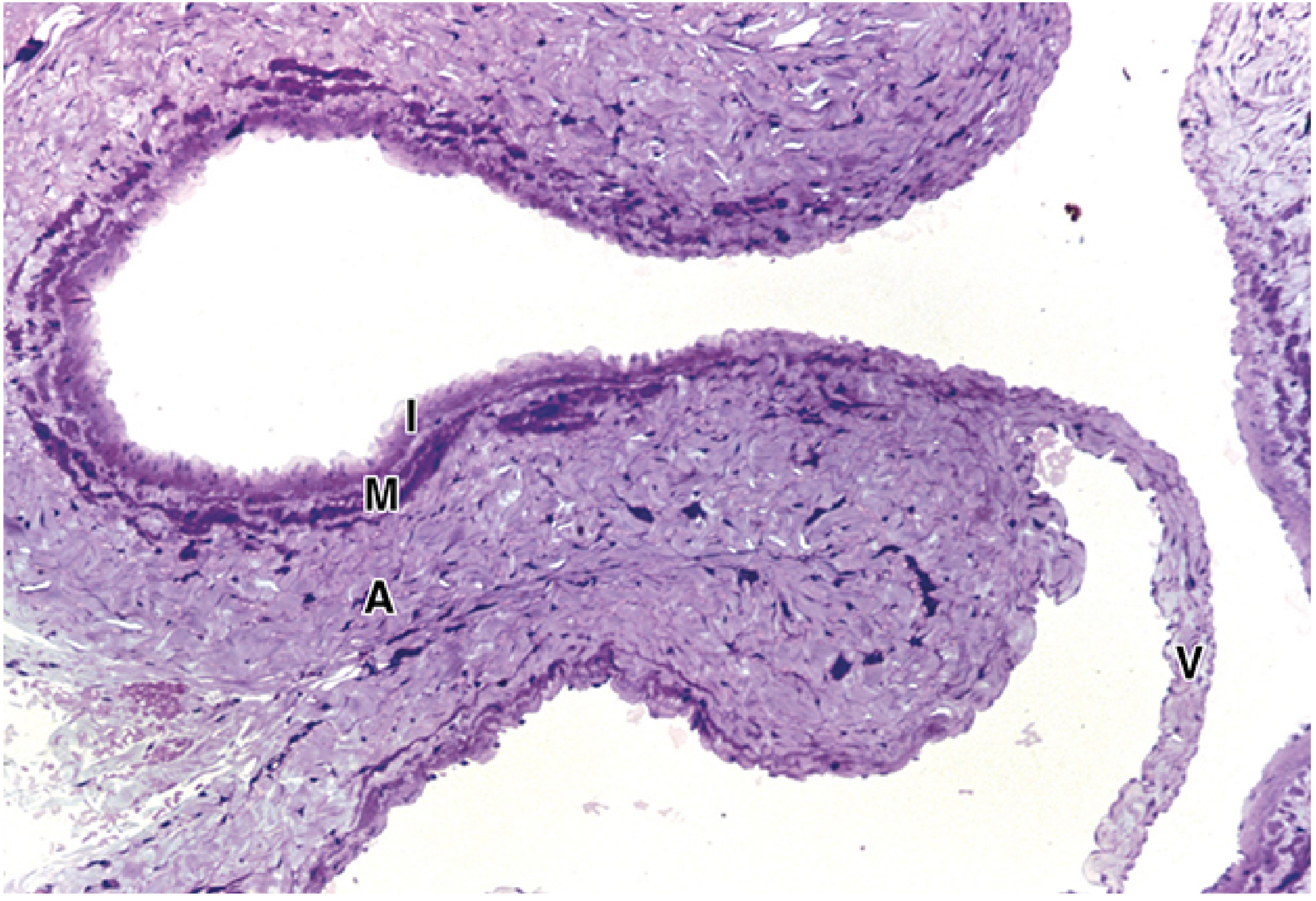
Types of veins
- Postcapillary venules: Endothelium with basal lamina and pericytes. Important site of leukocyte migration and fluid leakage during inflammation.
- Muscular venules: Have 1-2 layers of smooth muscle, forming an early tunica media.
- Small veins: Usually less than 1 mm diameter; thin wall with only a few smooth-muscle layers.
- Medium veins: Most named veins; have all three tunics but a comparatively thin media and relatively thick adventitia.
- Large veins: Examples include the venae cavae and portal vein. They have a thin media and especially thick adventitia containing longitudinal smooth muscle. - Histology: A Text and Atlas with Correlated Cell and Molecular Biology, pp. 1110-1112
Vein versus artery in histology
| Feature | Vein | Artery |
|---|---|---|
| Lumen | Wide, irregular, often collapsed | Smaller, rounder, usually patent |
| Wall | Thin | Thick |
| Tunica media | Thin, few smooth-muscle layers | Thick, many smooth-muscle layers |
| Adventitia | Often thickest layer | Usually less prominent than media in muscular arteries |
| Elastic laminae | Poorly defined | Usually distinct, especially internal elastic lamina |
| Valves | May be present | Absent |
Heart histology
heart histology myocardium endocardium epicardium cardiac muscle histology NCBI
histology heart wall endocardium myocardium epicardium cardiac muscle intercalated discs micrograph
cardiac histology
Heart histology
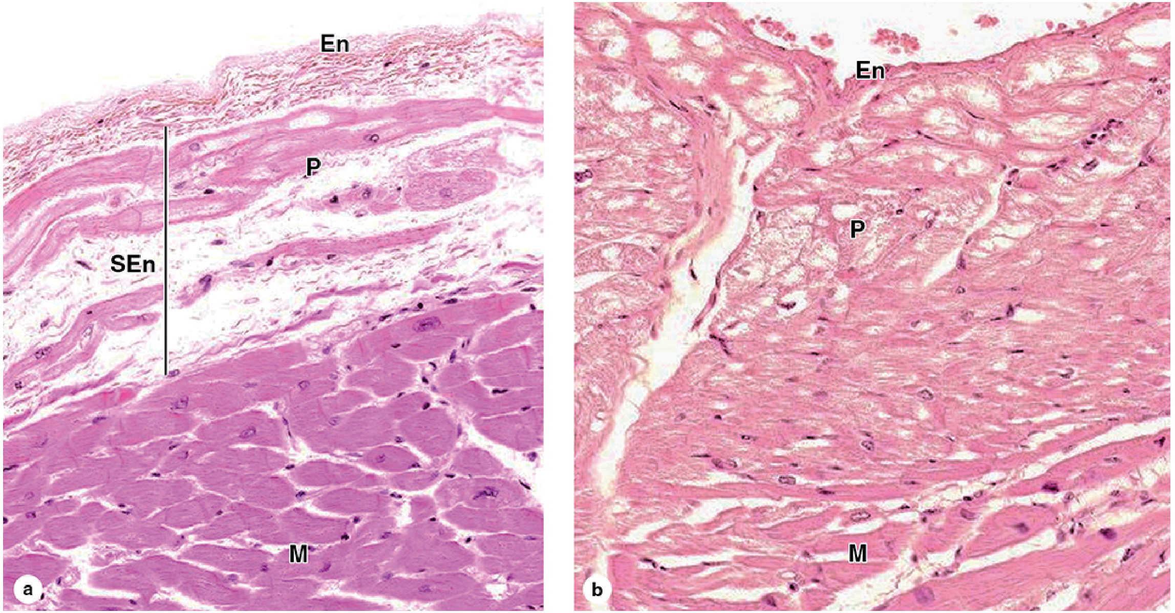
1. Endocardium
- Endothelium: simple squamous epithelium facing the blood.
- Subendothelial fibroelastic connective tissue: collagen and elastic fibers, with occasional smooth muscle cells.
- Subendocardial layer: loose connective tissue continuous with the myocardium. It contains blood vessels, nerves, and in the ventricles, Purkinje fibers.
2. Myocardium
- Short, cylindrical, and branched
- One central nucleus, occasionally two
- Striated, though striations may be less obvious than in skeletal muscle
- Connected end to end by dark transverse lines called intercalated discs
- Rich in capillaries and connective-tissue support
- Fascia adherens: mechanical attachment of actin filaments
- Desmosomes: prevent cells from pulling apart during contraction
- Gap junctions: permit rapid ionic and electrical communication, so the myocardium functions as a coordinated unit
3. Epicardium
- Surface mesothelium: simple squamous epithelium
- Underlying loose connective tissue
- Variable adipose tissue, especially in grooves of the heart
- Large coronary arteries, cardiac veins, lymphatics, and nerves
Histological identification of cardiac muscle
| Feature | Cardiac muscle |
|---|---|
| Fibers | Short and branched |
| Nuclei | One, centrally located, sometimes two |
| Striations | Present |
| Intercalated discs | Present, dark transverse lines |
| Control | Involuntary |
| Connective tissue | Rich capillary network between fibers |
Cardiac valves
- Fibrosa: dense collagen, provides strength
- Spongiosa: loose connective tissue rich in proteoglycans, acts as a shock absorber
- Ventricularis in semilunar valves, or atrialis in atrioventricular valves: elastic-fiber-rich layer
Quick slide answer
Veins deep histology about every layer fiber tissue cells types description funtion dif from artert picture
labeled histology comparison muscular artery and medium vein tunica intima media adventitia H and E
vein histology layers tunica intima media adventitia artery comparison source
venous wall histology
Veins: deep histology
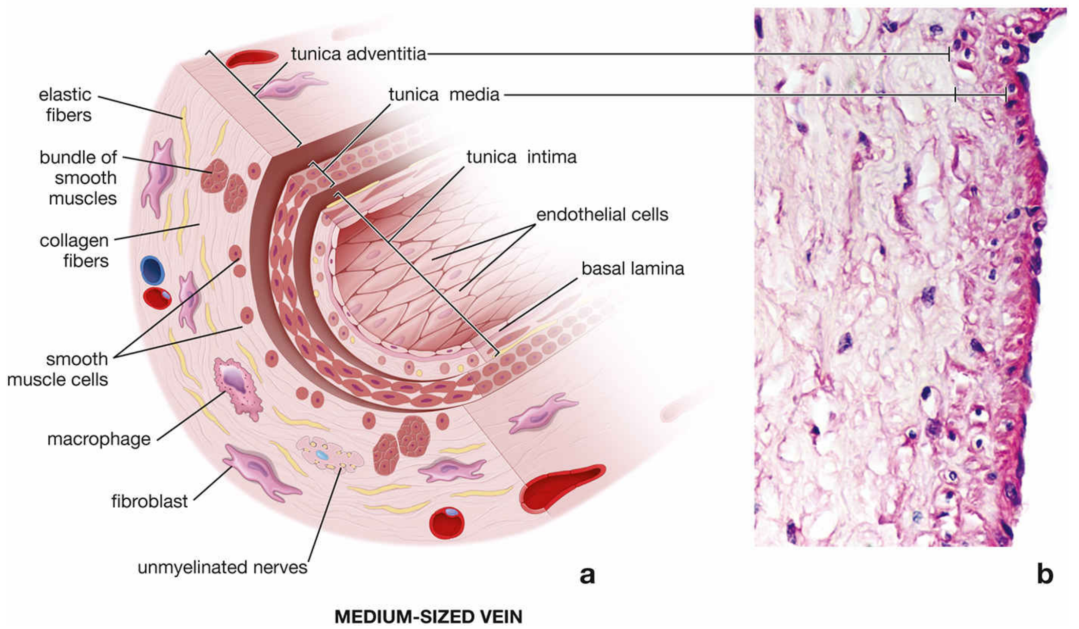
Overall structure: LIMA
- Lumen
- Intima
- Media
- Adventitia
1. Tunica intima
Components
- Endothelium: one layer of flat, simple squamous endothelial cells facing the blood.
- Basal lamina beneath the endothelial cells.
- A very thin subendothelial connective-tissue layer.
- Occasional smooth-muscle cells within the subendothelial connective tissue.
- A thin, discontinuous internal elastic membrane may occur, but is usually inconspicuous.
Cells and fibers
| Component | Description and role |
|---|---|
| Endothelial cells | Flat cells aligned in the direction of blood flow. They form a smooth, non-thrombogenic surface and regulate permeability, coagulation, leukocyte movement, and vascular tone. |
| Fibroblasts | Sparse connective-tissue cells that produce extracellular matrix. |
| Smooth-muscle cells | Occasional in the deeper intima of larger veins; contribute modestly to wall tone. |
| Collagen fibers | Provide tensile support. |
| Elastic fibers | Present in small quantity; allow limited stretch and recoil. |
Function
- Provides a smooth blood-contacting surface.
- Limits inappropriate clot formation.
- Acts as a selective barrier between blood and the vessel wall.
- Forms venous valves in many medium-sized and large veins.
Venous valves
2. Tunica media
Components
- Usually only a few layers of circular or spiral smooth-muscle cells
- Collagen fibers
- Fine elastic fibers
- In some medium veins, a few longitudinal smooth-muscle cells near the outer part of the media
Cells and fibers
| Component | Histological appearance | Function |
|---|---|---|
| Smooth-muscle cells | Elongated cells with central cigar-shaped nuclei | Mild vasoconstriction and vasodilation; helps regulate venous capacitance and return |
| Collagen | Pale eosinophilic extracellular material in H&E sections | Prevents overdistension |
| Elastic fibers | Fine, less abundant than in arteries | Allows distension and limited recoil |
| Fibroblasts | Scattered spindle-shaped nuclei | Produce and maintain extracellular matrix |
Function
3. Tunica adventitia
Components
- Abundant type I collagen fibers
- A network of elastic fibers
- Numerous fibroblasts
- In large veins, conspicuous longitudinal bundles of smooth muscle
- Vasa vasorum, small vessels that nourish the outer wall of very large veins
- Nervi vasorum, autonomic nerve fibers that influence smooth muscle and venous tone
- Sometimes adipose tissue in the surrounding connective tissue
Function
- Anchors the vein to adjacent structures.
- Resists excessive distension and tearing.
- Accommodates autonomic nerves and small nourishing vessels.
- In large veins, longitudinal smooth muscle helps support the wall and assists venous return.
Histology by size of vein
| Vessel | Main histological features |
|---|---|
| Postcapillary venule | Endothelium, basal lamina, and pericytes. No true muscular media. Principal site of leukocyte migration during inflammation. |
| Muscular venule | 1-2 layers of smooth muscle form a thin media. |
| Small vein | Diameter about 0.1-1 mm; all three tunics identifiable. Media has 2-3 smooth-muscle layers; adventitia is relatively thick. |
| Medium vein | Most named deep veins. Thin intima, few smooth-muscle layers in media, thick collagenous adventitia. Valves often present. |
| Large vein | Examples: venae cavae, portal vein. Thin media; adventitia is very thick and has longitudinal smooth-muscle bundles. |
- Histology: A Text and Atlas with Correlated Cell and Molecular Biology, pp. 1111-1116
Vein versus artery: histological differences
| Feature | Vein | Muscular artery |
|---|---|---|
| Blood pressure | Low | High |
| Lumen | Wide, irregular, often collapsed in slides | Smaller, round, usually remains open |
| Wall | Thin relative to lumen | Thick relative to lumen |
| Tunica intima | Thin; internal elastic lamina poorly defined or absent | More distinct; prominent internal elastic lamina |
| Tunica media | Thin, few smooth-muscle layers; little elastin | Thick, many circular smooth-muscle layers; more elastic tissue |
| Tunica adventitia | Usually thicker than media | Usually thinner than media |
| Valves | Present in many medium-sized limb veins | Absent |
| Main function | Blood reservoir and low-pressure return to the heart | Distribution of blood under high pressure; regulation of tissue perfusion |
| Shape in section | Collapsed, flattened, or irregular | Round or oval with a thick wall |
Why these differences exist
How to identify a vein on an H&E slide
- Large irregular lumen
- Thin wall relative to lumen diameter
- Thin media with few smooth-muscle layers
- Adventitia thicker than media
- No prominent wavy internal elastic lamina
- Possible valve leaflet projecting into lumen
- An accompanying artery nearby with a thicker, more circular muscular wall
Short exam answer
More deep about veins
venous wall histology endothelium venous valves vasa vasorum review
venous wall histology
Veins: deeper histology and functional anatomy
1. General microscopic appearance
- Wide, irregular or collapsed lumen
- Relatively thin wall compared with lumen diameter
- Thin tunica media
- Thick tunica adventitia, often the largest layer
- Poorly defined internal elastic lamina
- Sometimes an intimal fold forming a valve
- Often found beside an artery in a neurovascular bundle
2. Tunica intima in depth
- Endothelium
- Basement membrane
- Thin subendothelial connective tissue
- Occasional smooth-muscle cells in larger veins
- Sometimes a delicate, discontinuous internal elastic lamina
A. Endothelium
Endothelial cell functions
| Function | Explanation |
|---|---|
| Antithrombotic surface | Healthy endothelium prevents platelets and clotting factors from forming an unnecessary thrombus. |
| Selective barrier | Controls movement of water, solutes, proteins, and cells between blood and tissue. |
| Leukocyte recruitment | In inflammation, endothelial cells express adhesion molecules that allow white blood cells to roll, adhere, and migrate out of the blood. |
| Control of tone | Releases substances that influence underlying smooth muscle and venous diameter. |
| Repair and signaling | Participates in vessel remodeling, inflammation, and formation of new vessels. |
B. Subendothelial layer
- Fine collagen fibers
- Sparse elastic fibers
- Fibroblasts
- Occasional smooth-muscle cells, particularly in medium and large veins
3. Venous valves
Structure of a valve leaflet
- Endothelium on both surfaces
- Fine connective-tissue core
- Collagen fibers for tensile strength
- Elastic fibers for flexibility and recoil
Function of valves
4. Tunica media in depth
Components
- Several layers of smooth-muscle cells
- Collagen fibers
- Delicate elastic fibers
- Fibroblasts
- In some medium veins, longitudinal smooth muscle near the adventitial border
Smooth-muscle cells
- Venoconstriction: decreases venous capacitance and transfers blood to the central circulation.
- Venodilation: increases blood storage capacity.
- Wall support: limits sudden distension.
Collagen and elastin in the media
- Collagen prevents excessive stretch and gives tensile strength.
- Elastic fibers permit distension and partial recoil.
- Veins contain less elastin and less smooth muscle than arteries because they are exposed to much lower pressure.
5. Tunica adventitia in depth
Components
| Component | Function |
|---|---|
| Type I collagen fibers | Tensile strength and prevention of overdistension |
| Elastic fibers | Flexibility and recoil |
| Fibroblasts | Synthesize collagen, elastic components, and ground substance |
| Longitudinal smooth muscle | Prominent in large veins; supports wall and may aid return of blood |
| Vasa vasorum | Small blood vessels supplying the outer vessel wall |
| Nervi vasorum | Autonomic nerve fibers that control vessel tone |
| Macrophages and immune cells | Surveillance, repair, and inflammatory responses |
Fibroblasts
- Collagen
- Elastin-related extracellular components
- Ground substance containing proteoglycans and glycosaminoglycans
Collagen fibers
- Resists stretching and tearing
- Anchors the vein to surrounding tissue
- Prevents rupture when venous pressure rises
- Is more prominent than muscle in the venous wall
Elastic fibers
6. Large veins: special structure
Thin media and very thick adventitia.
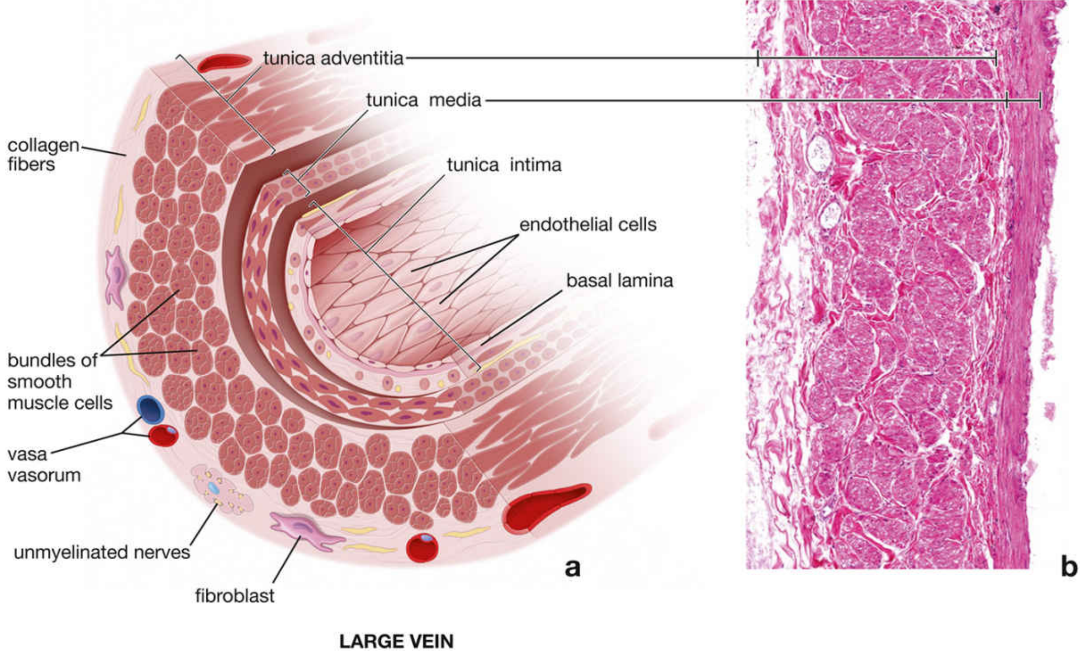
Large vein layers
- Endothelium and basal lamina
- Small amount of subendothelial connective tissue
- Some smooth muscle cells
- Boundary with media may be difficult to see
- Thin
- Circular smooth-muscle cells
- Collagen fibers
- Elastic fibers
- Fibroblasts
- The dominant layer
- Dense collagen and elastic fiber network
- Large longitudinal bundles of smooth muscle
- Vasa vasorum and autonomic nerves
7. Vasa vasorum and nervi vasorum
Vasa vasorum
- They deliver oxygen and nutrients.
- They remove metabolic waste.
- Large veins often have more vasa vasorum than arteries because venous luminal blood has lower oxygen content.
Nervi vasorum
- Most are sympathetic fibers.
- They release norepinephrine.
- They influence smooth muscle and venous tone.
- Their activation reduces venous capacity and increases venous return.
8. Small veins, venules, and postcapillary venules
A. Postcapillary venules
Histology
- Endothelial cells
- Basal lamina
- Pericytes
- No true tunica media
Pericytes
- Support of the vessel wall
- Regulation of capillary and venular blood flow
- Vessel repair and remodeling
- Contribution to new vessel formation
- Potential mesenchymal progenitor-cell activity
Why postcapillary venules are important
- Fluid leakage in acute inflammation
- White blood cell adhesion
- Diapedesis, meaning migration of leukocytes through the vessel wall into tissue
B. Muscular venules
- Possess 1-2 layers of smooth muscle
- Thus have the beginning of a true tunica media
- Have a thin adventitia
- Pericytes are generally absent
C. Small veins
- All three tunics can be identified.
- Media typically has 2-3 smooth-muscle layers.
- Adventitia is relatively prominent.
D. Medium veins
- All three tunics visible
- Thin intima
- Thin media
- Thick adventitia
- Valves common, especially in lower limbs
9. Vein versus artery: deeper comparison
| Feature | Vein | Muscular artery |
|---|---|---|
| Main hemodynamic environment | Low pressure, low resistance, high volume | High pressure, pulsatile flow |
| Lumen | Large and irregular; often collapsed | Smaller, relatively circular |
| Wall-to-lumen ratio | Low | High |
| Intima | Thin, IEL absent or weak | IEL usually prominent and wavy |
| Media | Thin, few smooth-muscle layers | Thick, many concentric smooth-muscle layers |
| Elastic tissue | Relatively sparse | More abundant, especially in elastic arteries |
| Adventitia | Usually thickest layer | Usually thinner than media |
| Valves | Common in limb veins | Absent |
| Compliance | High | Lower |
| Main role | Capacitance reservoir and return to heart | Distribution and pressure regulation |