
development of piatiyutari gland or mbbs student for6 mars question
The pituitary gland (hypophysis) develops from two distinct embryological sources that fuse together. This is a classic MBBS embryology topic. Let me pull the detailed content from the medical library.
Searching sources...
Reading File
Finding Sources
Finding Sources
Excellent results. Now let me read the key textbook sections - Langman's embryology (most exam-relevant), Junqueira's histology, and Ganong's physiology in parallel.
Reading File
Reading File
Reading File
I have rich content from three major textbooks plus images. Let me also fetch the developmental diagram from Histology (Junqueira) and read the Junqueira pituitary section more fully.
Loading Image
Reading File
I now have comprehensive information from multiple textbooks plus a clear diagram. Let me compile the full answer.
Development of the Pituitary Gland (Hypophysis)
MBBS Embryology - 6 Marks Answer
Key Concept: Dual Origin
The pituitary gland is unique because it develops from two completely different embryological sources that grow toward each other and fuse.
| Component | Embryological Origin | Structure Formed |
|---|---|---|
| Rathke's pouch | Oral ectoderm (stomodeum / roof of primitive mouth) | Adenohypophysis (anterior pituitary) |
| Infundibulum | Neuroectoderm (floor of diencephalon / 3rd ventricle) | Neurohypophysis (posterior pituitary) |
Step-by-Step Development
Week 3
- Rathke's pouch appears as an upward evagination (outpocketing) of the ectoderm of the stomodeum (primitive oral cavity), just in front of the oropharyngeal membrane.
- Simultaneously, the floor of the diencephalon sends a downward projection called the infundibulum.
- Both structures grow toward each other.
End of Week 6
- The connection of Rathke's pouch with the oral cavity disappears (the stalk is obliterated).
- Rathke's pouch is now in close contact with the infundibulum and lies completely enclosed within the cranium.
By End of Month 2
- Rathke's pouch loses its connection with the oral cavity entirely.
- Cells in the anterior wall of Rathke's pouch proliferate rapidly → form the anterior lobe (pars distalis) - the bulk of the adenohypophysis.
- The posterior wall of Rathke's pouch becomes the thin pars intermedia (rudimentary in humans).
- A small extension of the anterior wall grows upward around the infundibular stalk → pars tuberalis.
- The infundibulum gives rise to the infundibular stalk and pars nervosa (posterior lobe / neurohypophysis).
Final Parts of the Pituitary Gland
Adenohypophysis (from Rathke's pouch - oral ectoderm):
- Pars distalis - largest part; bulk of anterior lobe; from thickened anterior wall of Rathke's pouch
- Pars tuberalis - wraps around the infundibular stalk; from anterior wall extension
- Pars intermedia - thin strip adjacent to posterior lobe; from posterior wall of Rathke's pouch
Neurohypophysis (from infundibulum - neuroectoderm of diencephalon):
- Pars nervosa (posterior lobe) - composed of neuroglial cells (pituicytes) + nerve fibers from hypothalamus
- Infundibular stalk - connects pituitary to hypothalamus
Developmental Diagram
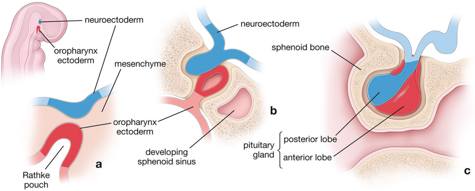
Fig: (a) 6-week embryo: Rathke's pouch (red) grows up from oropharynx; neuroectoderm (blue) grows down from diencephalon. (b) 10 weeks: Rathke's pouch nearly detached from oropharynx, merging with infundibulum. (c) Mature pituitary: anterior lobe (red) and posterior lobe (blue) within sella turcica of sphenoid bone.
Location
The fully formed pituitary gland lies in the sella turcica (the "Turkish saddle") - a bony fossa of the sphenoid bone. It is connected to the hypothalamus by the infundibular stalk.
Clinical Correlation - Craniopharyngioma ⭐
This is a high-yield exam point!
If the stalk of Rathke's pouch is not completely obliterated, remnants of oral epithelium persist along the path of migration. These epithelial remnants can give rise to a craniopharyngioma - a benign but locally invasive suprasellar tumor derived from squamous epithelium of Rathke's pouch remnants. It is the most common suprasellar tumor in children.
Summary Table for Quick Revision
| Feature | Adenohypophysis | Neurohypophysis |
|---|---|---|
| Origin | Oral ectoderm (Rathke's pouch) | Neuroectoderm (diencephalon) |
| Appears | ~Week 3 | ~Week 3-4 |
| Parts | Pars distalis, pars tuberalis, pars intermedia | Pars nervosa, infundibular stalk |
| Cell type | Endocrine secretory cells | Pituicytes (modified astrocytes) + axon terminals |
| Hormones | GH, TSH, ACTH, FSH, LH, Prolactin | ADH, Oxytocin (produced in hypothalamus, stored here) |
Sources: Langman's Medical Embryology, Junqueira's Basic Histology 17e, Ganong's Review of Medical Physiology 26e, Histology: A Text and Atlas (Ross)
This is a shared conversation. Sign in to Orris to start your own chat.