
Femoral hernia
Reading File
Searching sources...
Finding Sources
Searching PubMed
"femoral hernia"[MeSH Terms] AND management
Reading File
Reading File
Reading File
Reading File
Reading File
Reading File
Loading Image
Loading Image
Loading Image
Here is a comprehensive overview of femoral hernia drawn from authoritative surgical textbooks.
Femoral Hernia
Definition
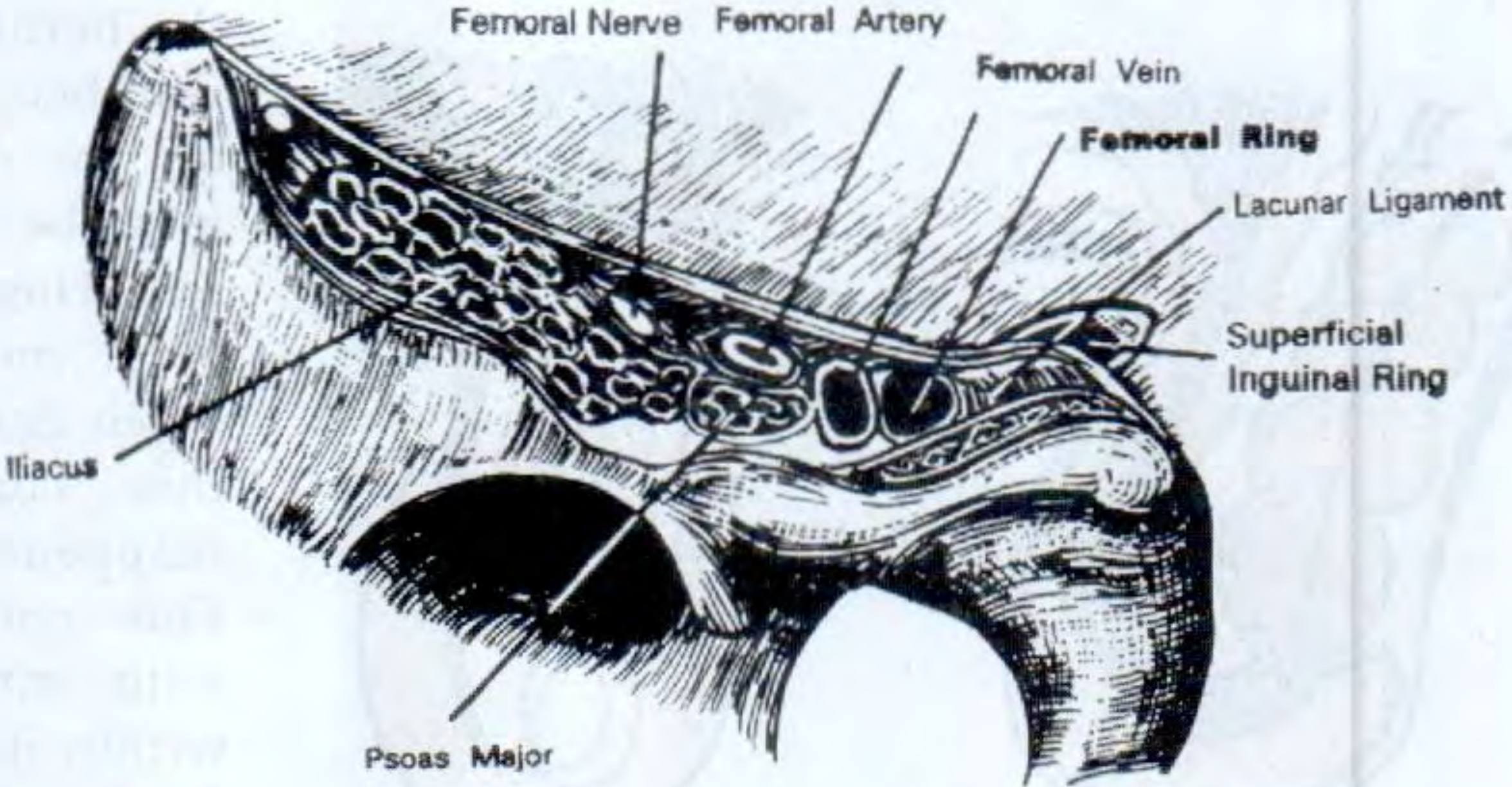
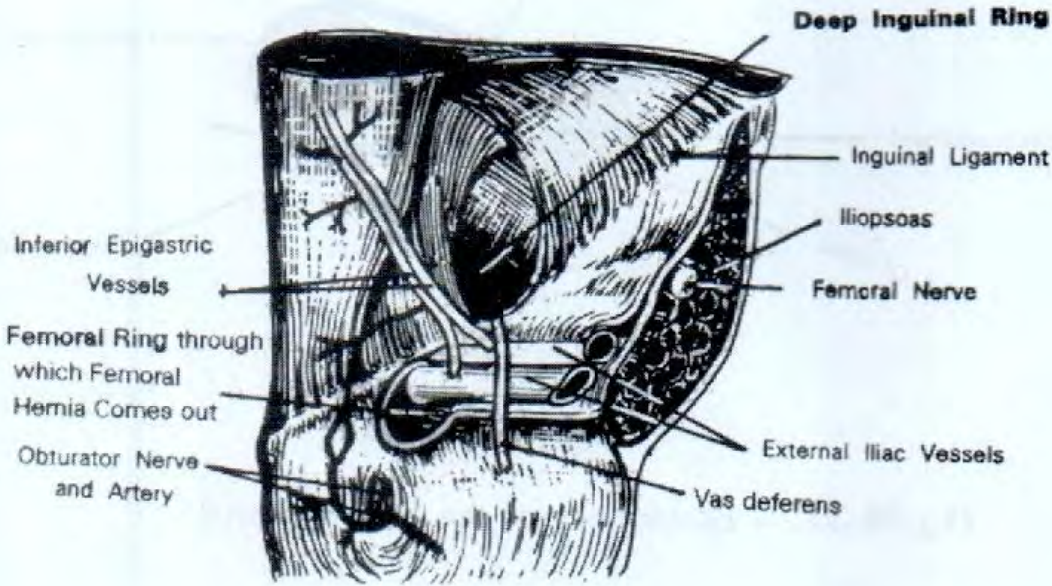
A femoral hernia is a protrusion of extraperitoneal tissue, peritoneum, and sometimes abdominal contents through the femoral canal - the small space just medial to the femoral vein below the inguinal ligament. - S Das Manual on Clinical Surgery, p. 611
Anatomy of the Femoral Canal
The boundaries of the femoral canal are:
| Boundary | Structure |
|---|---|
| Anterior / Superoanterior | Inguinal ligament |
| Posterior / Inferoposterior | Pelvic bone covered by iliopectineal (Astley Cooper's) ligament / pectineus muscle |
| Medial | Lacunar ligament (Gimbernat's ligament) - a sharp, unyielding curved edge |
| Lateral | Femoral vein |
The contents of the canal are fat and the node of Cloquet (lymphatic tissue). The external iliac artery and vein pass below the inguinal ligament to become the common femoral vessels; medial to the vein is where the hernia exploits its space.
Anatomical diagrams:
Shape of the Hernia - "Retort" Shape
The hernia remains narrow while in the femoral canal, then expands considerably once it escapes through the saphenous opening (1.5 inches below and lateral to the pubic tubercle) into the loose areolar tissues. It then curves back upward, giving it the classic retort shape - the bulbous extremity actually expands upward, even above the inguinal ligament. - S Das, p. 611
Epidemiology
- Sex: More common in women (2:1 ratio); but even in women, the commonest groin hernia remains inguinal hernia
- Age: Rare before 20 years; highest incidence over 50 years; particularly seen in thin, elderly women (female pelvis increases femoral canal size)
- Side: Right side affected twice as often as the left; bilateral in 20% of cases
- Emergency presentation: ~50% present as an emergency with strangulation - the femoral canal is a rigid, unyielding opening
- Bailey and Love 28th Ed., p. 1092; S Das, p. 611
Clinical Features
- Often a small (1-2 cm), painless swelling below and lateral to the pubic tubercle
- Easily missed on examination - inadequate exposure of the groin area is the main cause of diagnostic delay
- Cough impulse is felt at the saphenous opening, ~4 cm below and lateral to the pubic tubercle
- Often becomes rapidly irreducible and loses its cough impulse due to tightness of the neck
- As it enlarges, it reflects superiorly and can be mistaken for a direct inguinal hernia
Differentiation from Inguinal Hernia
| Feature | Femoral Hernia | Inguinal Hernia |
|---|---|---|
| Relation to pubic tubercle | Lateral to and below | Medial to and above |
| Relation to inguinal ligament | Below (initially) | Above |
| Invagination (finger) test | Inguinal canal is empty | Inguinal canal contains hernia |
| Occlusion test | Prevented by pressure over saphenous opening / femoral canal | Indirect stopped by occlusion of deep inguinal ring |
| Strangulation risk | Very high (rigid walls) | Lower |
S Das, p. 611
Differential Diagnosis
- Inguinal hernia (most important and commonly confused)
- Saphena varix - disappears on lying down; fluid thrill (not true expansile impulse) on coughing; Schwartz's test positive
- Enlarged lymph nodes - no impulse; search for infection in drainage area
- Psoas abscess - painless, no impulse, cross-fluctuation with iliac fossa; diminishes with hip flexion
- Femoral artery aneurysm - expansile pulsation
- Psoas bursa - cystic, diminishes with hip flexion, associated with hip osteoarthritis
- Lipoma
- Femoral hernia hydrocele (very rare - neck plugged by omentum/adhesions)
- Bailey and Love 28th Ed. Summary Box 64.12; S Das, p. 613
Investigations
- Routine cases: no specific investigations required
- If uncertainty: ultrasonography or CT
- Emergency with bowel obstruction: plain X-ray (often shows obstruction)
- CT is now routine for bowel obstruction to exclude malignancy - it can also identify a femoral hernia missed clinically
- Key rule: All patients with unexplained small bowel obstruction should undergo careful examination for a femoral hernia
- Bailey and Love 28th Ed., p. 1093
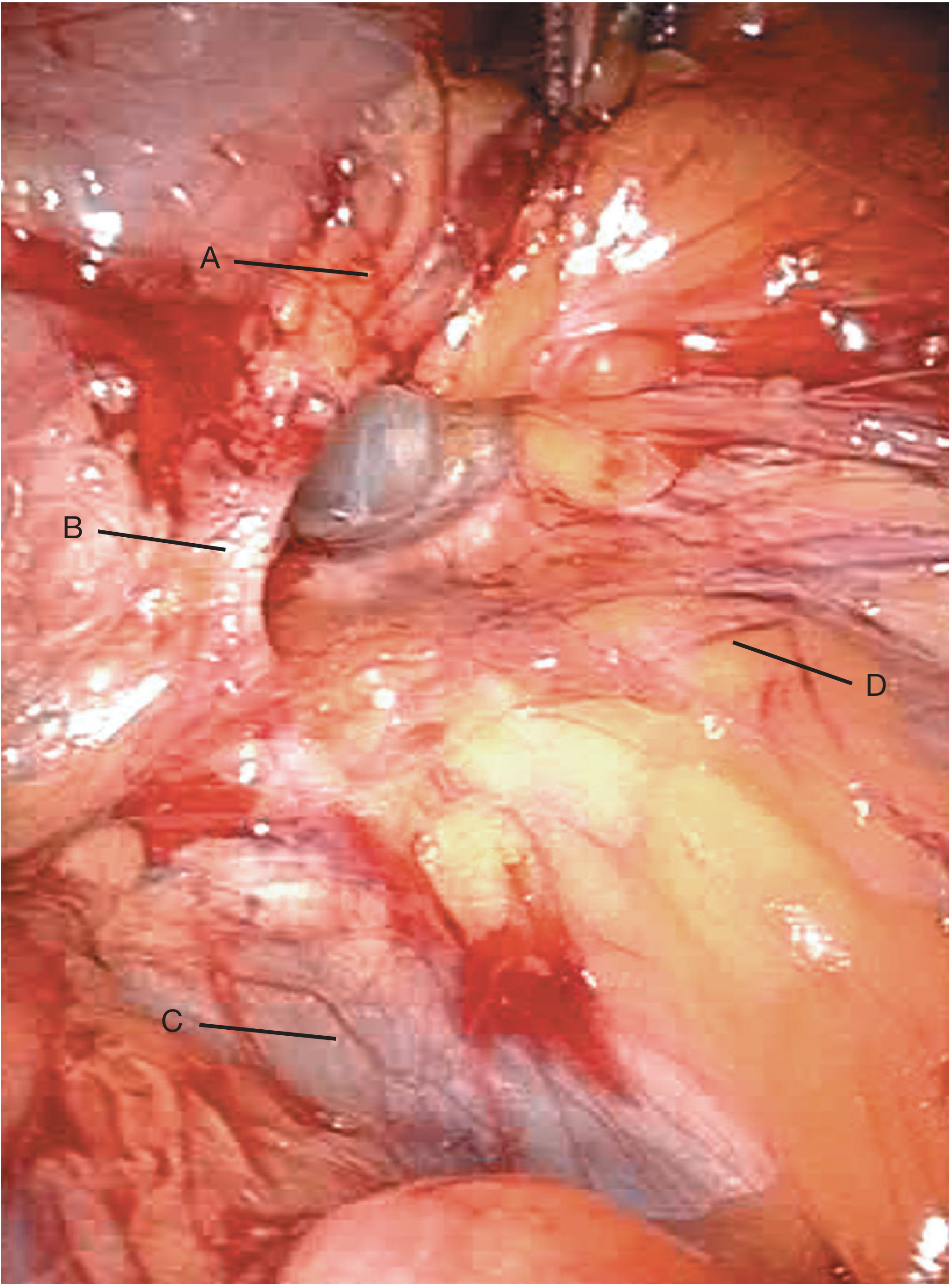
Laparoscopic View
Surgery
Surgery is the only treatment. Due to the high risk of strangulation, all cases should be treated with urgency. There are three open approaches and a laparoscopic option.
1. Low Approach (Lockwood)
- Simplest technique; incision directly over the hernia
- Suitable only when bowel resection is NOT anticipated
- Can be done under local anaesthesia
- Sac contents and sac reduced; sutures placed between inguinal ligament (above) and pectineal ligament (below)
- A small incision in the medial lacunar ligament aids reduction - but beware of an aberrant obturator artery ("corona mortis") just deep to it
- The femoral vein (lateral) must be protected throughout
- Some surgeons use a mesh plug for added reinforcement
- Limitation: Bowel resection is impossible - the anastomosis cannot be returned through the narrow femoral canal
2. Inguinal Approach (Lotheissen)
- Incision into the inguinal canal (like Bassini's or Lichtenstein's)
- Spermatic cord or round ligament mobilised; transversalis fascia opened
- Enters extraperitoneal space; femoral hernia lies just below
- Hernia is reduced by pulling from above + pushing from below
- Defect closed with sutures, mesh plug, or flat mesh in the extraperitoneal plane
- Peritoneum can be opened to deal with contents if needed
- Protect the external iliac vein throughout
3. High Approach (McEvedy / Nyhus modification)
- Preferred in emergencies with high strangulation risk
- Transverse incision just above the inguinal canal, centred on the lateral rectus border
- Anterior rectus sheath incised; proceeds deep in the preperitoneal space
- Sac opened for inspection of contents; bowel can be replaced into peritoneum for 5 minutes to assess viability
- Allows generous peritoneal incision - facilitates bowel resection when needed
- Can be extended to repair bilateral femoral hernias through a single incision (Henry's approach)
4. Laparoscopic Repair (TEP/TAPP)
- Appropriate selected cases can be managed laparoscopically
- Provides excellent view of femoral canal (as shown in the laparoscopic image above)
Special Variant: Pre-vascular Femoral Hernia
A rare variety that descends posterior to the inguinal ligament but anterior to the femoral artery and vein (rather than through the femoral canal). It has a wide neck, is flattened, rarely strangulates, but is difficult to repair. - S Das, p. 612
Key Clinical Pearls
- The femoral canal's rigid, unyielding walls (especially the lacunar ligament medially) make strangulation far more common than in inguinal hernia
- Never exclude femoral hernia in a woman with unexplained bowel obstruction - examine below the inguinal ligament
- A femoral hernia pointing upward above the inguinal ligament can easily mimic a direct inguinal hernia
- The aberrant obturator artery (crown of death / "corona mortis") runs close to the medial wall - injury during Lockwood repair can be catastrophic
- Bowel resection requires either the McEvedy (high) or laparoscopic approach - the Lockwood approach is contraindicated when strangulated bowel is suspected
Sources: Bailey and Love's Short Practice of Surgery 28th Ed.; S Das Manual on Clinical Surgery 13th Ed.; Maingot's Abdominal Operations
Asthma practice of medicine from devidson book
Searching sources...
Finding Sources
I don't see Davidson's directly in the search results. Let me look for it specifically.
Finding Sources
Finding Sources
Finding Sources
Davidson's Principles and Practice of Medicine is not in the library. The closest equivalent internal medicine textbooks available are Harrison's Principles of Internal Medicine 22E (2025) and Goldman-Cecil Medicine. Let me pull asthma from Harrison's - which is the gold-standard equivalent - along with the respiratory medicine textbook for a comprehensive answer.
Finding Sources
Finding Sources
Reading File
Reading File
Reading File
Reading File
Reading File
Reading File
Reading File
Reading File
Reading File
Reading File
Reading File
Reading File
Loading Image
Loading Image
I now have comprehensive content from Harrison's (2025) and Goldman-Cecil Medicine. Note that Davidson's Principles and Practice of Medicine is not in the library, but Harrison's is the direct equivalent in clinical depth and style. Here is the full summary:
Note: Davidson's Principles and Practice of Medicine is not available in the textbook library. The content below is drawn from Harrison's Principles of Internal Medicine 22E (2025) and Goldman-Cecil Medicine - the standard equivalent internal medicine references for asthma.
Asthma
Definition
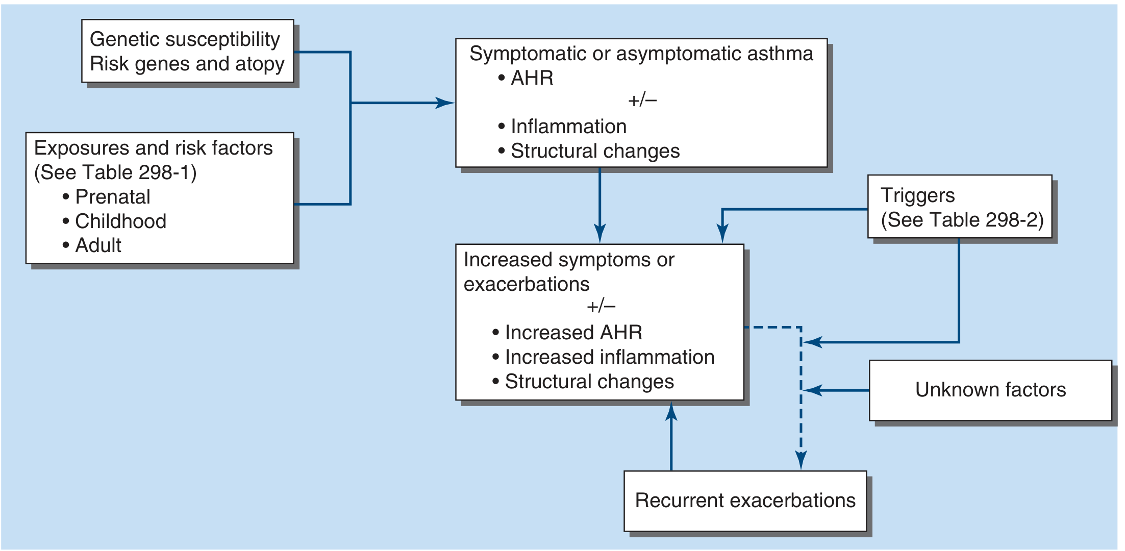
Asthma is a disease characterized by episodic airway obstruction and airway hyperresponsiveness (AHR), usually accompanied by airway inflammation. In most cases the obstruction is reversible, but in a subset of patients, a component may become irreversible. In a large proportion, airway inflammation is eosinophilic, but some patients present with differing inflammatory types, and in some there is no obvious inflammation. - Harrison's 22E, Ch. 298
Epidemiology
- Affects ~262 million people globally; prevalence in the US ~7.9%
- More prevalent in children (8.4%) than adults (7.7%); prevalence has plateaued since ~2015
- In children: boys > girls (2:1); after puberty, women > men (ratio 1.8:1)
- Asthma is more prevalent in Black Americans with greater case morbidity; highest US prevalence in Puerto Ricans
- 1.8 million US emergency department visits/year (2016 data)
- Asthma mortality declined globally from 0.44/100,000 (1993) to 0.19/100,000 (2006), attributed to increased ICS use
- Harrison's 22E
Asthma Development Pathway
Pathobiology
Genetics
- ~60% heritability (twin studies)
- Multiple genome-wide association loci identified - mostly immune mechanism genes
- Atopy is a major inherited risk factor
Pathology - Endotypes
Type 2 (Atopic/Allergic) - most common:
- Driven by IgE, Th2 cells, innate lymphoid cells type 2 (ILC2)
- Key cytokines: IL-4, IL-5, IL-13 (primary targets for biologic therapy), plus TSLP, IL-25, IL-33
- Key cells: mast cells, eosinophils, Th2 lymphocytes
- Structural changes: subepithelial fibrosis, smooth muscle hypertrophy, goblet cell metaplasia, mucus plugs
Non-Type 2 (Neutrophilic):
- Seen in severe, corticosteroid-resistant asthma
- Cytokines: IL-6, IL-17, TNF-α, IL-1β, IL-8
- Sometimes associated with atypical infections (Mycoplasma) - may respond to macrolides
Pauci-granulocytic asthma: pathologic changes without cell infiltration; etiology unclear
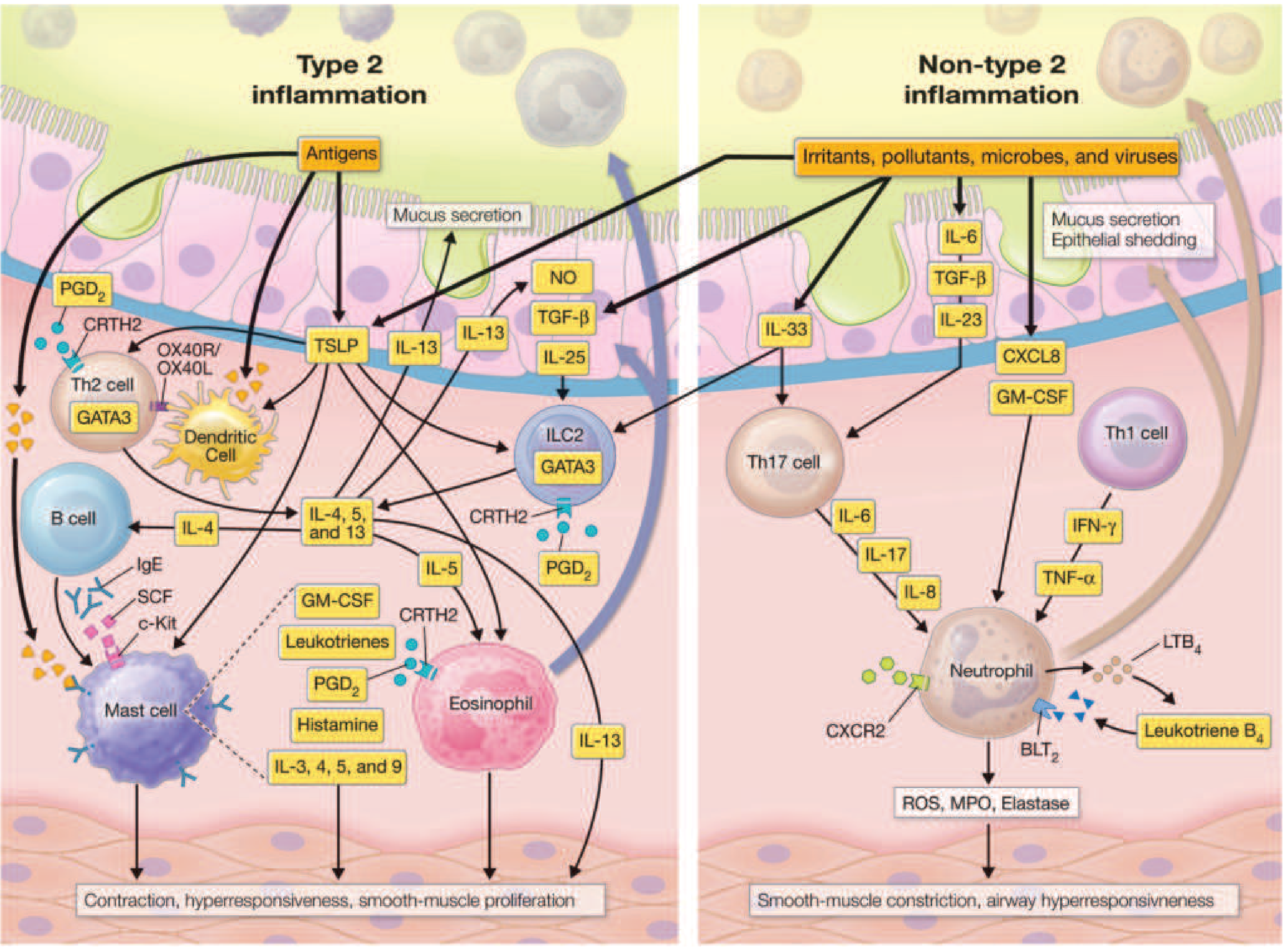
Mediators
| Class | Examples | Effects |
|---|---|---|
| Cytokines | IL-4, IL-5, IL-13, TSLP | Eosinophil recruitment, IgE production, mucus, AHR |
| Cysteinyl leukotrienes (LTC4, LTD4, LTE4) | From eosinophils + mast cells | Smooth muscle constriction, mucus secretion, microvascular leakage |
| Prostaglandins (PGD2) | From mast cells | Bronchoconstriction, Th2 recruitment |
| Histamine | From mast cells | Bronchoconstriction, edema |
| Nitric oxide | Epithelium | Vasodilation, FeNO used as biomarker |
Clinical Manifestations
- Episodic dyspnoea, wheezing, cough (can occur together or separately)
- Chest tightness and mucus production
- Symptoms resolve spontaneously or with therapy
- Exacerbations may be severe enough to require emergency care, hospitalization, or can be fatal
- Some patients have persistent wheezing/dyspnoea
Diagnosis
History + Examination
- Episodic, reversible symptoms; nocturnal or exercise-related component
- Personal or family history of atopy (allergic rhinitis, eczema)
- Exposure to triggers
Spirometry - Key finding: Obstructive pattern with reversibility
- FEV1/FVC ratio reduced (<0.75)
- ≥12% and ≥200 mL improvement in FEV1 after bronchodilator = significant reversibility
- PEFR variability >20% supports diagnosis
Bronchoprovocation testing
- Methacholine/histamine challenge: positive if PC20 FEV1 <8 mg/mL
- Used when spirometry is normal but diagnosis suspected
Arterial Blood Gases (in acute attack)
- Hypoxaemia + hypocapnia (hyperventilation) in moderate attacks
- Rising PaCO2 = sign of fatigue, impending respiratory failure - requires urgent escalation
- PaO2 usually 55-70 mmHg; SpO2 <95% warrants supplemental O2
Biomarkers
- FeNO (fractional exhaled NO): elevated in type 2 eosinophilic inflammation (>50 ppb = highly specific for eosinophilic asthma)
- Blood eosinophils: ≥300 cells/µL suggests type 2 endotype
- Serum IgE: elevated in atopic disease
- Sputum eosinophils: >2% confirms eosinophilic asthma
Comorbidities That Make Asthma Difficult to Control
- Chronic rhinosinusitis ± nasal polyposis - treat with intranasal corticosteroids; biologics increasingly useful for polyposis
- Obesity - 2-4x higher hospitalisation risk; bariatric surgery reduces exacerbations
- Gastroesophageal reflux disease (GERD) - treat symptomatic disease only; asymptomatic treatment shows no benefit
- Inducible laryngeal obstruction (vocal cord dysfunction) - diagnosed by laryngoscopy during symptoms
- COPD (Asthma-COPD overlap)
- Anxiety/depression - increased exacerbation rates
- Obstructive sleep apnea
Differential Diagnosis
- Heart failure ("cardiac asthma")
- COPD / α1-antitrypsin deficiency
- Airway obstruction from mass or foreign body
- Inducible laryngeal dysfunction
- Bronchiolitis obliterans
- Bronchiectasis
- Tracheobronchomalacia
Treatment - Stepwise Approach (GINA/NAEPP)
Key Principle: Anti-inflammatory Reliever (AIR) therapy
A major shift in guidelines: ICS/formoterol as the reliever at all steps (GINA approach), not just as a controller. This ensures patients always receive anti-inflammatory therapy when they use a reliever.
| Step | Controller | Reliever |
|---|---|---|
| Step 1 (intermittent) | None (or as-needed only) | ICS/formoterol as needed (GINA) OR albuterol/budesonide (US) |
| Step 2 (mild persistent) | Low-dose ICS daily | ICS/formoterol as needed; or SABA + ICS |
| Step 3 (moderate) | Low-dose ICS-LABA daily | ICS/formoterol as needed |
| Step 4 (moderate-severe) | High-dose ICS-LABA; ± tiotropium | ICS/formoterol |
| Step 5 (severe) | High-dose ICS-LABA + biologic therapy | SABA or ICS/formoterol |
Drug Classes
1. Inhaled Corticosteroids (ICS) - cornerstone of therapy
- Beclomethasone, budesonide, fluticasone, mometasone
- Suppress eosinophilic inflammation; reduce exacerbations and mortality
- Side effects: oropharyngeal candidiasis (rinse mouth after use), dysphonia; systemic effects at high doses
- LABAs should NOT be used as monotherapy (without ICS) in asthma
2. β2-Agonists
- SABAs (Short-acting): Albuterol (salbutamol) - onset 3-5 min, duration 4-6 h; primary rescue agent
- LABAs (Long-acting): Salmeterol (slow onset), Formoterol (rapid onset); ~12 h duration; only use with ICS in asthma
- Ultra-LABAs: Indacaterol, vilanterol (24 h); only in combination with ICS
- Safety: tremor, tachycardia, hypokalemia, type B lactic acidosis at high doses; frequent SABA use without ICS = increased mortality risk
3. Anticholinergics
- Tiotropium (LAMA) - add-on at step 4; particularly useful in asthma-COPD overlap
- Ipratropium - used in acute severe attacks
4. Leukotriene Modifiers
- Montelukast, zafirlukast (LTRAs) - alternative at step 2; useful in aspirin-exacerbated respiratory disease and exercise-induced asthma
- Warning: Montelukast carries FDA black-box warning for neuropsychiatric effects including suicidal ideation
5. Theophylline
- Weak bronchodilator; anti-inflammatory at low doses
- Narrow therapeutic index; many interactions; rarely used now
6. Biologics (Step 5 - severe eosinophilic asthma)
| Biologic | Target | Indication |
|---|---|---|
| Omalizumab | Anti-IgE | Moderate-severe allergic asthma, elevated IgE |
| Mepolizumab, Reslizumab | Anti-IL-5 | Severe eosinophilic asthma (eos ≥300) |
| Benralizumab | Anti-IL-5Rα | Severe eosinophilic asthma |
| Dupilumab | Anti-IL-4Rα (blocks IL-4 + IL-13) | Severe eosinophilic or OCS-dependent asthma |
| Tezepelumab | Anti-TSLP | Severe uncontrolled asthma (any phenotype) |
Acute Asthma Attack - Management
Mild-moderate:
- β2-agonist up to every 1 h; quadruple ICS dose
- If not improving after a few hours → urgent/emergency care
Emergency Department:
- Assess PEFR or FEV1
- Nebulised β2-agonist every 20 min
- Supplemental O2 to maintain SpO2 >94%
- If PEFR >60% predicted → likely responds to bronchodilators alone
- If PEFR <60% or not improving in 1-2 h → IV/oral corticosteroids
- Nebulised anticholinergics (ipratropium) - add-on bronchodilation
- IV magnesium sulfate (1-2 g) - smooth muscle relaxation
- Leukotriene receptor antagonist - sometimes used
Consider hospital admission if:
- PEFR persistently <60% after 4-6 h treatment
- Persistent severe tachypnoea
- Fatigue, rising PaCO2
In hospital:
- Continuous bronchodilator nebulisation
- Noninvasive positive pressure ventilation (NIPPV) to prevent intubation
- Helium-oxygen (Heliox) mixtures to reduce work of breathing
- Intubation and mechanical ventilation if deteriorating
Patients at Greater Risk for Asthma Mortality
- Previous near-fatal asthma (intubation/ICU)
- Hospitalisation or ED visit in past year
- Currently using or recently stopped oral corticosteroids
- Not currently using ICS
- Using >1 SABA canister/month
- Difficulty perceiving severity of obstruction
- Psychosocial problems, low socioeconomic status
- Food allergy in asthmatic patient
- Poor adherence to medication
Special Situations
Aspirin-exacerbated respiratory disease (AERD / Samter's triad):
- Asthma + nasal polyposis + aspirin/NSAID sensitivity
- Due to excess leukotriene production when COX-1 is inhibited
- Treat with LTRAs; aspirin desensitisation in some cases
Exercise-induced asthma:
- Pre-treatment with SABA or LTRA 15-30 min before exercise
- Warm-up reduces severity
Asthma in pregnancy:
- Active asthma is more dangerous to fetus than ICS use
- All standard asthma medications (ICS, SABAs, LABAs) are considered safe in pregnancy
- Oral corticosteroids if needed for exacerbations
Asthma-COPD Overlap (ACO):
- Features of both; typically older patients, smokers, incomplete reversibility
- Treated with ICS + LABA as cornerstone
Elderly patients with asthma:
- Often underdiagnosed due to misattribution to ageing or cardiac causes
- May have reduced perception of dyspnoea
- LTRA can be considered as alternative to ICS in mild disease in the elderly
Sources: Harrison's Principles of Internal Medicine 22E (2025), Ch. 298 (Elliot Israel); Goldman-Cecil Medicine, Ch. 75 (Drazen and Bel)
This is a shared conversation. Sign in to Orris to start your own chat.