
Muscles of forearm
Reading File
The forearm contains two groups of muscles: the anterior (flexor) compartment and the posterior (extensor) compartment, arranged in superficial and deep layers.
Searching sources...
Finding Sources
Finding Sources
Reading File
Reading File
Reading File
Finding Sources
Reading File
Reading File
Reading File
Reading File
Reading File
Reading File
Reading File
Reading File
Loading Image
Loading Image
Loading Image
Loading Image
Loading Image
Loading Image
Here is a complete, well-organized overview of the muscles of the forearm based on the THIEME Atlas of General Anatomy and Musculoskeletal System and Gray's Anatomy for Students.
Muscles of the Forearm
The forearm contains 20 muscles arranged in three groups: anterior (flexor) compartment, posterior (extensor) compartment, and the radial (lateral) group.
Anterior Compartment (Flexors & Pronators)
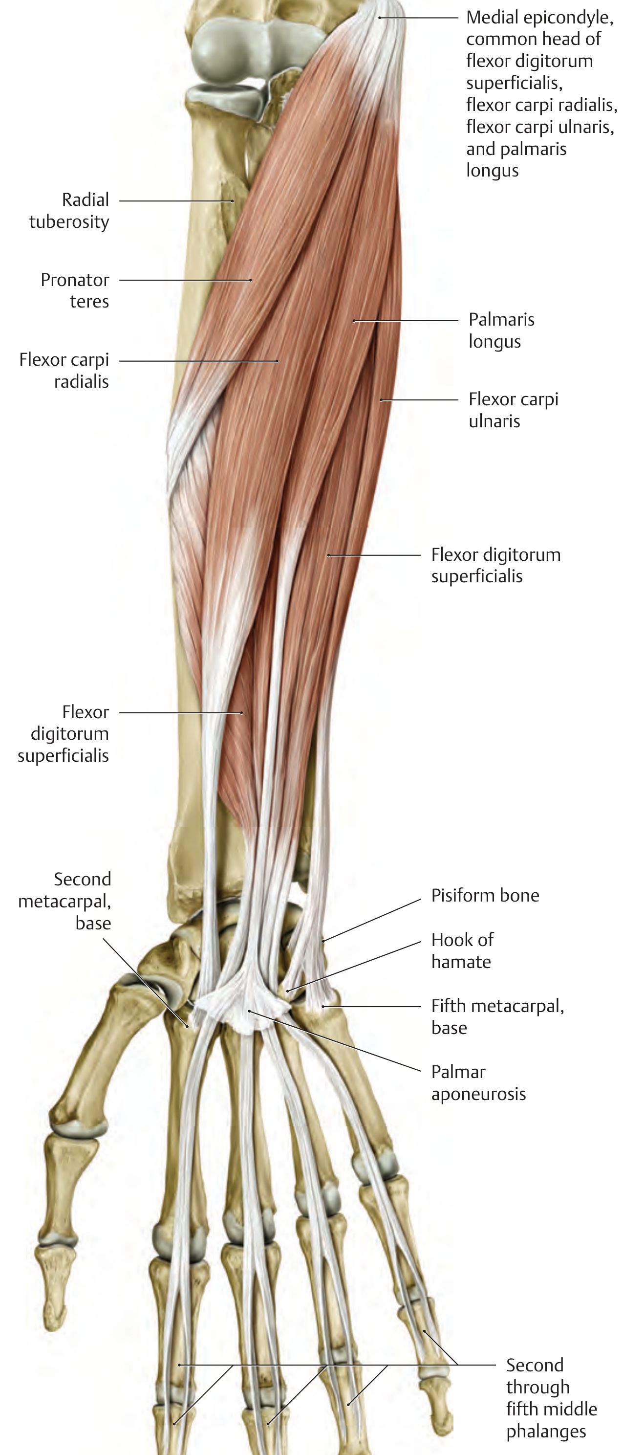
Superficial Layer (5 muscles)
All 5 originate from the common flexor origin - medial epicondyle of the humerus
| Muscle | Insertion | Action | Nerve |
|---|---|---|---|
| Pronator teres | Lateral surface of radius (mid-shaft) | Pronation; weak elbow flexion | Median (C6) |
| Flexor carpi radialis | Base of 2nd (& 3rd) metacarpal | Wrist flexion, radial deviation | Median (C6-C8) |
| Palmaris longus | Palmar aponeurosis | Wrist palmar flexion; tightens palmar aponeurosis | Median (C7-T1) |
| Flexor carpi ulnaris | Pisiform, hook of hamate, base of 5th metacarpal | Wrist flexion, ulnar deviation | Ulnar nerve (C8-T1) |
| Flexor digitorum superficialis (FDS) | Sides of middle phalanges of digits 2-5 | Flexes PIP & MCP joints of fingers; wrist flexion | Median (C7-T1) |
Note: FDS has two heads - humero-ulnar head (medial epicondyle + coronoid process) and radial head (oblique line of radius). The median nerve and ulnar artery pass between these two heads.
Intermediate Layer (1 muscle)
- Flexor digitorum superficialis - sometimes classified separately as it lies deep to the superficial group but superficial to the deep group.
Deep Layer (3 muscles)
| Muscle | Origin | Insertion | Action | Nerve |
|---|---|---|---|---|
| Flexor digitorum profundus (FDP) | Anterior & medial surfaces of ulna + interosseous membrane | Palmar surface of distal phalanges, digits 2-5 | Flexes DIP, PIP, MCP joints; wrist flexion | Median (AIN) for lateral half (index + middle); Ulnar nerve for medial half (ring + little) - both C8, T1 |
| Flexor pollicis longus | Anterior surface of radius + interosseous membrane | Palmar surface, distal phalanx of thumb | Flexes IP & MCP of thumb; wrist flexion | Median nerve (AIN) (C7, C8) |
| Pronator quadratus | Distal 1/4 of anterior ulna | Distal 1/4 of anterior radius | Pronation; stabilizes distal radioulnar joint | Median nerve (AIN) (C7, C8) |
AIN = Anterior Interosseous Nerve, a branch of the median nerve. It supplies all three deep muscles.
Radial Group (Lateral Compartment)
These three muscles lie on the lateral side and are innervated by the radial nerve:
| Muscle | Origin | Insertion | Action | Nerve |
|---|---|---|---|---|
| Brachioradialis | Lateral supracondylar ridge of humerus | Styloid process of radius | Elbow flexion; semipronation | Radial nerve (C5, C6) |
| Extensor carpi radialis longus (ECRL) | Lateral supracondylar ridge of humerus | Dorsal base of 2nd metacarpal | Wrist extension, radial deviation | Radial nerve (C6, C7) |
| Extensor carpi radialis brevis (ECRB) | Lateral epicondyle (common extensor head) | Dorsal base of 3rd metacarpal | Wrist extension, radial deviation | Radial nerve (C7, C8) |
Posterior Compartment (Extensors)
All posterior muscles are innervated by the radial nerve (deep branch / posterior interosseous nerve).
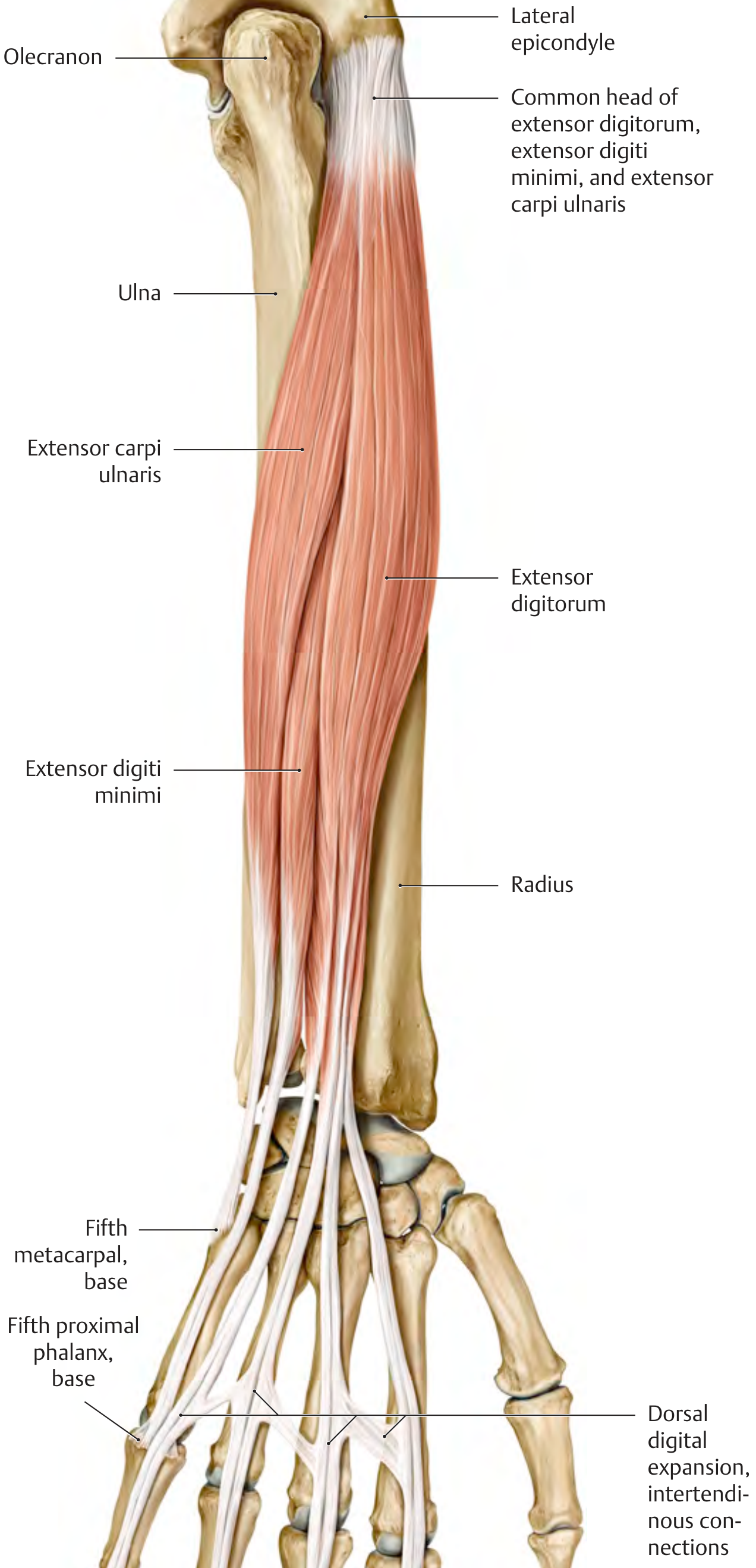
Superficial Layer (3 muscles)
All arise from the common extensor origin - lateral epicondyle of humerus
| Muscle | Insertion | Action | Nerve |
|---|---|---|---|
| Extensor digitorum | Dorsal digital expansion, digits 2-5 | Extends MCP, PIP, DIP of fingers 2-5; wrist extension | Radial nerve (C7, C8) |
| Extensor digiti minimi | Dorsal digital expansion, 5th digit | Extends little finger; wrist extension/ulnar deviation | Radial nerve (C7, C8) |
| Extensor carpi ulnaris | Base of 5th metacarpal | Wrist extension, ulnar deviation | Radial nerve (C7, C8) |
Deep Layer (5 muscles)
Arise from the radius, ulna, and interosseous membrane
| Muscle | Origin | Insertion | Action | Nerve |
|---|---|---|---|---|
| Supinator | Lateral epicondyle, supinator crest of ulna, radial collateral ligament, anular ligament | Radius (between radial tuberosity and pronator teres insertion) | Supination of forearm | Radial nerve (C7, C8) |
| Abductor pollicis longus (APL) | Posterior surfaces of radius & ulna + interosseous membrane | Base of 1st metacarpal | Thumb abduction; radial deviation of wrist | Radial nerve (C7, C8) |
| Extensor pollicis brevis (EPB) | Posterior surface of radius + interosseous membrane | Base of proximal phalanx of thumb | Extends MCP & CMC of thumb; radial deviation | Radial nerve (C7, C8) |
| Extensor pollicis longus (EPL) | Posterior surface of ulna + interosseous membrane | Base of distal phalanx of thumb | Extends IP & MCP of thumb; adducts thumb; wrist extension | Radial nerve (C7, C8) |
| Extensor indicis | Posterior surface of ulna + interosseous membrane | Dorsal digital expansion, 2nd digit | Extends index finger independently; wrist extension | Radial nerve (C7, C8) |
Key Points & Clinical Mnemonics
Mnemonic for superficial flexors (medial to lateral): "Please Forget Pain From Carpal" - Pronator teres, FCU, Palmaris longus, FDS, FCR
The "snuff box" muscles: APL and EPB form its anterior wall; EPL forms its posterior wall. These three tendons emerge from the posterior forearm as the "anatomical snuff box" at the wrist.
Nerve summary:
- Median nerve supplies all anterior muscles EXCEPT flexor carpi ulnaris and the medial half of FDP (those are ulnar nerve)
- Ulnar nerve supplies FCU + medial half of FDP (ring and little fingers)
- Radial nerve supplies all posterior muscles + radial group
Dual innervation of FDP: The lateral portion (index + middle fingers) is supplied by the anterior interosseous nerve (branch of median); the medial portion (ring + little fingers) is supplied by the ulnar nerve. This is clinically significant - isolated AIN palsy causes inability to flex the distal phalanx of the index finger and thumb (the "OK sign" test).
Pronation is performed by two muscles:
- Pronator teres (proximal; also aids elbow flexion)
- Pronator quadratus (distal; primary pronator, stabilizes distal radioulnar joint)
Supination is performed by two muscles:
- Supinator (forearm alone)
- Biceps brachii (most powerful when elbow is flexed 90°)
Sources: THIEME Atlas of General Anatomy and Musculoskeletal System, pp. 334-340; Gray's Anatomy for Students, pp. 899-902
This is a shared conversation. Sign in to Orris to start your own chat.