
Describe the lateral wall of nose for a 10 marks question with clinical anatomy
Reading File
Finding Sources
Finding Sources
Reading File
Finding Sources
Reading File
Finding Sources
Loading Image
Loading Image
Finding Sources
Reading File
Finding Sources
Finding Sources
Reading File
Reading File
Loading Image
Finding Sources
I now have everything needed for a comprehensive 10-mark answer. Let me compile it.
Lateral Wall of the Nose
(Gray's Anatomy for Students, Scott-Brown's Otorhinolaryngology, Cummings Otolaryngology)
Introduction
The lateral wall of the nose is the most complex wall of the nasal cavity. It is formed by contributions from multiple bones and cartilages, carries three scroll-like projections (conchae), and provides the drainage pathway for almost all the paranasal sinuses and the nasolacrimal duct. It is of immense clinical importance in rhinology, endoscopic sinus surgery, and the management of epistaxis.
Bony Framework
The lateral wall receives bony support from the following bones (Fig. 8.242A - Gray's Anatomy for Students):
| Bone | Contribution |
|---|---|
| Nasal bone | Anterosuperior part |
| Frontal process of maxilla | Anterolateral support |
| Lacrimal bone | Small contribution anteriorly |
| Ethmoidal labyrinth (superior + middle concha, uncinate process) | Central - forms the key functional area |
| Inferior concha (separate bone) | Lower third of lateral wall |
| Perpendicular plate of palatine bone | Posterior inferior part |
| Medial pterygoid plate of sphenoid | Posteriormost part |
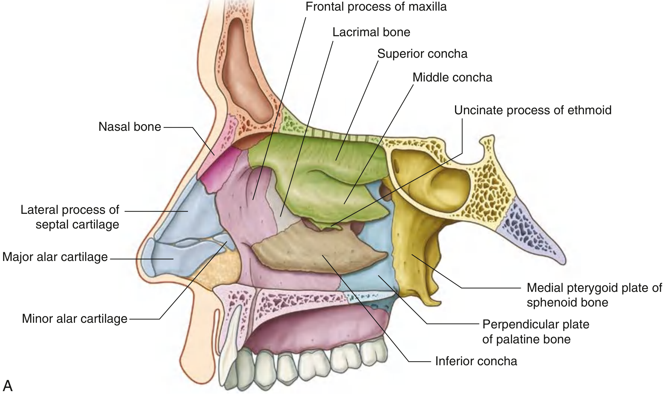
In the external (cartilaginous) nose, the lateral wall is supported by:
- The lateral process of the septal cartilage
- The major alar cartilage (lateral crus)
- The minor alar cartilages
Nasal Conchae (Turbinates)
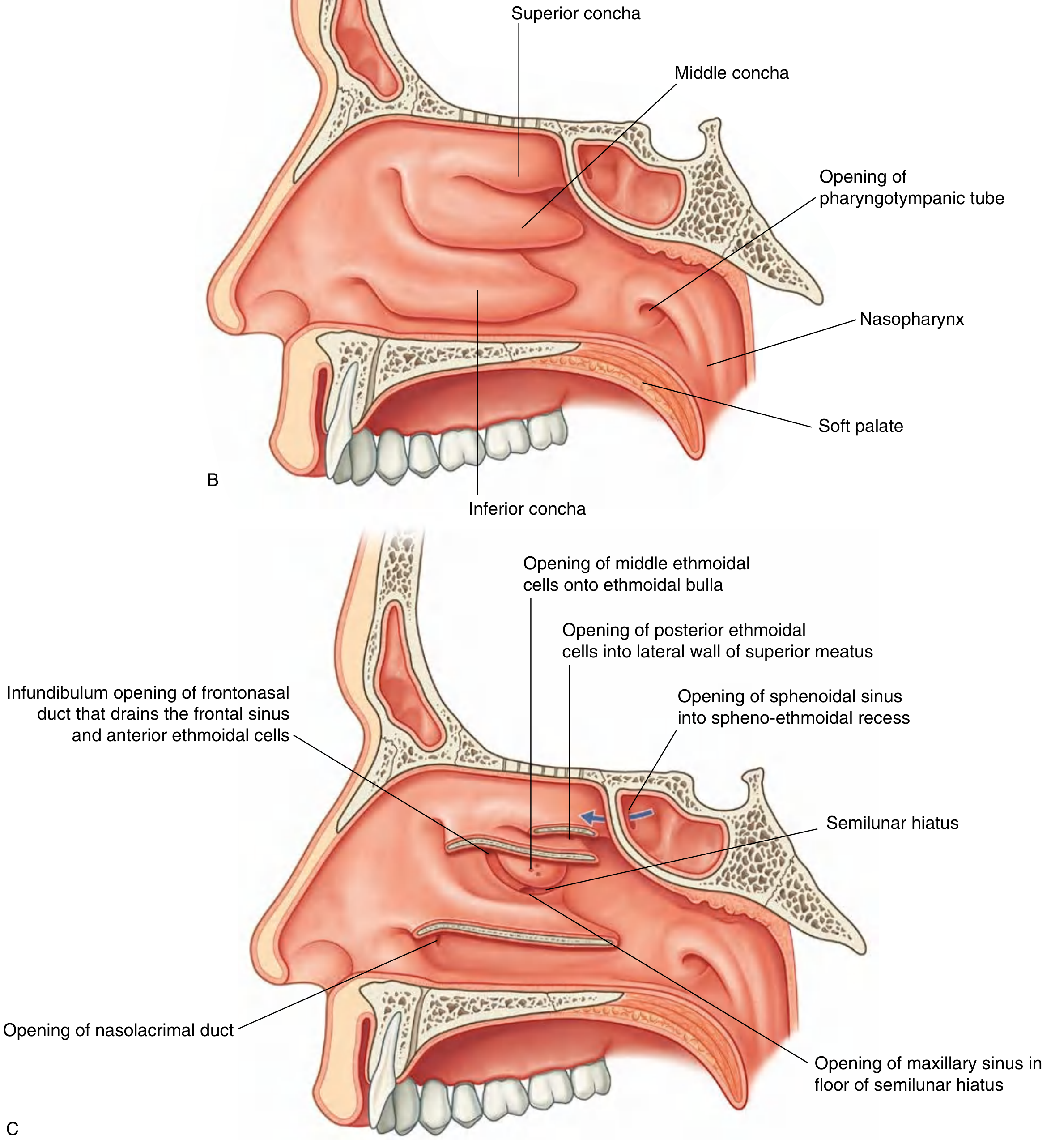
The surface of the lateral wall is thrown into three (occasionally four) scroll-like folds called conchae:
- Inferior concha - a separate independent bone; largest concha
- Middle concha - part of the ethmoidal labyrinth
- Superior concha - part of the ethmoidal labyrinth
- Supreme concha - occasionally present above the superior concha
The conchae extend medially across the nasal cavity, dividing it into four air channels:
- Inferior meatus
- Middle meatus
- Superior meatus
- Spheno-ethmoidal recess (above superior concha)
Key Features of the Middle Meatus
The middle meatus is the most important meatus clinically. It contains several key structures:
-
Ethmoidal bulla: A dome-shaped elevation formed by the underlying middle ethmoidal cells, lying just below the attachment of the middle concha and just anterior to its midpoint.
-
Semilunar hiatus: A curved gutter (groove) inferior to the ethmoidal bulla, formed by the mucosa spanning the gap between the ethmoidal bulla above and the uncinate process below.
-
Ethmoidal infundibulum: The anterior upward-curving extension of the semilunar hiatus, connecting it to the frontonasal duct and then the frontal sinus. It is a key drainage pathway.
-
Uncinate process: A sickle-shaped bony projection of the ethmoid, forming the inferior boundary of the semilunar hiatus.
Openings on the Lateral Wall
The lateral wall carries the openings of nearly all paranasal sinuses and the nasolacrimal duct:
| Structure | Opening Site |
|---|---|
| Nasolacrimal duct | Inferior meatus - under the anterior lip of the inferior concha |
| Maxillary sinus | Semilunar hiatus - usually just inferior to the center of the ethmoidal bulla (near its own roof) |
| Frontal sinus | Via frontonasal duct through ethmoidal infundibulum into anterior semilunar hiatus, middle meatus |
| Anterior ethmoidal cells | Frontonasal duct or ethmoidal infundibulum |
| Middle ethmoidal cells | On or just above the ethmoidal bulla |
| Posterior ethmoidal cells | Lateral wall of the superior meatus |
| Sphenoidal sinus | Spheno-ethmoidal recess (posterior roof) - NOT on the lateral wall |
Blood Supply
The lateral wall has a dual blood supply from both the internal and external carotid systems (Fig. 8.246 - Gray's Anatomy for Students):
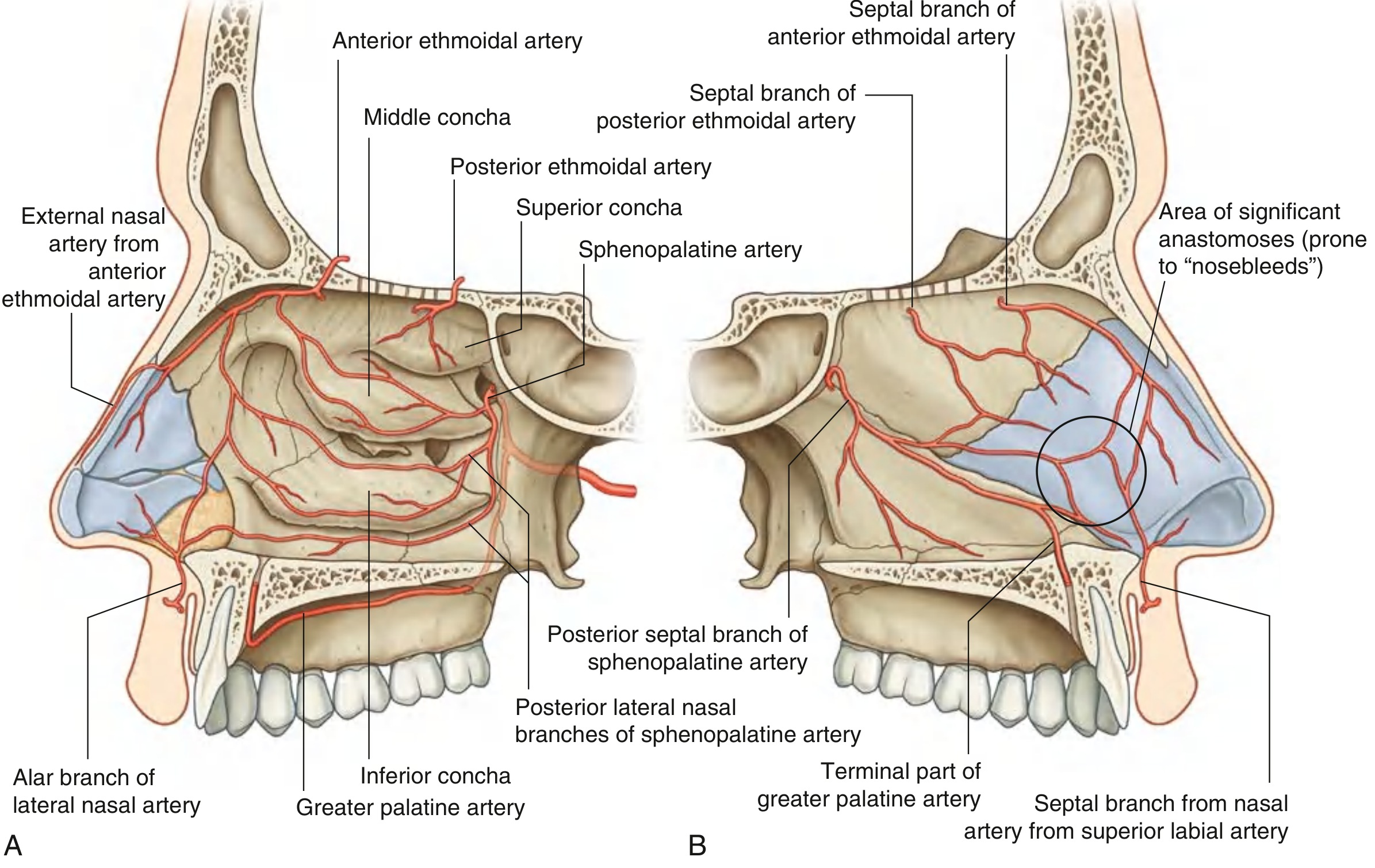
From external carotid artery (via maxillary artery):
- Sphenopalatine artery - the dominant supply. It is the terminal branch of the maxillary artery in the pterygopalatine fossa. It enters the nasal cavity through the sphenopalatine foramen (located just posterior to the posterior wall of the maxillary sinus) and gives posterior lateral nasal branches to a large part of the lateral wall.
- Greater palatine artery - enters the floor of the nasal cavity through the incisive canal from below; supplies the anterior floor and medial wall.
- Superior labial artery and lateral nasal artery - from the facial artery, supply the anterior part.
From internal carotid artery (via ophthalmic artery):
- Anterior ethmoidal artery - supplies the anterosuperior lateral wall
- Posterior ethmoidal artery - supplies the posterosuperior lateral wall
All these vessels anastomose freely. On the nasal septum (medial wall), this anastomosis is particularly rich at Little's area (Kiesselbach's plexus) in the anterior inferior septum.
Nerve Supply
(Scott-Brown's Otorhinolaryngology, Vol 1)
The nerve supply has both autonomic and sensory components:
Sensory (Trigeminal):
- Lateral wall and turbinates: Supplied by the posterolateral nasal nerves from V2 (maxillary nerve), arising from the sphenopalatine foramen.
- Anterosuperior lateral wall: Supplied by the anterior and posterior ethmoidal nerves from V1 (ophthalmic nerve).
Autonomic:
- Parasympathetic (via vidian nerve and greater superficial petrosal nerve → sphenopalatine ganglion): Regulates nasal secretions.
- Sympathetic (via deep petrosal nerve → vidian nerve → sphenopalatine ganglion): Regulates vascular tone and turbinate congestion.
Mucosa
Three types of epithelium line the lateral wall:
- Squamous epithelium - nasal vestibule
- Olfactory epithelium - superior concha, upper middle concha, and upper septum
- Respiratory epithelium (ciliated pseudostratified columnar) - remainder of the nasal cavity
The submucosa, especially over the conchae, is described as "erectile" or "cavernous" because it can enlarge or shrink depending on blood flow, forming the basis of the nasal cycle.
Clinical Anatomy
1. Ostiomeatal Complex (OMC)
The OMC is a functional unit on the lateral wall comprising the maxillary sinus ostium, ethmoidal infundibulum, semilunar hiatus, ethmoidal bulla, and the anterior middle meatus. It represents the final common drainage pathway for the frontal, maxillary, and anterior ethmoidal sinuses. Blockage of the OMC - by deviated septum, polyps, or turbinate hypertrophy - causes recurrent rhinosinusitis. Coronal CT is the gold standard imaging to assess the OMC. Functional endoscopic sinus surgery (FESS) is aimed at restoring OMC patency.
2. Epistaxis
- Anterior epistaxis (>90% of cases) arises from Little's area / Kiesselbach's plexus on the anteroinferior nasal septum, where branches of the sphenopalatine artery, anterior ethmoidal artery, greater palatine artery, and superior labial artery anastomose.
- Posterior epistaxis arises mainly from the Woodruff's plexus, located posterior to the inferior turbinate, formed by branches of the sphenopalatine and ascending pharyngeal arteries. It is more severe and difficult to control.
- Surgical or endoscopic sphenopalatine artery ligation (ESPAL) is effective for refractory posterior epistaxis.
3. Inferior Turbinate Hypertrophy
The inferior turbinate is the largest concha and the most common cause of nasal obstruction. Its submucosa has abundant cavernous tissue. Turbinate reduction procedures (submucosal diathermy, laser, partial turbinectomy) target this tissue.
4. Nasolacrimal Duct
The nasolacrimal duct opens into the inferior meatus under the anterior lip of the inferior concha. Obstruction leads to epiphora (watering eye). Dacryocystorhinostomy (DCR) creates a new opening from the lacrimal sac into the nasal cavity, bypassing the obstruction.
5. Maxillary Sinus Antrostomy
The natural ostium of the maxillary sinus opens high on the medial wall (the lateral wall of the nose), near the roof of the sinus, into the semilunar hiatus. This unfavorable position for gravity drainage makes the maxillary sinus prone to sinusitis. FESS enlarges this ostium (antrostomy) to improve drainage.
6. Ethmoidal Nerve Block / Sphenopalatine Ganglion Block
Knowledge of the nerve supply of the lateral wall is used for intranasal nerve blocks: the anterior ethmoidal nerve can be blocked at the anterior border of the cribriform plate, and the sphenopalatine ganglion can be blocked via the greater palatine foramen or transnasally through the sphenopalatine foramen, for headache and rhinitis.
7. Concha Bullosa
The middle concha may develop pneumatization (an air cell within it), called concha bullosa. This is a common anatomical variant seen on CT, and when large, it can obstruct the middle meatus and contribute to sinusitis.
Summary Table
| Feature | Details |
|---|---|
| Bones | Nasal bone, frontal process of maxilla, lacrimal bone, ethmoidal labyrinth, inferior concha, perpendicular plate of palatine bone, medial pterygoid plate |
| Conchae | Superior, middle, inferior (+ occasionally supreme) |
| Meatuses | Inferior, middle, superior, spheno-ethmoidal recess |
| Key middle meatus structures | Ethmoidal bulla, semilunar hiatus, ethmoidal infundibulum, uncinate process |
| Sinus openings | Frontal + anterior ethmoids + maxillary → middle meatus; Posterior ethmoids → superior meatus; Sphenoid → spheno-ethmoidal recess |
| Nasolacrimal duct opening | Inferior meatus |
| Dominant artery | Sphenopalatine artery (via sphenopalatine foramen) |
| Sensory nerves | V2 posterolateral nasal nerves (dominant) + V1 ethmoidal nerves (anterosuperior) |
| Clinical importance | Epistaxis, ostiomeatal complex and sinusitis, nasal obstruction, DCR, FESS |
Sources: Gray's Anatomy for Students (9780323934237); Scott-Brown's Otorhinolaryngology Head & Neck Surgery Vol 1 (9781138094611); Cummings Otolaryngology Head and Neck Surgery (9780323612173)
This is a shared conversation. Sign in to Orris to start your own chat.