Explain nasal polyp etiology and clinical features like donkey
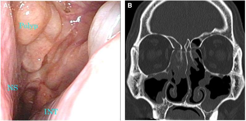
nasal polyp clinical appearance endoscopy
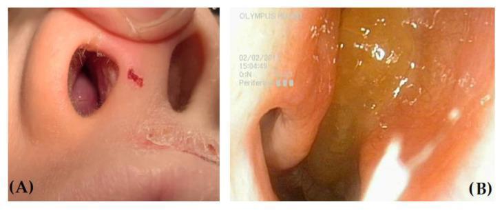
This clinical comparison features two images illustrating differential findings in the nasal cavity via anterior rhinoscopy and endoscopy. Image A shows a child's nose with a markedly hypertrophic right inferior turbinate, appearing as a fleshy, smooth, pinkish mass firmly attached to the lateral nasal wall; the external nares show signs of atopic dermatitis with dry, peeling skin. Image B provides a close-up endoscopic view of a nasal polyp (NP). The polyp is characterized by a yellowish, translucent, and gelatinous appearance with a rounded, grape-like morphology, distinct from the surrounding mucosa. This comparison is educational for differentiating benign inflammatory masses like nasal polyps from edematous turbinate hypertrophy commonly seen in pediatric patients with allergic rhinitis. Key diagnostic features include the polyp's glistening, wet surface reflections versus the solid, uniform texture of the hypertrophic turbinate. Medical specialty: Otolaryngology (ENT). Target audience: Medical students and practitioners.
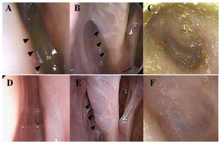
A comparison of endoscopic images from the nasal cavity and ear canal illustrating therapeutic response in a patient with eosinophilic chronic rhinosinusitis (ECRS) and eosinophilic otitis media (EOM). Panels A-C show findings after 13 months of mepolizumab (anti-IL-5) treatment. (A) Endoscopy of the left olfactory cleft reveals a persistent nasal polyp (arrowheads). (B) The nasal cavity and middle turbinate appear edematous, coated with a whitish exudate (arrowheads), with visible discharge from the maxillary sinus ostium (arrow). (C) Otoscopy shows the ear canal and eardrum obscured by copious dried exudate. Panels D-F show the same sites after 1 month of dupilumab (anti-IL-4Ṛ) treatment. (D) The olfactory cleft is clear with no polyps visible. (E) The nasal mucosa and middle turbinate (arrowhead) appear normal with resolution of inflammation and discharge. (F) The ear canal and eardrum have a normal, clear appearance. The series demonstrates the superior efficacy of dual IL-4/IL-13 inhibition over IL-5 inhibition in resolving inflammatory exudates and polyposis in this clinical scenario.
Nasal Polyps - Etiology and Clinical Features (Explained Like You're a Donkey)
What ARE These Things?
ETIOLOGY - Why Does Your Nose Grow Grapes?
1. The Main Driver: Chronic Inflammation (Type 2)
- IL-4, IL-5, IL-13 - these are the main troublemakers
- IgE - the allergy antibody that makes mast cells and eosinophils show up
2. The Trigger Suspects
A. Allergic Rhinitis (Hay Fever)
B. Staphylococcus aureus - The Sneaky Bacteria
C. Viral Infections
D. Allergens and Environmental Toxins
E. B Cells and Immunoglobulins - The Local Antibody Factory
F. T Regulatory Cell Dysfunction (Treg Failure)
G. Geographic / Ethnic Variation
H. Special Associations
| Association | Notes |
|---|---|
| Asthma | Up to 30-50% of nasal polyp patients have asthma |
| Samter's Triad | Nasal polyps + asthma + aspirin sensitivity - the full triple-threat |
| Cystic Fibrosis | Polyps in CF are driven by a Th1/Th17 neutrophilic pattern (different mechanism) |
| Allergic Fungal Rhinosinusitis | Fungal antigens drive intense type 2 inflammation |
CLINICAL FEATURES - What Does the Donkey With Polyps Actually Experience?
1. Nasal Obstruction / Blockage
2. Anosmia / Hyposmia (Loss of Smell)
3. Nasal Discharge / Rhinorrhoea
4. Facial Pressure / Headache
5. Voice Change (Hyponasality)
6. Appearance on Examination
- On anterior rhinoscopy: glistening, grey/pale or yellowish-translucent, smooth, grape-like masses visible in the nasal cavity
- Classically arise from the middle meatus (the area between the middle and inferior turbinates)
- They are insensate (no pain when probed - a key feature that distinguishes them from turbinate hypertrophy, which is painful to touch)
- Mobile, soft, non-tender, non-vascular (won't bleed on gentle probing unlike other masses)
- Endoscopically: rounded, gelatinous, wet-surfaced polyps in the olfactory cleft and middle meatus
7. Recurrence After Treatment
Quick Summary Table
| Feature | Detail |
|---|---|
| Core cause | Chronic Th2-skewed eosinophilic inflammation of the sinonasal mucosa |
| Key cytokines | IL-4, IL-5, IL-13, IgE |
| Key cells | Eosinophils, mast cells, Th2 lymphocytes, plasma cells |
| Bacteria involved | S. aureus (superantigens + biofilms) |
| Top symptom | Bilateral nasal obstruction |
| Hallmark exam finding | Grey/translucent, grape-like, insensate masses from middle meatus |
| Key associations | Asthma, aspirin sensitivity (Samter's triad), CF, allergic fungal sinusitis |
| Smell loss | Very common (olfactory cleft blockage) |
| Recurrence | Very high - especially in eosinophilic type |
- Cummings Otolaryngology Head and Neck Surgery - CRS pathogenesis, eosinophilic inflammation, T-cell subtypes, B-cell/BAFF/IgE mechanisms
- Scott-Brown's Otorhinolaryngology Head & Neck Surgery Vol 1 - CRS with nasal polyps treatment outcomes
Give answer for nasal polyp for ent subject from standard textbook
nasal polyp endoscopy clinical photograph
nasal polyp gross appearance pale gelatinous grape
Nasal Polyps - ENT Standard Textbook Answer
Definition
Epidemiology
| Parameter | Data |
|---|---|
| Annual incidence | 1-20 per 1000 population |
| Adult prevalence | 1-4% |
| Children | ~0.1% |
| Sex ratio | More common in males (2-4:1) |
| Age | Declines after 60 years |
| Race | No racial predilection (in general population) |
Pathogenesis
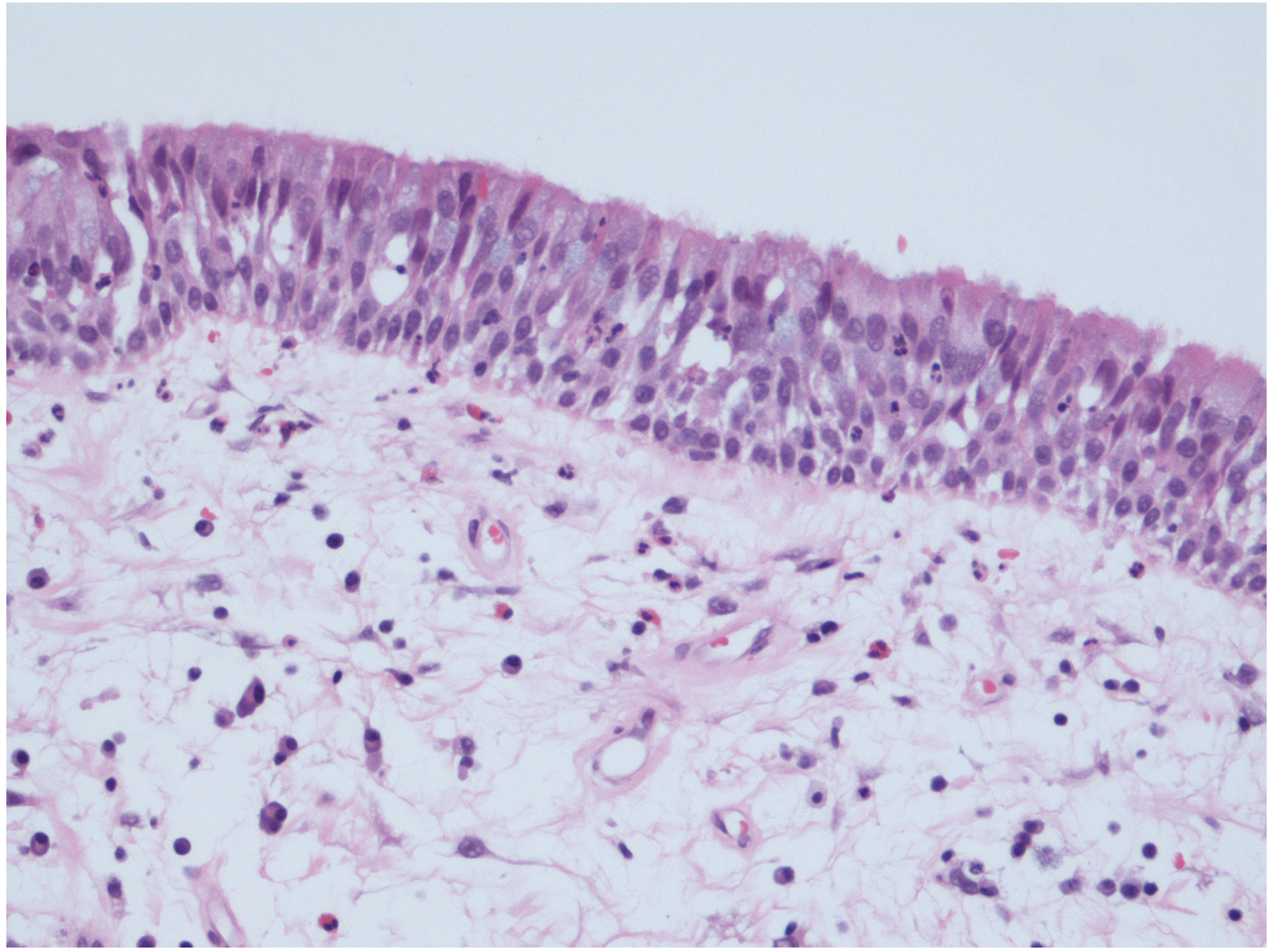
- Loose oedematous connective tissue stroma
- Inflammatory cells (predominantly eosinophils)
- Fluid accumulation
- Covered by pseudostratified, columnar, ciliated epithelium
Inflammatory Endotype (Key)
- A robust T-helper 2 (Th2) response
- Eosinophilic infiltration
- Decreased T regulatory (Treg) function
- Abundance of IL-5 cytokine
Etiology / Predisposing Factors
1. Allergy (Allergic Rhinitis)
- Prevalence of allergy in nasal polyp patients ranges from 10% to 64% in different studies
- Only 0.4-4.5% of patients with allergic rhinitis actually have nasal polyps - similar to the general population - suggesting allergy is not directly causative
- Elevated serum IgE and positive skin prick tests to inhalant allergens are found in the majority of polyp patients
- Importantly, local tissue IgE in polyps is often polyclonal and independent of total serum IgE or allergy skin prick test results
- Mucosal polyclonal IgE can induce mast cell degranulation independently of serum IgE levels
2. Bacteria - Staphylococcus aureus Superantigens
- The main bacterial species implicated in polyp exacerbation is Staphylococcus aureus
- 42.5% of S. aureus strains isolated from nasal polyp patients harbour at least one classic enterotoxin gene
- These toxins behave as superantigens: unlike conventional antigens, they do not need antigen processing
- Superantigens activate up to 20% of the body's T cells (vs. only 0.001-0.0001% for regular antigens)
- This results in massive cytokine secretion, increased eosinophilic inflammation, and formation of IgE antibodies
- In Western CRSwNP this manifests as severe eosinophilic disease; this effect is NOT seen in Asian CRSwNP patients (who have predominantly Th1/Th17 patterns)
3. Bacterial Biofilms
- Bacteria enclose themselves within a self-developed matrix of polysaccharides, rendering them inaccessible to antibiotics and host immune responses
- Nasal polyp cases with biofilm presence are associated with a more severe disease form and worse postoperative outcome
4. Fungi (Controversial)
- Fungi (particularly Alternaria) are detectable in the nasal cavity of all patients - those with CRS and controls
- The fungal hypothesis proposed an exaggerated host response to airborne fungi as the underlying cause
- Current evidence does NOT support fungi as a uniquely important or universal agent driving CRS
- However, fungi play a definite role in Allergic Fungal Rhinosinusitis (AFRS), a specific subtype characterized by type 1 hypersensitivity, type 2 cytokine expression, and nasal polyps
5. Epithelial Barrier Dysfunction
- The mechanical barrier (mucociliary clearance + tight junctions between epithelial cells) is the first line of defence
- Genetic and acquired defects in mucociliary flow with increased mucus viscosity are associated with a high incidence of CRS
- Intrinsic protective anti-protease activity is reduced in some forms of CRS with type 2 inflammation
- A permeable epithelial barrier allows environmental antigens access to the immune system, initiating and perpetuating the inflammatory cycle
6. Viruses
- Viral upper respiratory infections disrupt the epithelial barrier
- Recurrent viral infections secondary to allergic rhinitis may lead to acquired ostiomeatal obstruction and mucociliary stasis
- Early-life viral infections may predispose to subsequent CRS development, analogous to early-onset asthma
7. Environmental Toxins
- Cigarette smoke has the strongest epidemiologic association with CRS, acting through reactive oxygen/nitrogen species, mucociliary dysfunction, biofilm promotion, and pro-inflammatory cytokine induction
Associated Diseases
Asthma
- Prevalence of CRSwNP is ~7% in asthmatic patients vs. 4% in general population
- 30-71% of nasal polyp patients have asthma
- There is a direct association between asthma severity and nasal polyp presence
- Patients with late-onset asthma: 10-15% develop nasal polyps, generally developing asthma first, then polyps within 9-13 years
- Asthma and nasal polyps share: eosinophilia, mucus cell hyperplasia, oedema, thickened basal membrane, and increased cysteine leukotrienes
Samter's Triad / AERD (Aspirin-Exacerbated Respiratory Disease)
- Bronchial asthma
- Nasal polyposis
- Aspirin sensitivity (NSAIDs intolerance)
- First described by Widal in 1922; formally recognized by Samter in 1967
- Prevalence: up to 0.3% in general population; 5-10% of adult asthmatics
- Prevalence of nasal polyps + asthma + aspirin sensitivity increases after 40 years of age
- These patients are typically non-atopic
- Polyps with aspirin-induced asthma develop faster - about 2 years from symptom onset (vs. 9-13 years in non-aspirin asthma)
- CRSwNP prevalence in AERD: 30-60%
Cystic Fibrosis
- Nasal polyps in CF are driven by a Th1/Th17 neutrophilic pattern - a different pathophysiological mechanism from the typical Western eosinophilic polyp
- Treated differently as the underlying endotype is distinct
Other Associations
- Allergic Fungal Rhinosinusitis (AFRS)
- Primary Ciliary Dyskinesia
- Eosinophilic Granulomatosis with Polyangiitis (EGPA, formerly Churg-Strauss)
Clinical Presentation
| Symptom | Notes |
|---|---|
| Nasal obstruction | Bilateral; the hallmark and most common symptom. Persistent and progressive. |
| Watery rhinorrhoea | Early symptom; progresses to thick post-nasal discharge |
| Hyposmia / Anosmia | Blockage of the olfactory cleft. Very common. Associated taste disturbance. |
| Post-nasal drip | Thick discharge draining posteriorly |
| Headache / facial pressure | From sinus obstruction and pressure build-up; occasionally |
| Mouth breathing | From massive polyps obstructing nasal cavity or nasopharynx |
| Obstructive sleep symptoms | With large/massive polyps |
| Hyponasal voice | From loss of nasal resonance |
- Proptosis
- Hypertelorism
- Diplopia (from alteration of craniofacial structure)
Examination Findings
Anterior Rhinoscopy
- Smooth, pale, translucent, grape-like masses visible in the nasal cavity
- Typically arise from the middle meatus (osteomeatal complex region)
- Bilateral in most cases (unilateral polyps should prompt exclusion of other pathologies)
- Insensate on probing (no pain - distinguishes from turbinate hypertrophy, which is painful and bleeds)
- Soft, mobile, non-tender, non-vascular (do not bleed on gentle probing)
Nasal Endoscopy
- Polyps in the middle meatus and olfactory cleft
- Translucent, yellowish or greyish, gelatinous, wet-surfaced masses
- Mucopurulent discharge from middle meatus may be present
- Oedema of middle meatus mucosa
Differential Diagnosis of Nasal Masses
| Condition | Key Differentiating Feature |
|---|---|
| Turbinate hypertrophy | Attached to lateral wall, painful/bleeds on probing, does not move |
| Antrochoanal polyp | Unilateral, origin from maxillary antrum, extends to choana |
| Inverted papilloma | Unilateral, bleeds easily, tendency for malignant transformation |
| Nasal encephalocele | Midline, pulsatile, increases with crying/Valsalva, intracranial connection |
| Juvenile angiofibroma | Young males only, bleeds profusely, origin from sphenopalatine foramen |
| Malignancy | Unilateral, hard, bleeds, with pain and facial swelling |
Grading of Nasal Polyps (Mackay & Lund Scoring)
| Grade | Description |
|---|---|
| 0 | No polyps |
| 1 | Polyps confined to middle meatus |
| 2 | Polyps below inferior border of middle turbinate |
| 3 | Polyps reaching inferior turbinate or medial wall |
| 4 | Complete obstruction of nasal cavity |
Investigations
| Investigation | Purpose |
|---|---|
| Nasal endoscopy | Gold standard for diagnosis; grading; site of origin |
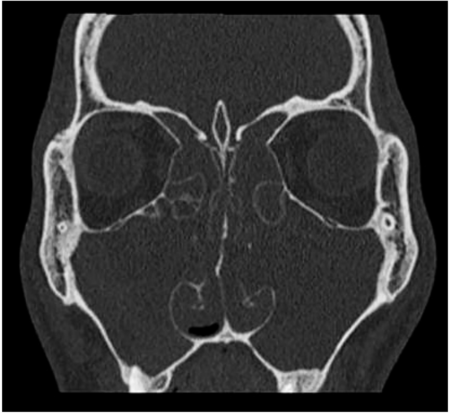
| CT sinuses (coronal) | Extent of disease, surgical planning; shows sinus opacification |
| MRI | Differentiates polyp from tumour; soft tissue detail |
| Allergy testing | IgE levels, skin prick tests - identify atopic contributors |
| Spirometry | Assess concomitant asthma |
| Sweat chloride / CFTR gene | Rule out cystic fibrosis, especially in children |
| Aspirin challenge | Confirm AERD/Samter's triad |
| Histopathology | Exclude malignancy; confirm eosinophilic vs. neutrophilic pattern |
Summary Box
- Nasal polyps = oedematous grape-like protrusions from osteomeatal complex
- Core mechanism: chronic Th2-eosinophilic inflammation (in 80% of Western cases)
- Exact cause unknown; bacteria (S. aureus superantigens), allergy, epithelial barrier defects all implicated
- Key symptoms: bilateral obstruction + anosmia + rhinorrhoea
- Hallmark exam sign: pale, insensate, non-bleeding, mobile masses from middle meatus
- Epistaxis = NOT a feature; its presence demands further workup
- Key associations: asthma (30-71%), Samter's triad (AERD), AFRS, cystic fibrosis
- Bilateral = rule; unilateral = investigate for other pathology
- Scott-Brown's Otorhinolaryngology Head & Neck Surgery Vol 1, Chapter 95 (Nasal Polyps, pp.1085-1088)
- Cummings Otolaryngology Head and Neck Surgery, Chapter 41 (Etiology and Pathogenesis of CRS, pp.824-831)
Clinical features and management of antrochonal polyp
Antrochoanal Polyp (ACP) - Clinical Features and Management
Definition
Key Distinguishing Features from Ordinary Nasal Polyps
| Feature | Antrochoanal Polyp | Ordinary Nasal Polyp (CRSwNP) |
|---|---|---|
| Number | Solitary | Multiple |
| Laterality | Unilateral | Bilateral |
| Origin | Maxillary antrum | Ethmoid sinuses / osteomeatal complex |
| Extension | Choana → nasopharynx | Nasal cavity only |
| Age | Young adults, adolescents, children | Adults (peak 30-60 years) |
| Association with allergy | Not strongly linked | Often linked |
| Association with asthma | Not typical | Common (30-71%) |
Structure / Gross Pathology
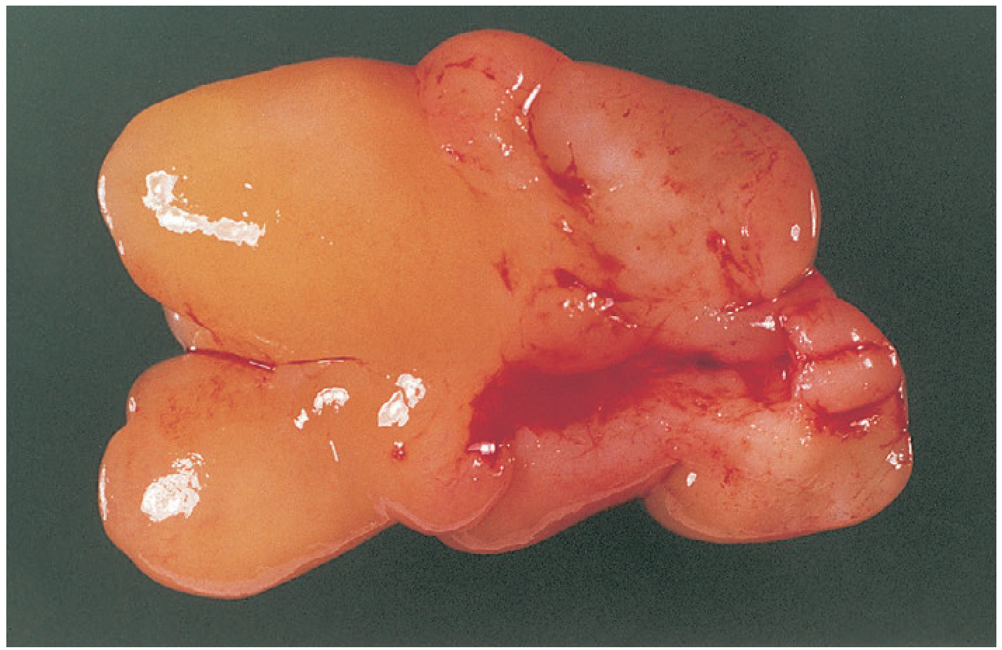
- Antral part - a cystic portion within the maxillary sinus (often the dominant, larger part)
- Isthmus - a narrow neck passing through the accessory ostium or posterior fontanelle of the maxillary sinus
- Nasal/choanal part - the fleshy, pedunculated portion that fills the nasal cavity and extends posteriorly to the choana
Gross Appearance
Clinical Features
Symptoms
- The dominant presenting symptom
- Progressive, unilateral (this is the key differentiating point from CRSwNP polyps)
- Becomes increasingly severe as the polyp enlarges
- Watery or mucoid, typically unilateral
- Post-nasal drip may be present
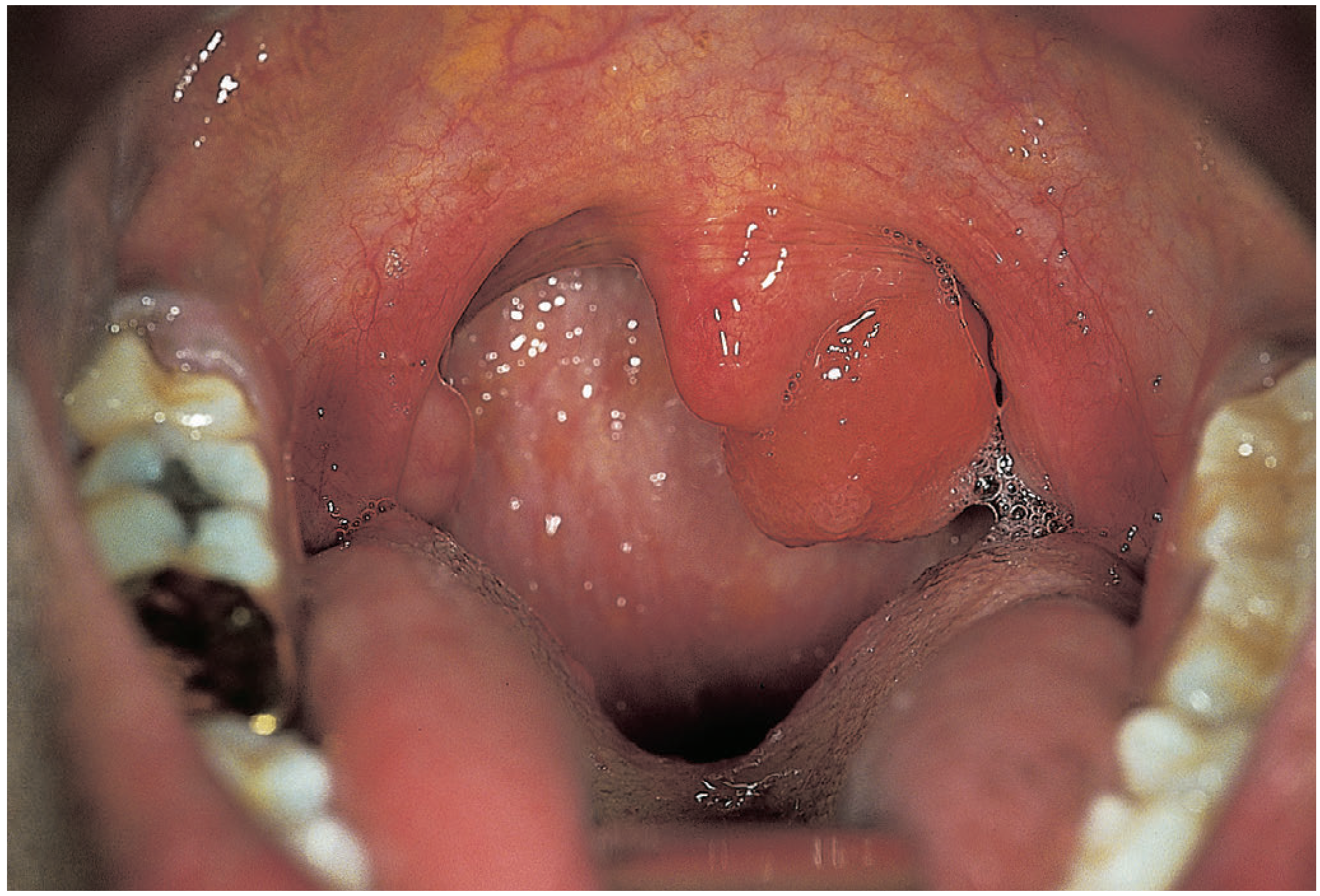
- As the polyp prolapses posteriorly into the nasopharynx and oropharynx, patients may feel a mass hanging in the back of the throat
- They may notice a smooth fleshy lump on opening the mouth
- From obstruction of the nasal airway on the affected side
- Less prominent than in bilateral CRSwNP disease
- Especially in children, where the nasopharynx is proportionally smaller and more easily obstructed
- Can cause obstructive sleep symptoms
- From nasopharyngeal obstruction
Signs
- A single, smooth, pale/grey-white, soft, mobile, glistening mass filling one side of the nasal cavity
- Non-tender on probing, non-bleeding (avascular surface)
- Typically occupying the posterior part of the nasal cavity more than the anterior
- The choanal component visible as a smooth mass in the nasopharynx / postnasal space
- In large polyps: visible in the oropharynx on opening the mouth
- Large polyps may be seen as a fleshy pale mass hanging from the soft palate region / oropharynx
Imaging
CT Sinuses (Gold Standard)
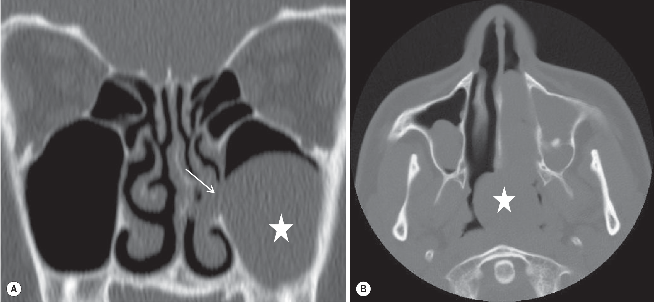
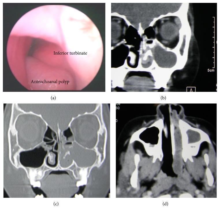
- Opacification of the ipsilateral maxillary sinus (homogeneous soft tissue density)
- Widening of the accessory ostium or posterior fontanelle
- Soft tissue mass extending through the widened ostium into the middle meatus → filling the nasal cavity → passing through the choana → filling the postnasal space
- The antral component is typically larger and often cystic
- No bone destruction (distinguishes from malignancy)
Important Note on Imaging
Differential Diagnosis
| Condition | Key Differentiating Feature |
|---|---|
| CRSwNP (ordinary polyps) | Bilateral, multiple, arise from ethmoid/OMC |
| Juvenile Nasopharyngeal Angiofibroma | Males only; highly vascular; bleeds profusely on probing; CT shows intense enhancement |
| Inverted Papilloma | Adults; unilateral; tendency to malignant transformation; CT shows bony remodelling |
| Meningocele/Encephalocele | Children; midline; pulsatile; connects to intracranial space; high-resolution CT skull base needed |
| Malignancy | Hard, irregular; bone destruction on CT; pain; epistaxis |
| Nasal septum deviation | Bilateral, no mass lesion |
Management
Principle
Surgical Options
1. Functional Endoscopic Sinus Surgery (FESS) - First-Line / Preferred
- Uncinectomy and middle meatal antrostomy (widening of the natural maxillary ostium)
- Endoscopic delivery and removal of the nasal portion from the posterior choana
- Endoscopic removal of the antral cystic component through the widened middle meatal antrostomy using curved powered instruments or grasping forceps
- A small, temporary inferior meatal antrostomy may be created to improve access to the antral portion
- Mega-antrostomy (extension of middle meatal antrostomy to the floor of the nasal cavity by resecting the mid-segment of the inferior turbinate) - an option for difficult cases
- Curved powered instruments (microdebriders) and angled endoscopes are very helpful in removing the maxillary component
2. Caldwell-Luc Approach (Open) - Reserved for Difficult/Recurrent Cases
- An external approach via a sublabial incision accessing the maxillary antrum through the anterior wall
- Used historically as the standard approach but now largely replaced by FESS
- Indications: incomplete removal at FESS, recurrence, or situations where endoscopic access is insufficient
- Traditional Caldwell-Luc (stripping of maxillary sinus mucosa) is NOT necessary for ACP - only the polyp/cystic component needs removal (Cummings Otolaryngology)
3. Sublabial / Transcanine / Inferior Meatus Approach
- Used in selected patients as a supplementary approach to access the antral portion
- Can be combined with endoscopy for difficult cases
Endoscopic Surgical Specimen
Recurrence
- Low recurrence when the antral cystic component is completely removed at surgery
- High recurrence when only the nasal portion is avulsed (simple polypectomy), leaving the antral root intact
- This is the most common reason for surgical failure - incomplete removal of the maxillary sinus component
Summary Box
Antrochoanal polyp:
- Solitary, unilateral, dumbbell-shaped benign polyp
- Origin: maxillary antrum → posterior fontanelle → nasal cavity → choana → nasopharynx (± oropharynx)
- Commonest in young adults / adolescents / children
- Key symptom: progressive unilateral nasal obstruction + sensation of mass in throat
- NOT associated with bilateral disease, asthma, or aspirin sensitivity
- Pale, avascular, insensate, non-bleeding on probing
- CT: dumbbell soft tissue mass from antrum through ostium to postnasal space, no bony destruction
- Always exclude inverted papilloma / malignancy (biopsy / imaging)
- Treatment: FESS - complete removal including antral cystic component is essential to prevent recurrence
- Caldwell-Luc reserved for recurrence or failed FESS
- Bailey and Love's Short Practice of Surgery, 28th Ed., Chapters 51 & 52
- Cummings Otolaryngology Head and Neck Surgery, Chapter on Surgical Indications in Rhinosinusitis, p.69
- Grainger & Allison's Diagnostic Radiology, Chapter 61 (Sinonasal Radiology), p.1603
Clinical features and management of ethmoidal polyposis
ethmoidal polyposis nasal endoscopy CT coronal bilateral polyps