Bones of lower limb
Reading File
Finding Sources
Searching Images
bones of lower limb anatomy diagram
Finding Sources
Finding Sources
Reading File
Reading File
Reading File
Loading Image
Finding Sources
Finding Sources
Finding Sources
Reading File
Reading File
Reading File
Reading File
Reading File
Reading File
Loading Image
Loading Image
Loading Image
Loading Image
Loading Image
Loading Image
Loading Image
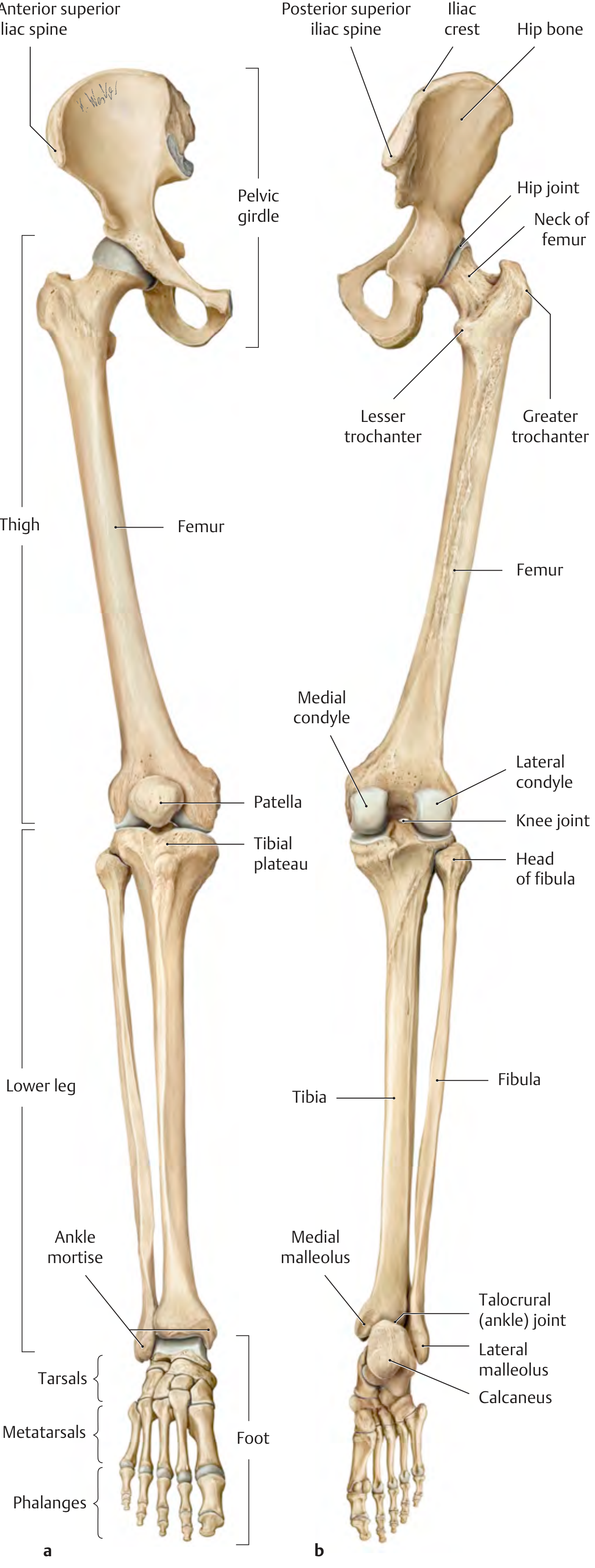
Here is a comprehensive overview of the bones of the lower limb, based on the THIEME Atlas of Anatomy: General Anatomy and Musculoskeletal System.
Overview of the Skeleton of the Lower Limb
The lower limb skeleton consists of two parts:
- Pelvic girdle — connects the free limb to the axial skeleton
- Free limb — thigh, leg, and foot
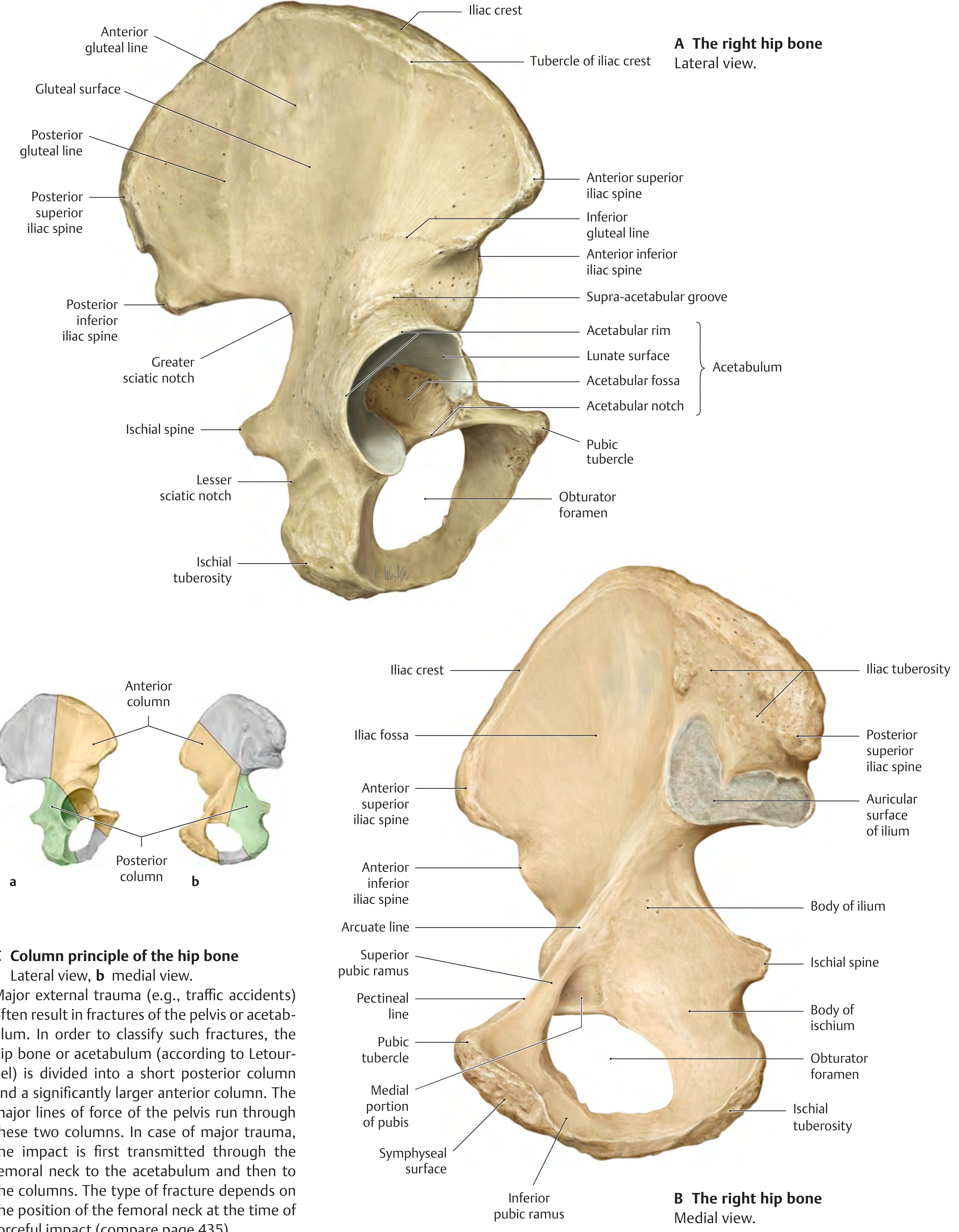
1. Pelvic Girdle — Hip Bone (Os Coxae / Innominate Bone)
Each hip bone (os coxa) is formed by the fusion of three bones at the acetabulum:
| Bone | Contribution to Acetabulum | Key Features |
|---|---|---|
| Ilium | 2/5 | Iliac crest, ASIS, AIIS, PSIS, PIIS, iliac fossa, gluteal surface (with anterior, posterior, and inferior gluteal lines), auricular surface |
| Ischium | 2/5 | Ischial tuberosity, ischial spine, greater & lesser sciatic notches, body of ischium |
| Pubis | 1/5 | Pubic tubercle, superior & inferior pubic rami, symphyseal surface, pectineal line, obturator foramen |
- The triradiate (Y-shaped) cartilage fuses at 14–16 years
- The two hip bones + sacrum + pubic symphysis form the bony pelvic ring
2. Thigh — Femur
The femur is the longest and strongest bone in the body.
Proximal Femur
- Head — articulates with acetabulum (hip joint); has a fovea for the ligament of the head
- Neck — connects head to shaft; CCD angle (centrum-collum-diaphysis angle) = ~126° in adults (150° at birth, decreases with age)
- Greater trochanter — lateral, site for gluteal muscle attachment
- Lesser trochanter — posteromedial, site for iliopsoas attachment
- Intertrochanteric line (anterior) and intertrochanteric crest (posterior)
Shaft
- Posterior ridge = linea aspera (medial and lateral lips)
- Gives rise to the medial and lateral supracondylar ridges
Distal Femur
- Medial and lateral condyles — articulate with tibial plateau
- Intercondylar fossa/notch (posteriorly)
- Patellar surface (femoral trochlea) — anteriorly, for patella
- Medial and lateral epicondyles
- Adductor tubercle — on medial epicondyle
Clinical note: Coxa vara (↓ CCD angle) → increased tensile stress; Coxa valga (↑ CCD angle) → increased compressive stress.
3. Patella
- Largest sesamoid bone in the body
- Embedded in the quadriceps tendon
- Articulates with the femoral trochlea
- Has a larger lateral facet and smaller medial facet (variants: Wiberg classification)
- Protects the knee joint and improves the mechanical advantage of quadriceps
4. Leg — Tibia and Fibula
Both bones articulate proximally (synovial tibiofibular joint) and distally (tibiofibular syndesmosis), connected throughout by the crural interosseous membrane.
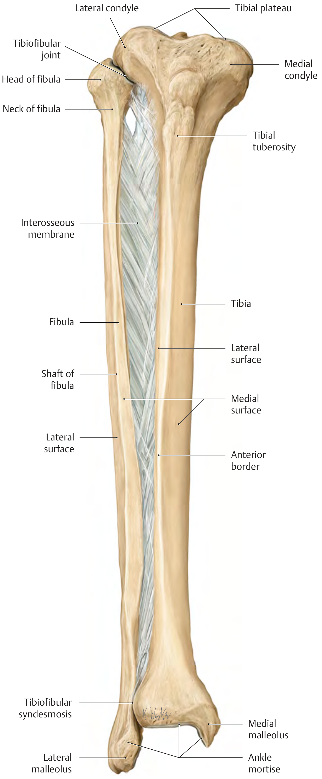
Tibia (medial, weight-bearing bone)
| Region | Features |
|---|---|
| Proximal | Medial & lateral condyles, tibial plateau (5–7° posterior slope), tibial tuberosity (patellar ligament insertion), Gerdy's tubercle (IT band) |
| Shaft | Anterior border (subcutaneous, palpable), medial surface, lateral surface |
| Distal | Medial malleolus, inferior articular surface (ankle mortise) |
- Tibial torsion: transverse axis of ankle mortise is rotated ~23° laterally relative to the tibial plateau → physiologic toe-out
Fibula (lateral, non-weight-bearing)
| Region | Features |
|---|---|
| Proximal | Head of fibula, neck of fibula, styloid process |
| Shaft | Shaft of fibula, lateral surface |
| Distal | Lateral malleolus (forms lateral wall of ankle mortise) |
- Primary role: muscle attachment and ankle stability (via syndesmosis)
5. Foot — 26 Bones (+ 2 Sesamoids)
The foot is divided into three functional regions:
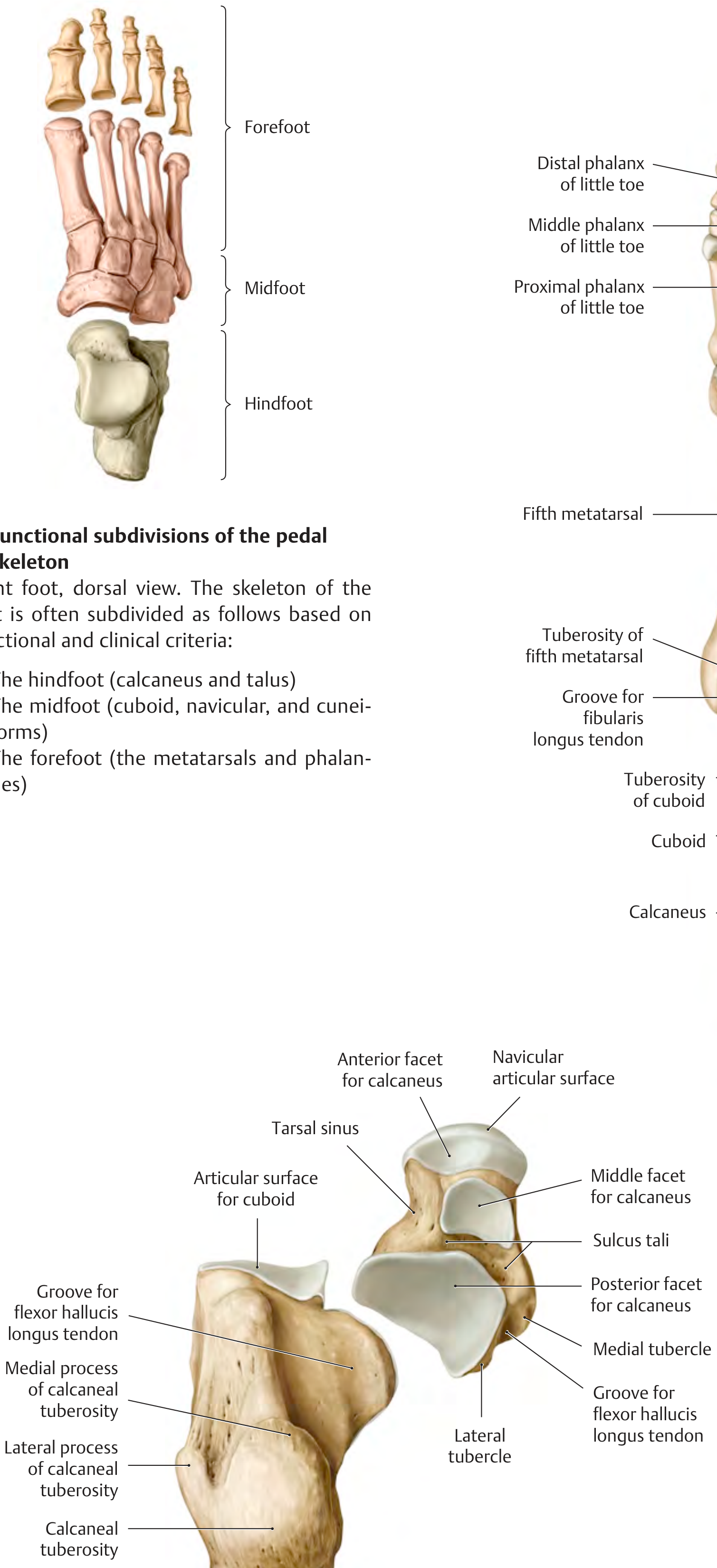
A. Hindfoot — 2 Tarsal Bones
| Bone | Key Features |
|---|---|
| Talus | Trochlea (superior articular surface for ankle), head (articulates with navicular), neck; no muscle attachments; has tarsal sinus separating subtalar joint chambers |
| Calcaneus | Largest tarsal bone; calcaneal tuberosity (Achilles insertion); sustentaculum tali (shelf supporting talus, ~1.5 cm below medial malleolus); articular facets for talus and cuboid |
B. Midfoot — 5 Tarsal Bones
| Bone | Notes |
|---|---|
| Navicular | Medial column; tuberosity is site of tibialis posterior insertion |
| Cuboid | Lateral column; groove for fibularis longus tendon |
| Medial cuneiform | Largest cuneiform |
| Intermediate cuneiform | Smallest cuneiform |
| Lateral cuneiform | Articulates with 3rd metatarsal |
C. Forefoot — 19 Bones
| Segment | Count | Details |
|---|---|---|
| Metatarsals | 5 | Each has a base, shaft, and head; 5th metatarsal has a prominent tuberosity (styloid process) — common avulsion fracture site |
| Phalanges | 14 | Great toe: 2 (proximal + distal); toes 2–5: 3 each (proximal, middle, distal) |
| Sesamoids | 2 | Under 1st metatarsal head (within flexor hallucis brevis tendons) |
Accessory tarsal bones (inconstant ossicles) exist and must be distinguished from fractures. The most clinically significant is the os tibiale externum (external tibial bone), which can cause pain with tight footwear.
Summary Table
| Region | Bone(s) |
|---|---|
| Pelvic girdle | Hip bone (ilium + ischium + pubis) |
| Thigh | Femur |
| Knee | Patella |
| Leg | Tibia, Fibula |
| Hindfoot | Talus, Calcaneus |
| Midfoot | Navicular, Cuboid, 3 Cuneiforms |
| Forefoot | 5 Metatarsals, 14 Phalanges (+ 2 sesamoids) |
| Total | ~31 bones per limb |
Source: THIEME Atlas of Anatomy — General Anatomy and Musculoskeletal System, pp. 429–446
This is a shared conversation. Sign in to Orris to start your own chat.