Explain plaque Psoriasis for b pharm student level

Reading File
Finding Sources
Searching Images

plaque psoriasis skin lesion clinical photo

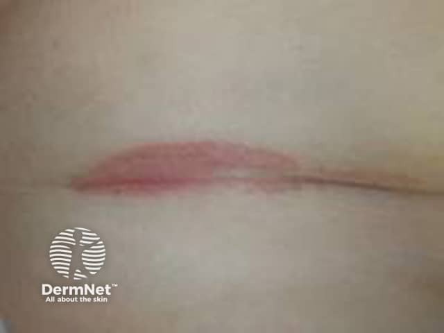
Clinical photography of the antecubital fossa region shows a single erythematous plaque within a flexural skin fold. The lesion is well-demarcated, with a smooth pink-to-erythematous surface and minimal scales evident, reflecting inverse/ flexural psoriasis features in moist environments where scaling is less conspicuous. The plaque lies centrally within the fold, with intact surrounding skin and no obvious crusting or excoriation. Lighting emphasizes a uniform coloration without effusion or ulceration. The regional skin may appear slightly edematous or glistening from moisture, a common appearance in intertriginous psoriasis. In clinical practice, such lesions are often symmetric or multipart and may co-occur with plaques on extensor surfaces; history of chronic plaque psoriasis or family history supports the diagnosis. Differential considerations include irritant dermatitis, candidal intertrigo, or seborrheic dermatitis; biopsy would reveal epidermal hyperplasia with parakeratosis and neutrophilic microabscesses typical of plaque psoriasis, though histology is not demonstrated here. The image is intended for dermatology education, aiding recognition of flexural psoriasis, differentiation from dermatitis, and guiding topical therapy, including high-potency corticosteroids or vitamin D analogs with occlusion, while avoiding excessive moisture. This photo supports image-based teaching, clinical pattern recognition, and correlation with patient history in practice and research.

Clinical photography of the antecubital fossa region shows a single erythematous plaque within a flexural skin fold. The lesion is well-demarcated, with a smooth pink-to-erythematous surface and minimal scales evident, reflecting inverse/ flexural psoriasis features in moist environments where scaling is less conspicuous. The plaque lies centrally within the fold, with intact surrounding skin and no obvious crusting or excoriation. Lighting emphasizes a uniform coloration without effusion or ulceration. The regional skin may appear slightly edematous or glistening from moisture, a common appearance in intertriginous psoriasis. In clinical practice, such lesions are often symmetric or multipart and may co-occur with plaques on extensor surfaces; history of chronic plaque psoriasis or family history supports the diagnosis. Differential considerations include irritant dermatitis, candidal intertrigo, or seborrheic dermatitis; biopsy would reveal epidermal hyperplasia with parakeratosis and neutrophilic microabscesses typical of plaque psoriasis, though histology is not demonstrated here. The image is intended for dermatology education, aiding recognition of flexural psoriasis, differentiation from dermatitis, and guiding topical therapy, including high-potency corticosteroids or vitamin D analogs with occlusion, while avoiding excessive moisture. This photo supports image-based teaching, clinical pattern recognition, and correlation with patient history in practice and research.

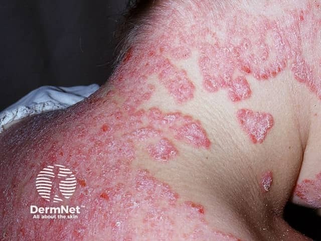
Clinical photography of facial skin demonstrates classic psoriatic plaques involving the malar region and surrounding cheeks. The lesions are erythematous to violaceous, well-demarcated, and markedly scaly with silvery-white scale, consistent with plaque-type psoriasis (psoriasis vulgaris). Involved areas show surface coarseness, thickening (acanthosis) and mild induration, while adjacent skin remains relatively spared. Perilesional inflammation creates a ring of erythema, and some plaques exhibit superficial fissuring. The distribution is facial rather than scalp, trunk, or limbs, suggesting involvement limited to the head-and-face region in this image. No vesiculation or purulence is evident. The appearance may mimic seborrheic dermatitis, eczema, or contact dermatitis; however, the well-circumscribed plaques with thick scale and the typical red-silver presentation strongly favor psoriasis. Skin photo shows high-contrast textural differences between lesional and non-lesional skin. Clinically, facial psoriasis can be challenging due to cosmetic and functional concerns, including itching and social impact. This image supports differential diagnosis discussions in dermatology education and serves as a reference for recognizing facial plaque psoriasis, assessing severity, and guiding treatment planning (topical steroids, vitamin D analogs, or phototherapy). It is useful for teaching photodocumentation, lesion morphology, and pattern recognition in psoriasis spectrum. This image thus enhances reasoning for diagnosis, prognosis, and management planning.

Clinical photography of facial skin demonstrates classic psoriatic plaques involving the malar region and surrounding cheeks. The lesions are erythematous to violaceous, well-demarcated, and markedly scaly with silvery-white scale, consistent with plaque-type psoriasis (psoriasis vulgaris). Involved areas show surface coarseness, thickening (acanthosis) and mild induration, while adjacent skin remains relatively spared. Perilesional inflammation creates a ring of erythema, and some plaques exhibit superficial fissuring. The distribution is facial rather than scalp, trunk, or limbs, suggesting involvement limited to the head-and-face region in this image. No vesiculation or purulence is evident. The appearance may mimic seborrheic dermatitis, eczema, or contact dermatitis; however, the well-circumscribed plaques with thick scale and the typical red-silver presentation strongly favor psoriasis. Skin photo shows high-contrast textural differences between lesional and non-lesional skin. Clinically, facial psoriasis can be challenging due to cosmetic and functional concerns, including itching and social impact. This image supports differential diagnosis discussions in dermatology education and serves as a reference for recognizing facial plaque psoriasis, assessing severity, and guiding treatment planning (topical steroids, vitamin D analogs, or phototherapy). It is useful for teaching photodocumentation, lesion morphology, and pattern recognition in psoriasis spectrum. This image thus enhances reasoning for diagnosis, prognosis, and management planning.

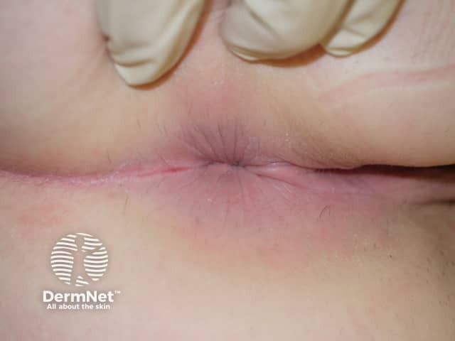
Clinical photograph of external genital skin showing an erythematous, well-demarcated plaque in an intertriginous fold. The lesion is pink to red, with a smooth surface and minimal peripheral scaling within moist skin areas; a slight fissure is seen along the crease. The surrounding skin is mildly inflamed but otherwise unremarkable. This appearance is characteristic of genital psoriasis, most compatible with inverse psoriasis in moist genital folds where scaling is less prominent than on extensor surfaces. Differential diagnoses include candidal balanitis or vulvitis, irritant contact dermatitis, lichen sclerosus, eczema, and less commonly erosive lesions. Clinical context such as past or concurrent psoriasis elsewhere, absence of itch or burning, and response to topical steroids can help confirmation. Histopathology would typically show epidermal hyperplasia with parakeratosis, regular elongation of rete ridges, and a dermal perivascular lymphocytic infiltrate if biopsy were performed; in practice, biopsy is not required for typical genital psoriasis. The diagnostic significance lies in recognizing genito-pubic psoriasis to guide therapy and avoid misdiagnosis as infection. This image is useful for education on genital psoriasis recognition, differential diagnosis, and management considerations including topical corticosteroids, vitamin D analogs, and calcineurin inhibitors, with referrals for systemic therapy if extensive disease. Clinical photo for reference.

Clinical photograph of external genital skin showing an erythematous, well-demarcated plaque in an intertriginous fold. The lesion is pink to red, with a smooth surface and minimal peripheral scaling within moist skin areas; a slight fissure is seen along the crease. The surrounding skin is mildly inflamed but otherwise unremarkable. This appearance is characteristic of genital psoriasis, most compatible with inverse psoriasis in moist genital folds where scaling is less prominent than on extensor surfaces. Differential diagnoses include candidal balanitis or vulvitis, irritant contact dermatitis, lichen sclerosus, eczema, and less commonly erosive lesions. Clinical context such as past or concurrent psoriasis elsewhere, absence of itch or burning, and response to topical steroids can help confirmation. Histopathology would typically show epidermal hyperplasia with parakeratosis, regular elongation of rete ridges, and a dermal perivascular lymphocytic infiltrate if biopsy were performed; in practice, biopsy is not required for typical genital psoriasis. The diagnostic significance lies in recognizing genito-pubic psoriasis to guide therapy and avoid misdiagnosis as infection. This image is useful for education on genital psoriasis recognition, differential diagnosis, and management considerations including topical corticosteroids, vitamin D analogs, and calcineurin inhibitors, with referrals for systemic therapy if extensive disease. Clinical photo for reference.

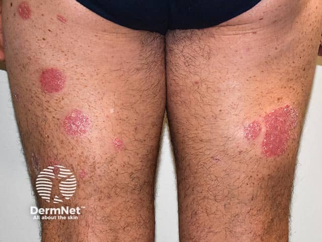
Clinical photography of bilateral lower legs demonstrates features typical of plaque psoriasis. Modality: Clinical photograph; technique: Standard color digital image captured in visible light without dermoscopic magnification or contact; staining and contrast agents not used. Anatomical localization: skin of the legs, pretibial and extensor regions, bilateral (anterior aspects). Visual impression: well-circumscribed, erythematous plaques with diffuse or loosely adherent silvery scales; plaques are raised with well-defined borders and variable size, some larger patches measuring several centimeters in diameter. The distribution is symmetric and predominantly on extensor surfaces, consistent with plaque psoriasis vulgaris; surrounding skin may show mild xerosis or epidermal scaling. Pathology cannot be confirmed from a photo alone, but clinical correlation with history and examination would favor psoriasis; differential diagnoses include eczema/atopic dermatitis, tinea corporis, pityriasis rosea, and nummular dermatitis. Clinical significance: this image aids diagnosis, patient education, and monitoring of treatment response over time; it supports documentation for dermatology records and can be used in educational or research contexts to illustrate chronic plaque psoriasis phenotype. Potential clinical use cases: baseline assessment, response evaluation to topical therapies, phototherapy planning, and AI-driven lesion recognition datasets. Ethical note: ensure patient consent for image use in training datasets.

Clinical photography of bilateral lower legs demonstrates features typical of plaque psoriasis. Modality: Clinical photograph; technique: Standard color digital image captured in visible light without dermoscopic magnification or contact; staining and contrast agents not used. Anatomical localization: skin of the legs, pretibial and extensor regions, bilateral (anterior aspects). Visual impression: well-circumscribed, erythematous plaques with diffuse or loosely adherent silvery scales; plaques are raised with well-defined borders and variable size, some larger patches measuring several centimeters in diameter. The distribution is symmetric and predominantly on extensor surfaces, consistent with plaque psoriasis vulgaris; surrounding skin may show mild xerosis or epidermal scaling. Pathology cannot be confirmed from a photo alone, but clinical correlation with history and examination would favor psoriasis; differential diagnoses include eczema/atopic dermatitis, tinea corporis, pityriasis rosea, and nummular dermatitis. Clinical significance: this image aids diagnosis, patient education, and monitoring of treatment response over time; it supports documentation for dermatology records and can be used in educational or research contexts to illustrate chronic plaque psoriasis phenotype. Potential clinical use cases: baseline assessment, response evaluation to topical therapies, phototherapy planning, and AI-driven lesion recognition datasets. Ethical note: ensure patient consent for image use in training datasets.

Reading File
Finding Sources
Finding Sources
Reading File
Reading File
Finding Sources
Reading File
Reading File
Finding Sources
Reading File
Finding Sources
Loading Image
Finding Sources
Finding Sources
Reading File
Reading File
I now have comprehensive information from Fitzpatrick's Dermatology, Robbins Pathology, Goodman & Gilman's Pharmacology, and Family Medicine textbooks. Let me compose a well-structured B.Pharm-level explanation.

Plaque Psoriasis - B.Pharm Level Notes


1. Definition and Overview

Plaque psoriasis (psoriasis vulgaris) is a chronic, immune-mediated inflammatory skin disease characterized by discrete, well-demarcated erythematous (red) plaques covered with adherent silvery-white scales. It is the most common form of psoriasis, accounting for 80-90% of all psoriasis cases.
  • Prevalence: 1-2% of the U.S. population
  • Age of onset: Two peaks - 16 to 22 years and 57 to 60 years
  • It is a systemic disease - skin inflammation is the most visible sign, but internal comorbidities exist
(Textbook of Family Medicine 9e; Fitzpatrick's Dermatology)

2. Etiology and Risk Factors

Psoriasis results from a combination of genetic and environmental factors:
FactorDetails
GeneticsPolygenic inheritance; specific HLA gene variants (HLA-Cw6 is most associated). If both parents have psoriasis, risk ~50%; one parent affected, risk ~16%
Immune dysregulationAberrant activation of T lymphocytes, dendritic cells
TriggersSkin trauma (Koebner phenomenon), infections (streptococcal pharyngitis triggers guttate psoriasis), stress, drugs (lithium, beta-blockers, antimalarials), smoking, alcohol
Withdrawal of systemic steroidsCan provoke pustular psoriasis
(Robbins, Cotran & Kumar Pathologic Basis of Disease; Textbook of Family Medicine 9e)

3. Pathogenesis (Mechanism)

This is the most pharmacologically important section for B.Pharm students.

Step-by-step Immune Cascade:

Step 1 - Antigen Presentation: An unknown trigger (possibly self-antigen or microbial antigen) activates antigen-presenting cells (APCs) - particularly dendritic cells in the skin.
Step 2 - T Cell Activation: APCs activate CD4+ Th1 cells and Th17 cells (and CD8+ cytotoxic T cells accumulate in the epidermis). This is the central immune abnormality.
Step 3 - Cytokine Release (the "cytokine soup"): Activated T cells and dendritic cells release a storm of pro-inflammatory cytokines:
CytokineSourceKey Effect
TNF-αDendritic cells, T cellsMaster pro-inflammatory cytokine; activates keratinocytes
IL-17 (key cytokine)Th17 cellsPromotes neutrophil recruitment and keratinocyte activation; drives hyperproliferation
IL-23Dendritic cellsRequired for Th17 cell development; composed of p19 + p40 subunits
IL-12APCsDrives Th1 differentiation
IFN-γTh1 cellsPro-inflammatory at keratinocyte level
IL-22T cells, innate lymphoid cellsDrives keratinocyte hyperplasia and migration
Step 4 - Keratinocyte Hyperproliferation: Cytokines drive markedly reduced basal keratinocyte transit times - normal skin cell turnover is ~28 days; in psoriasis it is reduced to 3-4 days. This results in thickened, poorly differentiated skin.
Step 5 - Vascular Remodeling: Capillary loops in the dermal papillae elongate, dilate, and touch the epidermis ("kissing" phenomenon). Leukocytes exit through these remodeled vessels into the superficial epidermis ("squirting papillae").
The importance of this cytokine cascade is proven by treatment: TNF-α inhibitors and IL-17 inhibitors produce excellent clinical responses.
(Fitzpatrick's Dermatology Vol. 1, 2; Robbins Pathology; Goodman & Gilman's Pharmacology)

4. Histopathology (Microscopic Features)

Know these for exams:
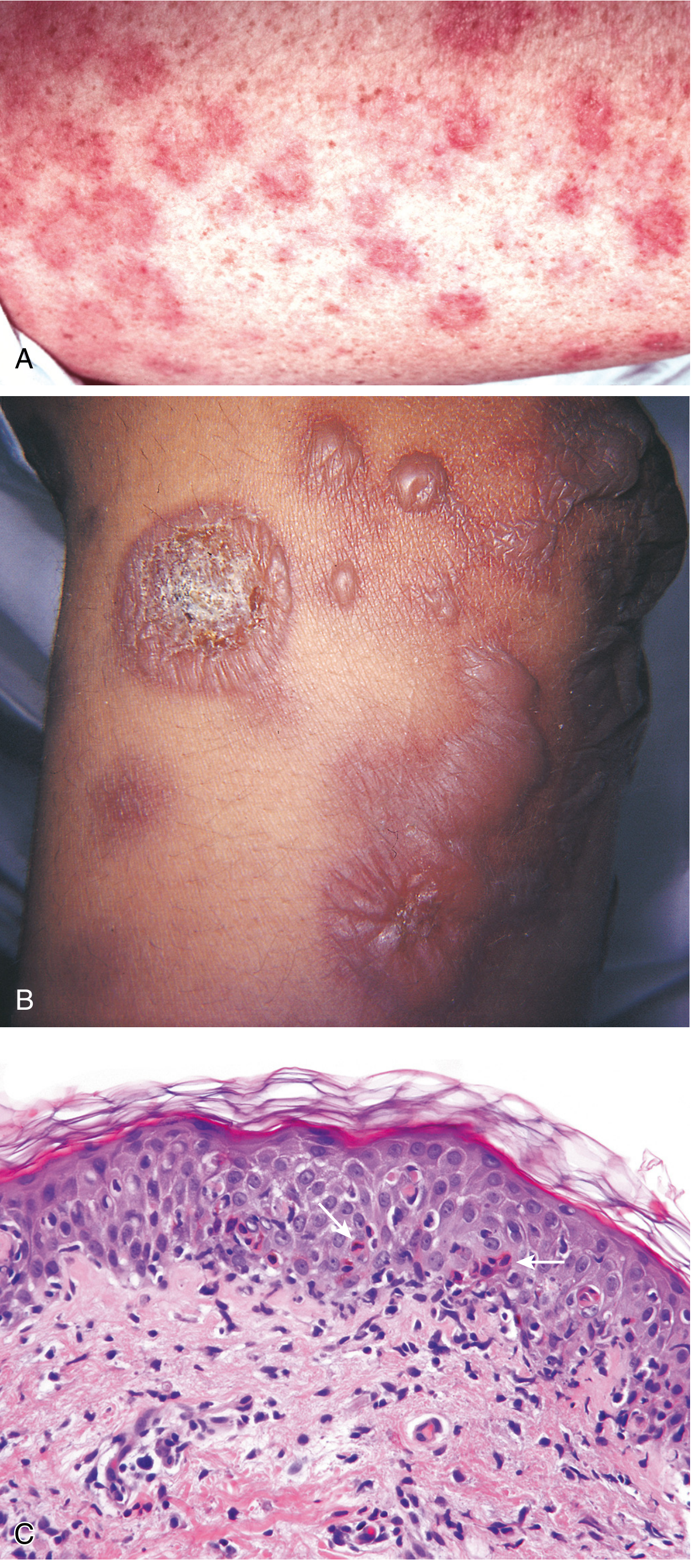
Psoriasis histopathology and clinical lesions - Robbins Pathology
Fig: Psoriasis - erythematous plaques (A/B) and histology showing epidermal hyperplasia with Munro microabscesses (C) - from Robbins, Cotran & Kumar Pathologic Basis of Disease
FeatureExplanation
AcanthosisEpidermal thickening; rete ridges elongated downward like "test tubes in a rack"
ParakeratosisRetention of nuclei in stratum corneum (incomplete keratinization)
Absent/thin stratum granulosumLost due to rapid cell turnover
Munro microabscessesSmall neutrophil aggregates in the parakeratotic stratum corneum - pathognomonic
Spongiform pustulesNeutrophil aggregates in superficial epidermis
Thinned suprapapillary platesEpidermis over dermal papillae tips is very thin
Dilated dermal papillary vesselsTortuous, elongated capillary loops
(Robbins, Cotran & Kumar Pathologic Basis of Disease)

5. Clinical Features

Lesion Description (Classic Morphology):

  • Well-demarcated, raised erythematous plaques
  • Covered with adherent silvery-white (micaceous) scales
  • Plaques vary from small papules to large confluent areas

Sites of Predilection:

  • Extensor surfaces - elbows and knees (most classic)
  • Scalp, lower back (lumbosacral area)
  • Umbilicus, intergluteal cleft
  • Nails, genitalia, face

Clinical Signs to Remember:

SignDescription
Auspitz signRemoval of scale reveals multiple minute bleeding points (dilated papillary vessels touch the thin suprapapillary epidermis)
Koebner phenomenonNew psoriatic plaques appear at sites of skin trauma (cuts, scratches, burns)
Candle sign (Grattage sign)Scraping a plaque produces silvery scales resembling candle wax

Types of Psoriasis (for context):

TypeKey Feature
Plaque (vulgaris)80-90%, classic silvery plaques on extensor surfaces
GuttateSmall drop-like lesions; often post-streptococcal in children
InverseFlexural areas (axilla, groin); minimal scale due to moisture
ErythrodermicGeneralized erythema and scales; medical emergency
PustularSterile pustules; can be generalized (von Zumbusch)
Nail psoriasisPitting, onycholysis, "oil spots"
Psoriatic arthritis~15% of patients; affects hands, feet, knees
(Textbook of Family Medicine 9e; Goldman-Cecil Medicine)
Clinical plaque psoriasis on legs showing erythematous plaques with silvery scales
Typical plaque psoriasis on legs: bilateral erythematous, well-demarcated plaques with silvery scales on extensor surfaces

6. Diagnosis

Diagnosis is primarily clinical (based on appearance and distribution). Investigations include:
  • Skin biopsy - confirms histopathology (Munro microabscesses, acanthosis, parakeratosis)
  • KOH preparation - to rule out fungal infection
  • No specific blood test for diagnosis; ESR, CRP may be elevated
  • PASI score (Psoriasis Area Severity Index) - used to grade severity for treatment decisions

7. Treatment (Pharmacology Focus)

Treatment follows a step-up approach based on severity:

Step 1 - Mild Psoriasis: Topical Agents

DrugMechanismNotes
Topical corticosteroids (e.g., clobetasol)Reduce inflammation; suppress immune cellsFirst-line; 68-89% show clear improvement with ultrahigh-potency (clobetasol)
Calcipotriol/Calcipotriene (Vitamin D analog)Inhibits keratinocyte proliferation; promotes differentiationComparable efficacy to potent steroids
Tazarotene (Retinoid)Normalizes keratinocyte differentiationSlightly more adverse effects
Combination (steroid + calcipotriol)Additive anti-inflammatory + antiproliferativeMost promising topical approach; fewer side effects
Coal tarAnti-inflammatory; reduces keratinocyte proliferationOld but still used; smelly
Tacrolimus (calcineurin inhibitor)Suppresses T-cell activationUsed for facial and inverse psoriasis

Step 2 - Moderate-to-Severe: Systemic (Non-biologic)

DrugMechanismKey Notes
Methotrexate (MTX)Antimetabolite (inhibits dihydrofolate reductase); antiproliferative and immunosuppressiveOral, 5-15 mg/week; effective for widespread disease; hepatotoxic with long-term use
Acitretin (Oral retinoid)Normalizes keratinocyte differentiationUsed for generalized/palmoplantar psoriasis not responding to topicals; teratogenic
CyclosporineCalcineurin inhibitor; selectively suppresses T lymphocytes (blocks IL-2 production)Remarkably effective; nephrotoxic with long-term use
Narrow-band UVB phototherapyInduces apoptosis of T cells; immunomodulatorySafer and more effective than broadband UVB
PUVA (Psoralen + UVA)Psoralen intercalates DNA; UVA activation inhibits cell proliferationEffective but risk of photodamage and skin cancer

Step 3 - Moderate-to-Severe: Biologic Agents (Targeted)

These are the most pharmacologically interesting drugs - they are monoclonal antibodies or fusion proteins that target specific cytokines in the pathogenic cascade:

Anti-TNF-α Agents:

DrugClassRoute
EtanerceptHuman p75 TNF receptor-Fc fusion proteinSC
AdalimumabHuman IgG1 monoclonal antibodySC
InfliximabChimeric IgG1 monoclonal antibodyIV
Certolizumab pegolPEGylated humanized Fab fragmentSC

Anti-IL-17 Agents (highly effective in psoriasis):

DrugTargetRoute
SecukinumabIL-17ASC
IxekizumabIL-17ASC
BrodalumabIL-17 receptor ASC
BimekizumabIL-17A and IL-17FSC

Anti-IL-12/23 and Anti-IL-23 Agents:

DrugTargetNotes
UstekinumabIL-12 and IL-23 (p40 subunit)PASI75 achieved in >66% at 45 mg and >75% at 90 mg by week 12
Guselkumab, Risankizumab, TildrakizumabIL-23 (p19 subunit)Newer IL-23 specific agents
(Goodman & Gilman's Pharmacology; Fitzpatrick's Dermatology)

8. Quick Summary Diagram

TRIGGER (trauma, infection, stress, drugs)
          ↓
  Dendritic cell activation
          ↓
  T cell activation → Th1 + Th17 cells
          ↓
Cytokines: TNF-α, IL-17, IL-23, IL-22, IFN-γ
          ↓
Keratinocyte hyperproliferation + Neutrophil recruitment
          ↓
PLAQUE: Thickened epidermis + Parakeratosis + Silvery scale + Erythema
          ↓
Clinical Signs: Auspitz sign, Koebner phenomenon

9. Key Points to Remember for Exams

  1. Plaque psoriasis = most common type (80-90%); silvery-white scales on erythematous plaques on extensor surfaces
  2. Key pathogenic cytokines: TNF-α, IL-17, IL-23 (these are the drug targets!)
  3. Histology hallmark: Munro microabscesses (neutrophils in stratum corneum) + acanthosis + parakeratosis
  4. Auspitz sign = pin-point bleeding on scale removal (due to papillary vessel remodeling)
  5. Koebner phenomenon = new lesions at trauma sites
  6. Keratinocyte turnover reduced from 28 days → 3-4 days
  7. Cyclosporine works by suppressing T cells - proved T cells are the central pathogenic cell
  8. Biologics work by blocking the cytokine cascade - each targets a specific step in pathogenesis
  9. Methotrexate 5-15 mg/week is the key systemic non-biologic for moderate-severe disease
  10. Teratogenic drugs in psoriasis: Acitretin (retinoid) - contraindicated in pregnancy

Sources: Fitzpatrick's Dermatology Vols. 1 & 2; Robbins, Cotran & Kumar Pathologic Basis of Disease; Goodman & Gilman's Pharmacological Basis of Therapeutics; Textbook of Family Medicine 9e; Goldman-Cecil Medicine
This is a shared conversation. Sign in to Orris to start your own chat.