
Telle about human skull
The human skull is the bony framework of the head, composed of 22 bones divided into the cranium (8 bones protecting the brain) and the facial skeleton (14 bones). Key features include the foramen magnum (where the brainstem exits), sutures (immovable joints between bones), and the mandible (the only movable skull bone). The skull protects the brain, houses the sense organs, and provides attachment points for muscles.
Searching sources...
Reading File
Finding Sources
Searching the Web
human skull anatomy bones cranium facial skeleton overview
Reading File
Finding Sources
Reading File
Finding Sources
Reading File
Loading Image
Loading Image
Reading File
Loading Image
Reading File
Here is a thorough overview of the human skull, drawn from Gray's Anatomy for Students and supplemented with high-quality anatomical diagrams.
The Human Skull
Overview
The skull has 22 bones (excluding the 6 ossicles of the ear). It is divided into:
- The cranium - all skull bones except the mandible
- The mandible - the only movable bone of the skull
The cranium is further subdivided into:
- Calvaria (skullcap) - the upper domed part covering the brain
- Cranial base - the floor of the cranial cavity
- Facial skeleton (viscerocranium) - the lower anterior portion
- Gray's Anatomy for Students, p. 992
The 8 Cranial Bones (Neurocranium)
These bones enclose and protect the brain:
| Bone | Count | Location |
|---|---|---|
| Frontal bone | 1 | Forehead and superior orbital rim |
| Parietal bones | 2 (paired) | Top and sides of the skull |
| Temporal bones | 2 (paired) | Sides and base; contain the ear canal |
| Occipital bone | 1 | Back and base; contains the foramen magnum |
| Sphenoid bone | 1 | Central base; butterfly-shaped |
| Ethmoid bone | 1 | Between the eyes; separates nasal cavity from brain |
The 14 Facial Bones (Viscerocranium)
| Bone | Count |
|---|---|
| Nasal bones | 2 |
| Lacrimal bones | 2 |
| Zygomatic (cheekbones) | 2 |
| Maxillae (upper jaw) | 2 |
| Palatine bones | 2 |
| Inferior nasal conchae | 2 |
| Vomer | 1 |
| Mandible (lower jaw) | 1 |
Anterior (Front) View of the Skull
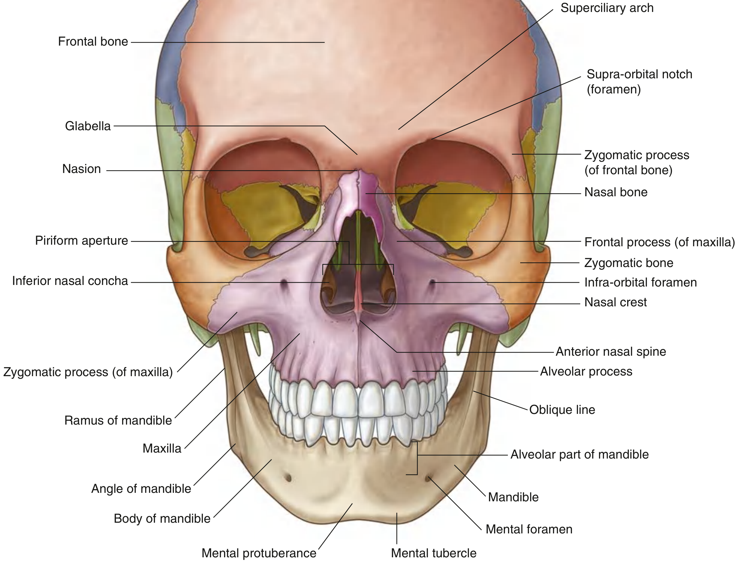
Key features visible from the front:
- Glabella - the smooth flat area between the eyebrows
- Nasion - the junction of the nasal and frontal bones
- Superciliary arches - bony ridges above the orbits (more pronounced in males)
- Supra-orbital foramen - transmits the supra-orbital nerve and vessels
- Piriform aperture - the bony entrance to the nasal cavity
- Infra-orbital foramen - transmits infra-orbital nerve and vessels
- Mental foramen - on the mandible; transmits mental nerve and vessels
- Mental protuberance - the chin prominence
Lateral (Side) View of the Skull
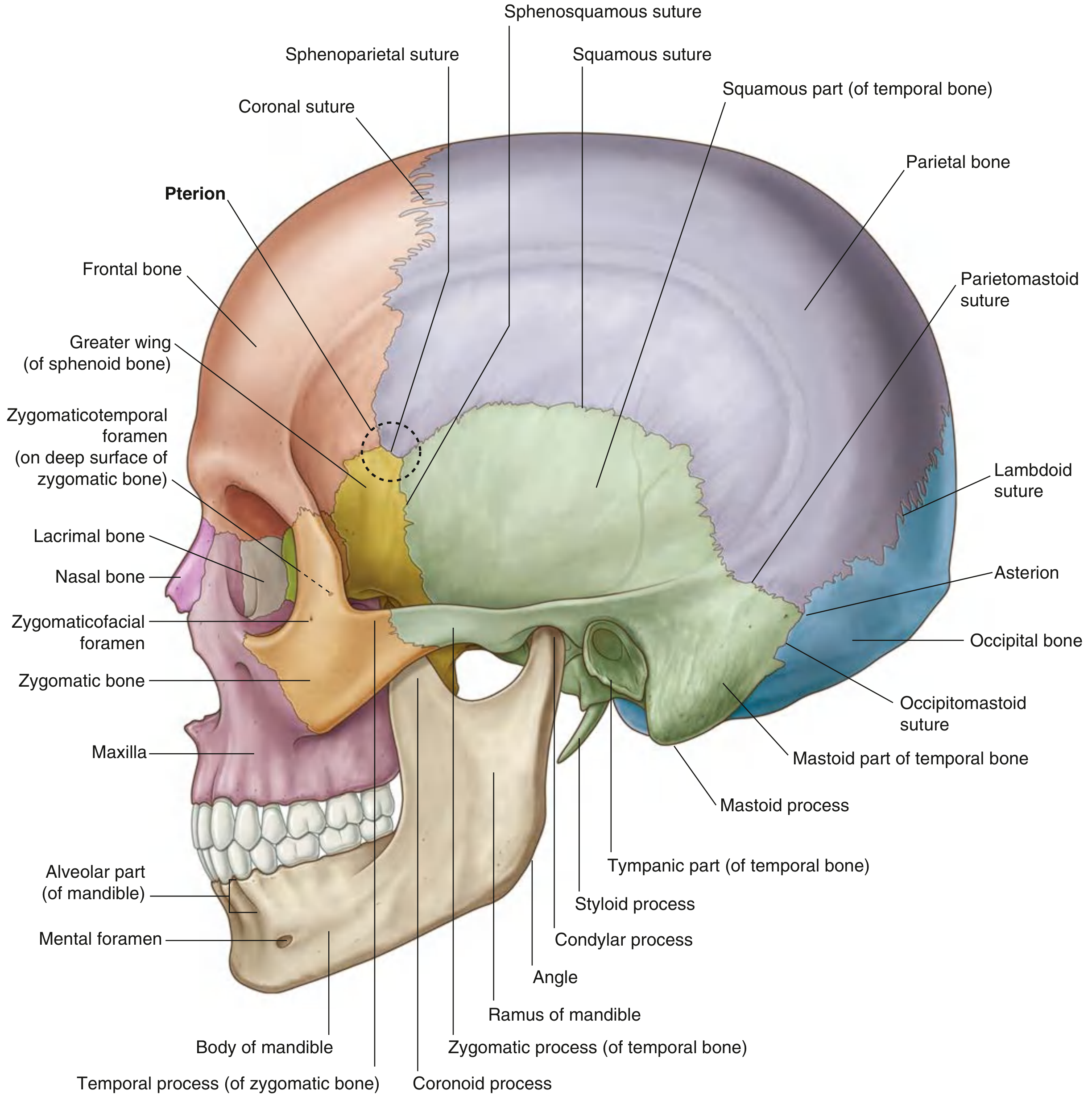
Key features:
- Pterion - the thin, H-shaped junction of frontal, parietal, sphenoid, and temporal bones. Clinically important because the middle meningeal artery runs beneath it - a blow here can cause an extradural (epidural) haematoma
- Coronal suture - frontal meets parietal bones
- Squamous suture - temporal meets parietal
- Lambdoid suture - parietal meets occipital
- Mastoid process - rounded projection behind the ear; attachment for sternocleidomastoid
- Styloid process - slender projection below the ear
- Zygomatic arch - the cheekbone arch formed by zygomatic and temporal bones
Sutures and Fontanelles
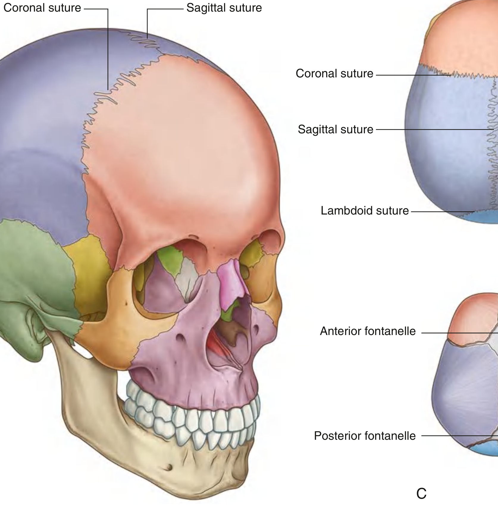
Sutures are immovable fibrous joints between skull bones. The major ones are:
| Suture | Bones Joined |
|---|---|
| Coronal | Frontal + parietal |
| Sagittal | Two parietal bones (midline) |
| Lambdoid | Parietal + occipital |
| Squamous | Parietal + temporal |
Key junctions:
- Bregma - where sagittal and coronal sutures meet (site of anterior fontanelle in infants)
- Lambda - where sagittal and lambdoid sutures meet (site of posterior fontanelle)
- Asterion - where lambdoid, parietomastoid, and occipitomastoid sutures meet
In fetuses and newborns, fontanelles (unossified membranous gaps) allow the skull to deform during birth and permit postnatal brain growth. The anterior fontanelle closes by around 18 months; the posterior by about 3 months.
Special Joints of the Skull
There are only three pairs of synovial joints in the head:
- Temporomandibular joints (TMJ) - between the mandible and temporal bone; the most important movable joints of the skull
- Malleus-incus joint (in the middle ear)
- Incus-stapes joint (in the middle ear)
All other skull bones are united by sutures.
The Cranial Base (Internal View)
The interior of the base of the skull is divided into three fossae:
| Fossa | Key Contents |
|---|---|
| Anterior cranial fossa | Frontal lobes of the brain; ethmoid bone (cribriform plate - where olfactory nerves pass) |
| Middle cranial fossa | Temporal lobes; pituitary fossa (sella turcica) on the sphenoid; numerous foramina for cranial nerves |
| Posterior cranial fossa | Brainstem and cerebellum; foramen magnum (where the brainstem exits to become the spinal cord) |
Mandible
The mandible consists of:
- Body - the horizontal part bearing the teeth (alveolar part above, base below)
- Ramus - the vertical part ascending posteriorly
- Angle - where body and ramus meet
- Condylar process - articulates with the temporal bone to form the TMJ
- Coronoid process - attachment for temporalis muscle
- Mental protuberance - the midline chin bump
The Calvaria's Unique Structure
The flat bones of the skullcap have a specialized three-layer sandwich structure:
- External table - outer compact (dense) bone
- Diploe - middle layer of spongy cancellous bone containing red bone marrow and diploic veins
- Internal table - inner compact bone (thinner and more fragile)
This structure provides strength while minimizing weight, and the diploe acts as a shock absorber.
Clinical Highlights
- Pterion fractures - can tear the middle meningeal artery, causing an extradural haematoma with a "lucid interval"
- Skull base fractures - may result in cerebrospinal fluid leaking from the nose (rhinorrhoea) or ear (otorrhoea)
- Craniosynostosis - premature fusion of cranial sutures, restricting brain growth; requires surgical correction
- Bulging anterior fontanelle in an infant suggests raised intracranial pressure
- Sunken fontanelle suggests dehydration
- Wormian (sutural) bones - small extra islands of bone occasionally seen along the lambdoid suture, a normal variant
Sources: Gray's Anatomy for Students; TeachMeAnatomy skull overview
This is a shared conversation. Sign in to Orris to start your own chat.