
Q1 — 20 marks Define inflammation and discuss in detail the process of phagocytosis and clearing of offending agents. Q2 — 20 marks Define thrombosis. Describe in detail the pathology of thrombosis and add a note on fate of thrombus. Q3 — 20 marks Describe in detail the laboratory diagnosis of cancer. Q4 — Short notes — 4 × 10 marks a) Fluorescence in situ hybridization (FISH) b) Transplant rejection and graft-versus-host disease c) Various pigments in the body and their special stains d) Classify briefly storage disorders; write a note on Gaucher disease
Reading File
This is a comprehensive pathology exam with 4 questions. Given the budget constraints, I'll run parallel searches across all topics simultaneously.
Finding Sources
Finding Sources
Finding Sources
Finding Sources
Now let me read the key sections from Robbins (the primary pathology textbook) in parallel:
Reading File
Reading File
Reading File
Finding Sources
Reading File
Reading File
Finding Sources
Finding Sources
Reading File
Reading File
Reading File
Finding Sources
Reading File
Finding Sources
Finding Sources
Reading File
Reading File
Reading File
Reading File
Reading File
Reading File
I now have sufficient material from Robbins to write comprehensive exam answers. Let me also get the sections on inflammation definition, thrombosis morphology, pigments/stains, and Gaucher's.
Reading File
Reading File
Reading File
I now have comprehensive material from Robbins & Kumar. Let me update todos and compile the full exam answers.---
Q1 — Inflammation and Phagocytosis (20 Marks)
Definition of Inflammation
Inflammation is a protective response of vascularized tissues to infections, damaged cells, or other noxious stimuli. Its purpose is to eliminate the initial cause of cell injury, clear out necrotic cells and tissues resulting from the original insult, and to initiate tissue repair. It is fundamentally a response of the living, vascularized connective tissue and involves the vessels, leukocytes, and plasma proteins. While inflammation is ordinarily protective, the inflammatory response itself can cause damage — it underlies many common and serious human diseases (e.g., rheumatoid arthritis, atherosclerosis, ARDS, septic shock).
Inflammation can be:
- Acute - rapid in onset, short duration; characterized by exudation of fluid, plasma proteins (edema), and predominance of neutrophils
- Chronic - longer duration; characterized by lymphocytes, macrophages, plasma cells, new vessel formation (angiogenesis), and fibrosis
Causes of Inflammation
- Infections - bacterial, viral, fungal, parasitic; microbial products trigger recognition
- Tissue necrosis - from ischemia, trauma, thermal/chemical injury; necrotic cells release danger signals (sterile inflammation)
- Foreign bodies - sutures, implants, splinters
- Immune reactions - autoimmune diseases, hypersensitivity reactions
Recognition of Offending Agents
The first step is recognition via pattern-recognition receptors (PRRs) on macrophages and dendritic cells:
- Toll-like receptors (TLRs) - recognize PAMPs (pathogen-associated molecular patterns) such as LPS of gram-negative bacteria, peptidoglycan of gram-positive bacteria, viral RNA, and unmethylated CpG DNA
- NOD-like receptors (NLRs) - cytoplasmic receptors that recognize microbial products and DAMPs; form the inflammasome which activates IL-1β and IL-18
- DAMPs (damage-associated molecular patterns) - molecules released from damaged cells, e.g., uric acid crystals, ATP, HMGB1, DNA - stimulate sterile inflammation
Process of Phagocytosis
Phagocytosis is the ingestion of particulate material (microbes, dead cells, foreign particles) by cells. The key phagocytes are:
| Property | Neutrophils | Macrophages |
|---|---|---|
| Origin | HSCs in bone marrow | HSCs/embryonic precursors |
| Life span in tissues | 1-2 days | Days to years |
| Key responses | Rapid degranulation, ROS burst | Prolonged; cytokine secretion |
| Nitric oxide | Low/none | iNOS-induced |
| NET formation | Yes | Little/none |
Step 1: Recruitment of Leukocytes (Leukocyte Extravasation)
Leukocytes must first reach the site. This occurs in postcapillary venules in four sequential steps:
a) Margination - Under normal laminar flow, RBCs are in the center and leukocytes at the periphery. As blood slows (stasis), leukocytes are displaced further toward the vessel wall.
b) Rolling - Loose, transient binding mediated by selectins:
- P-selectin and E-selectin on endothelium (stored in Weibel-Palade bodies; upregulated by histamine, thrombin, TNF, IL-1)
- L-selectin on leukocytes
- Selectins bind sialylated carbohydrate ligands (e.g., PSGL-1)
c) Firm adhesion - Mediated by integrins on leukocytes (e.g., LFA-1, MAC-1) binding to ICAMs (intercellular adhesion molecules) on endothelium. Integrins are activated to high-affinity state by chemokines (e.g., IL-8/CXCL8) displayed on the endothelial surface.
d) Transmigration (diapedesis) - Leukocytes squeeze between endothelial cells at intercellular junctions, mediated by PECAM-1 (CD31) expressed on both leukocytes and endothelial cells. They then breach the basement membrane using collagenases.
e) Chemotaxis - Leukocytes migrate along a chemical gradient toward the injury site, guided by chemotactic agents:
- Exogenous: bacterial products (e.g., formyl-methionyl peptides, N-fMLP)
- Endogenous: C5a (complement), leukotriene B4 (LTB4), IL-8 (CXCL8), platelet-activating factor (PAF)
Step 2: Recognition and Attachment (Opsonization)
Phagocytes recognize microbes via:
- Mannose receptors - bind terminal mannose residues of microbial glycoproteins
- Scavenger receptors - recognize oxidized LDL, microbial surfaces
Efficiency is greatly enhanced by opsonization - coating the target with molecules called opsonins for which phagocytes have specific receptors:
- IgG antibodies (Fc portion binds Fc-γ receptors on phagocytes)
- C3b (complement fragment; binds complement receptors CR1, CR3)
- Plasma lectins (mannose-binding lectin, collectins)
Step 3: Engulfment
After receptor binding, the phagocyte membrane pseudopods flow around the particle in a zipper-like manner. The particle is enclosed within a phagosome (membrane-bound vesicle).
Step 4: Killing and Degradation
The phagosome fuses with lysosomes to form a phagolysosome. Microbial killing uses:
A. Reactive Oxygen Species (ROS) - Oxidative Burst (Respiratory Burst)
- Triggered by the assembly of NADPH oxidase (phagocyte oxidase) on the phagosomal membrane
- O₂ + NADPH → superoxide (O₂⁻) → H₂O₂ (hydrogen peroxide)
- MPO (myeloperoxidase) from neutrophil granules: H₂O₂ + Cl⁻ → HOCl (hypochlorous acid) - the most potent microbicidal agent
- Also formed: hydroxyl radical (OH·), peroxynitrite (ONOO⁻)
B. Reactive Nitrogen Species (RNS)
- iNOS (inducible nitric oxide synthase) in macrophages catalyzes: arginine → nitric oxide (NO)
- NO reacts with ROS to form peroxynitrite (ONOO⁻), which destroys microbes
C. Lysosomal Enzymes
- Primary (azurophil) granules of neutrophils: myeloperoxidase (MPO), lysozyme, acid hydrolases, elastase, defensins
- Secondary (specific) granules: lactoferrin (iron chelator), collagenase, B12-binding proteins
- Lysozyme cleaves the muramic acid-acetylglucosamine bond in bacterial cell walls
- Defensins punch holes in microbial membranes
Step 5: Neutrophil Extracellular Traps (NETs)
An additional mechanism: neutrophils eject a web of nuclear chromatin, histones, and antimicrobial proteins (NETs) that trap and kill extracellular bacteria and fungi. This process may be associated with neutrophil death (NETosis).
Clearance of Offending Agents
Once the inciting stimulus is eliminated:
- Dead microbes and debris are phagocytosed by macrophages
- Neutrophils undergo apoptosis (lifespan 1-2 days in tissues) and are cleared by macrophages via efferocytosis - this actively promotes anti-inflammatory signals (release of TGF-β, IL-10)
- Short-lived inflammatory mediators (prostaglandins, leukotrienes, PAF, histamine) are rapidly degraded or their synthesis ceases
- Lipoxins, resolvins, and protectins are biosynthesized from arachidonic acid and omega-3 fatty acids; these are pro-resolution mediators that actively terminate the acute inflammatory response
- The net result is return to homeostasis, followed by tissue repair through regeneration or scarring
Q2 — Thrombosis: Definition, Pathology, and Fate of Thrombus (20 Marks)
Definition
Thrombosis is defined as the pathological formation of a blood clot (thrombus) within the cardiovascular system in a living individual. It is distinct from physiological hemostasis (which involves clot formation to stop bleeding from injured vessels). A thrombus is a solid mass formed from blood constituents that has the potential to obstruct blood flow, leading to tissue ischemia and infarction.
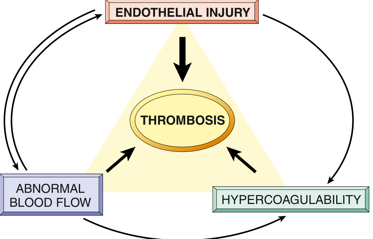
Pathology of Thrombosis - Virchow's Triad
The primary abnormalities that lead to intravascular thrombosis form the Virchow triad:
- Endothelial injury
- Abnormal blood flow (stasis or turbulence)
- Hypercoagulability of blood
These factors may act independently or together; Virchow's triad is illustrated below:
1. Endothelial Injury
This is the most important factor. Endothelial injury is almost always the cause of thrombus formation in the heart and arterial circulation (high flow states).
Causes of endothelial injury/dysfunction:
- Physical injury: trauma, surgery, catheter placement
- Hemodynamic stress: hypertension, turbulent flow at bifurcations
- Hypercholesterolemia, homocysteinemia
- Inflammatory cytokines (TNF, IL-1)
- Toxins from cigarette smoke
- Infectious agents
Prothrombotic changes in activated endothelium:
- Procoagulant changes: downregulation of thrombomodulin, endothelial protein C receptor, tissue factor pathway inhibitor (TFPI); upregulation of tissue factor (TF) → activates extrinsic coagulation pathway
- Antifibrinolytic effects: increased secretion of PAI (plasminogen activator inhibitors) → reduced fibrinolysis
- Exposure of subendothelial collagen and von Willebrand factor (vWF) → platelet adhesion and activation
Cardiac and arterial clots are typically platelet-rich ("white thrombi") because platelet activation is essential for thrombus formation under high shear conditions.
2. Abnormal Blood Flow
Turbulence (arterial and cardiac thrombosis):
- Disrupts laminar flow, causes endothelial injury
- Forms countercurrents and local pockets of stasis
Stasis (venous thrombosis - the dominant factor):
- Prevents dilution of activated clotting factors by fresh blood
- Retards the inflow of clotting factor inhibitors
- Promotes endothelial activation
Clinical situations with abnormal flow:
- Ulcerated atherosclerotic plaques → turbulence
- Aneurysms → local stasis
- Myocardial infarction → dyskinetic wall motion → stasis
- Atrial fibrillation → stasis in atria (classically in the left atrial appendage)
- Venous thrombosis in bed-ridden patients → stasis in leg veins
3. Hypercoagulability
An alteration in the coagulation pathways that predisposes to thrombosis.
Primary (genetic) hypercoagulable states:
- Factor V Leiden mutation - point mutation renders Factor V resistant to cleavage by activated protein C (most common inherited cause of hypercoagulability)
- Prothrombin G20210A mutation - elevated prothrombin levels
- Antithrombin III deficiency - reduced inhibition of thrombin and Xa
- Protein C or Protein S deficiency - reduced inactivation of Factors Va and VIIIa
- MTHFR mutation → hyperhomocysteinemia → endothelial damage
Secondary (acquired) hypercoagulable states:
- Heparin-Induced Thrombocytopenia (HIT): autoantibodies to heparin-PF4 complexes → platelet activation → paradoxical thrombosis despite heparin
- Antiphospholipid antibody syndrome (lupus anticoagulant): antibodies to β₂-glycoprotein I; causes recurrent thromboses, miscarriages, valve vegetations
- Oral contraceptives, pregnancy → increased hepatic coagulation factor synthesis
- Disseminated cancers → release of procoagulant mucins
- Smoking, obesity → unknown mechanisms
Morphology of Thrombi
Arterial thrombi:
- Frequently occlusive
- Platelet-rich, pale/grey ("white thrombus")
- Typically on ruptured atherosclerotic plaques
- Lines of Zahn (alternating layers of fibrin/platelets and red cells) seen in cardiac/aortic thrombi - indicate formation during life
Venous thrombi (phlebothrombosis):
- Almost invariably occlusive
- Red, "stasis thrombus" - rich in enmeshed red cells within fibrin mesh
- Commonly in leg veins (90% of cases)
- Propagate toward the heart as long casts
Mural thrombi: in heart chambers or aortic lumen; seen in MI, arrhythmias, aneurysms
Vegetations: thrombi on heart valves - infective endocarditis (large, friable), non-bacterial thrombotic endocarditis (small, sterile), Libman-Sacks endocarditis (SLE)
Fate of Thrombus
If a patient survives the initial event, the thrombus evolves through four possible outcomes:
1. Propagation
- The thrombus enlarges through continued accretion of platelets and fibrin
- Increases the risk of vascular occlusion and embolization
- Most clinically dangerous early fate
2. Embolization
- Part or all of the thrombus dislodges and is transported to a distant site
- Venous thrombi → pulmonary emboli (PE) → right heart failure, hypoxia, sudden death
- Arterial/cardiac thrombi → systemic emboli → stroke, limb ischemia, bowel infarction
3. Dissolution (Lysis)
- Activation of fibrinolytic factors (plasmin cleaves fibrin)
- Effective only for newly formed thrombi - older thrombi resist lysis due to extensive fibrin polymerization
- This is the rationale for giving t-PA within hours of acute coronary thrombosis (narrow therapeutic window)
4. Organization and Recanalization
- Organization - ingrowth of endothelial cells, smooth muscle cells, and fibroblasts into the thrombus
- Recanalization - new capillary channels form within the organized thrombus, partially restoring blood flow through the original lumen
- Over time, the thrombus is incorporated into the vessel wall as vascularized connective tissue
- Occasionally the center undergoes enzymatic digestion (release of lysosomal enzymes from entrapped leukocytes) rather than organizing
Clinical significance: Thrombi are dangerous primarily because they obstruct arteries/veins and generate emboli. Venous thrombi are most dangerous for embolization; arterial thrombi are most dangerous for local infarction.
Q3 — Laboratory Diagnosis of Cancer (20 Marks)
Introduction
Every year the approach to laboratory diagnosis of cancer grows more sophisticated. The goals are: (1) accurate diagnosis and classification, (2) prognosis and prediction of therapeutic response, (3) detection of minimal residual disease, (4) identifying hereditary cancer syndromes, and (5) monitoring therapy. Multiple complementary methods are now used.
1. Morphologic Methods
These remain the cornerstone of cancer diagnosis.
a) Histopathology (Tissue Biopsy)
The most important method. The pathologist evaluates:
- Cellular features of malignancy: nuclear pleomorphism, hyperchromasia, prominent nucleoli, increased N:C ratio, atypical mitoses
- Architectural features: glandular pattern (carcinoma), sheets vs. nests vs. cords
- Invasion: beyond basement membrane (cardinal feature of malignancy)
- Grading: based on degree of differentiation; ranges from Grade 1 (well-differentiated) to Grade 3-4 (poorly differentiated/undifferentiated = anaplastic)
- Staging (TNM system): T1-T4 (primary tumor size/invasion), N0-N3 (nodal involvement), M0-M1 (metastases); staging has greater clinical value than grading
Types of biopsies:
- Excision biopsy: complete removal - gold standard
- Incision biopsy / core needle biopsy: representative sample
- Frozen section: rapid intraoperative diagnosis (within minutes) - used for surgical decision-making; excellent accuracy but inferior detail
- Fine-needle aspiration (FNA): minimally invasive; aspiration of cells from palpable masses (breast, thyroid, lymph nodes); rapid but limited by small sample
b) Cytology (Papanicolaou Smear)
- Shed or scraped cells examined for cytological features of malignancy
- Used for: cervical carcinoma (Pap smear), bronchogenic carcinoma (sputum cytology), bladder carcinoma (urinary cytology), effusions (ascitic, pleural, CSF), endometrial sampling
- Malignant cells are less cohesive, shed easily, show anaplastic features
- Most successful public health application: control of cervical cancer by Pap smear
2. Immunohistochemistry (IHC) and Flow Cytometry
Immunohistochemistry
Uses specific monoclonal antibodies conjugated to fluorescent dyes or enzymes to detect proteins in tissue sections:
| Marker | Utility |
|---|---|
| Cytokeratins (CK7, CK20) | Identify carcinoma (epithelial origin) vs. lymphoma |
| Vimentin | Sarcomas, lymphomas |
| CD45 (leukocyte common antigen) | Lymphoma vs. carcinoma |
| PSA (prostate-specific antigen) | Diagnose prostatic carcinoma in metastases |
| ER/PR (estrogen/progesterone receptors) | Breast cancer prognosis and hormone therapy eligibility |
| HER2/neu (ERBB2) | Breast/gastric cancer; eligibility for trastuzumab |
| Synaptophysin, chromogranin | Neuroendocrine tumors |
| S-100, HMB-45, Melan-A | Melanoma |
| TTF-1 | Thyroid and lung primary carcinomas |
| AFP | Hepatocellular carcinoma, yolk sac tumors |
Flow Cytometry
- Fluorescently labeled antibodies used to phenotype cells
- Used routinely in classification of leukemias and lymphomas (T-cell vs. B-cell, lymphoblastic vs. mature)
- Determines CD marker patterns (e.g., CD10, CD19, CD20 for B-cell; CD3, CD4, CD8 for T-cell)
3. Tumor Markers (Biochemical Assays)
Cannot be used for definitive diagnosis alone, but useful for screening, monitoring therapy, and detecting recurrence:
| Marker | Normal | Tumor Association |
|---|---|---|
| AFP (alpha-fetoprotein) | <8.4 ng/mL | Hepatocellular carcinoma, yolk sac tumor |
| CEA (carcinoembryonic antigen) | <3 ng/mL | Colorectal, gastric, lung, breast carcinomas |
| PSA (prostate-specific antigen) | <4 ng/mL | Prostate carcinoma (screening + monitoring) |
| CA-125 | <46 U/mL | Epithelial ovarian carcinoma |
| CA 19-9 | <35 U/mL | Pancreatic ductal adenocarcinoma |
| CA 15-3 | <30 U/mL | Breast carcinoma |
| hCG (beta-human chorionic gonadotropin) | - | Gestational trophoblastic disease, testicular tumors |
| LDH | - | Lymphomas, testicular tumors (prognosis) |
| Calcitonin | - | Medullary carcinoma of thyroid |
| 5-HIAA (urine) | - | Carcinoid tumors |
4. Molecular/Genetic Methods
Modern oncology relies heavily on molecular characterization for diagnosis, prognosis, and targeted therapy.
a) Cytogenetics and FISH
- Conventional karyotyping: detects chromosomal translocations, deletions, amplifications
- FISH: fluorescent probes detect specific chromosomal loci; detects gene amplification (e.g., HER2), translocations (e.g., BCR-ABL in CML), deletions; can be done on non-dividing (interphase) cells
b) PCR (Polymerase Chain Reaction)
- Detects minimal residual disease (e.g., BCR-ABL transcripts in CML after treatment)
- Detects viral oncogenes (HPV in cervical cancer)
- RT-PCR for gene expression profiling
c) DNA/RNA Sequencing (Next-Generation Sequencing - NGS)
- Somatic mutation profiling: identifies actionable driver mutations (e.g., EGFR, KRAS, BRAF, ALK, ROS1)
- Guides targeted therapy (e.g., EGFR mutation → erlotinib; BRAF V600E → vemurafenib)
- Germline sequencing: identifies hereditary predispositions (BRCA1/2, TP53 in Li-Fraumeni syndrome, APC in FAP, MLH1/MSH2 in Lynch syndrome)
d) Gene Expression Profiling (Microarrays, RNA-Seq)
- Classifies molecularly distinct subtypes (e.g., luminal A/B vs. HER2-enriched vs. basal-like breast cancers)
- OncotypeDX (21-gene signature) and MammaPrint (70-gene signature): predict recurrence risk in breast cancer and guide chemotherapy decisions
e) Methylation Analysis
- Methylation of MGMT promoter in glioblastoma → better response to alkylating agents (temozolomide)
- MLH1 promoter methylation in colorectal cancer → MSI (microsatellite instability)
f) Liquid Biopsy
- Detection of circulating tumor DNA (ctDNA), circulating tumor cells (CTCs), exosomes in blood, stool, sputum, urine
- Non-invasive; can detect minimal residual disease, treatment resistance mutations, early recurrence
- Growing clinical role for monitoring therapy
5. Electron Microscopy
- Now largely replaced by IHC and molecular methods
- Still used for: classification of undifferentiated tumors, confirming neuroendocrine origin (dense-core secretory granules), Langerhans cell histiocytosis (Birbeck granules)
Q4 — Short Notes (4 × 10 Marks)
(a) Fluorescence In Situ Hybridization (FISH)
Principle
FISH is a molecular cytogenetic technique that uses fluorescently labeled DNA probes to detect and localize specific DNA sequences on chromosomes or in cell nuclei in situ (in place, without disrupting cellular architecture). It was developed in the early 1990s and dramatically expanded the scope of cytogenetic analysis.
Steps in FISH
- Probe preparation: DNA clones (from BAC or PAC libraries) containing the sequence of interest are labeled with fluorescent dyes (fluorophores) by nick translation or random priming
- Specimen preparation: chromosome preparations (metaphase spreads) or interphase nuclei on glass slides
- Denaturation: both probe and target DNA are denatured (double-stranded → single-stranded) by heat or formamide
- Hybridization: the labeled probe is applied and incubated; it anneals (hybridizes) to its complementary target sequence
- Washing: unbound probe is washed off
- Visualization: fluorescence microscope; each fluorescent signal = one copy of the target sequence
Types of FISH Probes
| Probe Type | Detects |
|---|---|
| Locus-specific probes | Gene-level deletions, duplications, amplifications |
| Centromere probes (CEP) | Chromosome aneuploidy (gains/losses) |
| Whole chromosome painting probes | Structural rearrangements, translocations |
| Telomere probes | Cryptic subtelomeric deletions |
Applications in Pathology
- Oncology:
- HER2 amplification in breast/gastric cancer (eligibility for trastuzumab) - counted as HER2:CEP17 ratio
- BCR-ABL fusion (Philadelphia chromosome, t(9;22)) in CML and ALL
- ALK rearrangement in lung adenocarcinoma (eligibility for crizotinib)
- N-MYC amplification in neuroblastoma (poor prognosis)
- EGFR, ROS1, RET rearrangements in lung cancer
- Congenital anomalies:
- Detection of microdeletions: 22q11.2 (DiGeorge syndrome), 15q11 (Prader-Willi/Angelman), 7q11.23 (Williams syndrome)
- Prenatal diagnosis: on uncultured amniocytes for rapid aneuploidy detection (trisomy 21, 13, 18; X, Y)
Advantages
- Can be performed on non-dividing (interphase) cells - no need for cell culture
- Higher resolution than conventional karyotyping
- Can be used on FFPE (formalin-fixed paraffin-embedded) tissue sections
- Multiple probes with different colors (multicolor FISH) can be used simultaneously
Limitations
- Only detects targeted regions; not genome-wide
- Cannot detect point mutations or low-level mosaicism
- Balanced translocations without gene disruption may be missed
(b) Transplant Rejection and Graft-Versus-Host Disease (GVHD)
Transplant Rejection
Rejection is an immune-mediated destruction of the transplanted organ by the recipient's immune system due to recognition of transplant antigens (mainly HLA/MHC antigens) as foreign.
Mechanisms of Allorecognition
- Direct pathway: recipient T cells recognize intact donor MHC molecules on donor APCs (in graft) - major pathway of acute rejection
- Indirect pathway: recipient T cells recognize donor-derived peptides (processed from donor MHC) presented by recipient APCs - more important in chronic rejection
Types of Rejection
1. Hyperacute Rejection
- Occurs within minutes to hours of transplantation
- Caused by preformed antibodies against donor endothelium (ABO blood group antigens, HLA Class I)
- Diffuse thrombosis, widespread ischemic necrosis of the graft
- Prevented by pre-transplant cross-matching
2. Acute Rejection
- Occurs within days to weeks (can occur months later if poorly immunosuppressed)
- Two forms:
- Acute cellular rejection: CD4 T cells (via cytokines) and CD8 cytotoxic T cells attack donor parenchymal cells; tubulitis in kidney, bile duct destruction in liver
- Acute antibody-mediated (humoral) rejection: donor-specific antibodies cause endothelialitis, neutrophil infiltration, C4d deposition on capillary endothelium
- Responds to intensified immunosuppression (corticosteroids, anti-thymocyte globulin)
3. Chronic Rejection
- Occurs over months to years; major cause of long-term graft failure
- Both cellular and humoral mechanisms involved
- Characterized by intimal smooth muscle proliferation (transplant vasculopathy), interstitial fibrosis, tubular atrophy
- Poorly responsive to immunosuppression; may require re-transplantation
Graft-Versus-Host Disease (GVHD)
GVHD is the reverse of rejection - it occurs primarily after allogeneic hematopoietic stem cell transplantation (HSCT), when immunocompetent donor T cells recognize the recipient's tissues as foreign and mount an immune attack.
Setting
- Allogeneic HSCT for leukemias, lymphomas, aplastic anemia, severe thalassemia
- Recipient's marrow must first be destroyed by irradiation/chemotherapy (conditioning)
- Mature donor T cells contaminating the HSC preparation are the effectors
Pathogenesis
- Donor T cells (both CD4 and CD8) recognize recipient HLA alloantigens on recipient dendritic cells in secondary lymphoid organs
- They become activated, expand clonally, and migrate to target tissues
- Target organs (damaged by conditioning): skin (rash, desquamation), intestines (watery/bloody diarrhea), liver (hepatitis, cholestatic jaundice)
Clinical Features
| Feature | Acute GVHD (<100 days) | Chronic GVHD (>100 days) |
|---|---|---|
| Skin | Erythematous rash, bullae | Scleroderma-like fibrosis |
| GI | Diarrhea, vomiting | Malabsorption, esophageal stricture |
| Liver | Elevated bilirubin/enzymes | Biliary cirrhosis |
| Immunosuppression | Profound | Profound |
Beneficial Effect - Graft-Versus-Leukemia (GVL) Effect
- The same alloreactive donor T cells that cause GVHD also recognize and kill residual leukemia cells
- This GVL effect is crucial for the therapeutic success of HSCT in leukemia
- Total elimination of GVHD would increase relapse rates; a degree of controlled GVHD is acceptable
Prevention and Treatment
- HLA matching (more critical than solid organ transplantation, especially MHC Class I and II)
- T-cell depletion of the HSC graft (reduces GVHD but also GVL effect)
- Prophylaxis: cyclosporine + methotrexate; tacrolimus
- Treatment of established GVHD: high-dose corticosteroids, ruxolitinib (JAK inhibitor)
(c) Various Pigments in the Body and Their Special Stains
Pigments are colored substances that can accumulate in cells and tissues, either endogenously or from exogenous sources. They are important diagnostically.
Exogenous Pigments
| Pigment | Source | Appearance | Stain |
|---|---|---|---|
| Carbon (anthracosis) | Inhaled coal dust, pollution | Black, granular in macrophages; lung, lymph nodes | No special stain needed (black) |
| Silica | Occupational exposure | Silicotic nodules | Polarized light (birefringent) |
| Asbestos (ferruginous bodies) | Occupational | Golden-brown, beaded "dumbell" rods | Prussian blue (iron coat) |
| Tattoo pigments | Tattoo ink | Various colors in dermal macrophages | No special stain |
| Lead | Environmental | Basophilic stippling of RBCs | - |
Endogenous Pigments
1. Hemosiderin
- Derived from hemoglobin breakdown; insoluble iron-storage complex (ferritin aggregates)
- Golden-brown, granular pigment within macrophages
- Local accumulation: hemorrhage, bruising (the yellow-brown color of old bruise)
- Systemic accumulation: hemosiderosis (excess iron deposition without damage), hemochromatosis (organ damage)
- Stain: Prussian blue (Perls' stain) - hemosiderin stains blue
2. Hematin / Formalin Pigment
- Artifact from formalin fixation of blood-rich tissues
- Brown-black, granular, birefringent
- Stain: Not specifically; can be removed by alkaline alcohol
3. Bilirubin
- Yellow-green pigment from heme catabolism (non-iron portion of hemoglobin)
- Deposits in jaundice: bile thrombi in hepatic canaliculi, bilirubin crystals in gallbladder
- Inspissated bile in hepatocytes and bile ducts
- Stain: Hall's bile stain (stains bile duct plugs green); van Gieson's
4. Lipofuscin ("Wear-and-tear" pigment)
- Yellow-brown, finely granular pigment in cytoplasm of hepatocytes, cardiac myocytes, neurons
- Composed of oxidized lipids and proteins (lipid peroxidation products)
- Accumulates with aging and oxidative stress; not harmful per se
- Causes brown atrophy of organs (heart, liver)
- Stain: PAS positive; Sudan black B; autofluorescent on fluorescence microscopy
5. Melanin
- Brown-black pigment produced by melanocytes from tyrosine via tyrosinase
- Normally in skin, hair, eye (retinal pigment epithelium)
- Pathological accumulations: melanoma, Addison disease (increased ACTH → hyperpigmentation), Peutz-Jeghers syndrome (mucosal)
- Stain: Fontana-Masson stain (silver stain, stains melanin black); Schmorl's stain
- IHC: HMB-45, Melan-A, S-100 protein
6. Hemozoin (Malarial Pigment)
- Dark brown/black crystalline pigment derived from hemoglobin degradation by Plasmodium
- Found within infected RBCs and phagocytes in malaria
- Stain: Giemsa stain for blood films; appears brown-black
7. Ochronotic Pigment
- Dark brown-black homogentisic acid polymer deposited in connective tissue, cartilage, sclera
- Seen in alkaptonuria (homogentisic acid oxidase deficiency)
Summary Table - Stains for Endogenous Pigments
| Pigment | Special Stain | Color |
|---|---|---|
| Hemosiderin | Prussian blue (Perls') | Blue |
| Lipofuscin | PAS, Sudan black B | Pink (PAS), Black (Sudan) |
| Melanin | Fontana-Masson | Black |
| Bile/bilirubin | Hall's bile stain | Green |
| Calcium | von Kossa | Black |
| Amyloid | Congo red | Apple-green (polarized) |
| Fibrin | MSB (Martius-Scarlet-Blue), PTAH | Red/blue |
(d) Classification of Storage Disorders and Gaucher Disease
Classification of Storage (Lysosomal Storage) Disorders
Lysosomal storage disorders (LSDs) are caused by inherited deficiencies of lysosomal enzymes (or, less commonly, of proteins needed for transport of substances out of lysosomes). The result is accumulation of specific substrates within lysosomes.
Classification by stored substrate:
| Category | Enzyme Defect | Disease |
|---|---|---|
| Sphingolipidoses | β-glucocerebrosidase | Gaucher disease |
| Sphingomyelinase | Niemann-Pick disease (A, B) | |
| Hexosaminidase A | Tay-Sachs disease | |
| Galactocerebrosidase | Krabbe disease | |
| Arylsulfatase A | Metachromatic leukodystrophy | |
| α-galactosidase A | Fabry disease | |
| Mucopolysaccharidoses (MPS) | α-L-iduronidase | Hurler syndrome (MPS I) |
| Iduronate sulfatase | Hunter syndrome (MPS II, X-linked) | |
| Heparan sulfatase | Sanfilippo syndrome (MPS III) | |
| Glycogenoses | Acid maltase (α-1,4-glucosidase) | Pompe disease (Type II glycogenosis) |
| Oligosaccharidoses | α-mannosidase | Mannosidosis |
| Mucolipidoses | Multiple lysosomal hydrolases | I-cell disease (ML II) |
| Lipidoses | Acid lipase | Wolman disease |
| Acid ceramidase | Farber disease |
Inheritance: All LSDs are autosomal recessive except Hunter syndrome (MPS II) which is X-linked recessive and Fabry disease (also X-linked).
Gaucher Disease - A Detailed Note
Gaucher disease is the most common lysosomal storage disorder and the most common inherited disorder among Ashkenazi Jews (carrier frequency ~1:15).
Etiopathogenesis
- Enzyme deficiency: Deficiency of acid β-glucocerebrosidase (glucocerebrosidase) encoded by the GBA gene on chromosome 1q21
- Inheritance: Autosomal recessive
- Result: Accumulation of glucocerebroside (glucosylceramide) within lysosomes of macrophages throughout the body (Kupffer cells in liver, red pulp macrophages in spleen, bone marrow macrophages, pulmonary macrophages)
- The accumulating glucocerebroside comes primarily from the normal catabolism of senescent red and white blood cell membranes
Types of Gaucher Disease
| Type | Frequency | CNS involvement | Prognosis |
|---|---|---|---|
| Type 1 (Non-neuronopathic) | ~95% | None | Chronic; compatible with long life |
| Type 2 (Acute neuronopathic) | Rare | Severe, early-onset (infancy) | Death by age 2 |
| Type 3 (Subacute neuronopathic) | Intermediate | Present but slower | Variable |
Morphology
The Gaucher Cell:
- The hallmark lesion
- Large macrophage (20-100 μm) with abundant pale cytoplasm showing a characteristic "crumpled tissue paper" or "wrinkled tissue paper" appearance
- Cytoplasm: filled with elongated lysosomes packed with glucocerebroside; fibrillary pattern due to tubular storage structures
- Eccentric, small nucleus
- PAS positive (glucocerebroside is a glycolipid)
- Electron microscopy: elongated, curved (spindle-shaped) lysosomes
Organ involvement:
- Spleen: Most dramatically enlarged (can reach 10 kg); splenomegaly is the cardinal clinical feature; packed with Gaucher cells in the red pulp
- Liver: Hepatomegaly (Gaucher cells in sinusoids); fibrosis in severe/long-standing disease
- Bone marrow: Replacement of hematopoietic marrow → cytopenias (anemia, thrombocytopenia, leukopenia)
- Bone: Cortical thinning, medullary expansion → "Erlenmeyer flask deformity" of distal femur on X-ray; osteonecrosis (avascular necrosis of femoral head); pathological fractures
- Lungs: Gaucher cells in alveolar capillaries → respiratory failure in severe cases
- CNS: In Types 2 and 3
Clinical Features (Type 1)
- Massive splenomegaly (often the presenting feature)
- Hepatomegaly (less prominent than spleen)
- Pancytopenia (due to hypersplenism + marrow infiltration): anemia, thrombocytopenia (bleeding), neutropenia
- Bone pain, pathological fractures, avascular necrosis
- Growth retardation in children
- No neurological involvement (Type 1)
Diagnosis
- Enzyme assay: Decreased β-glucocerebrosidase activity in leukocytes or fibroblasts (definitive)
- Gene mutation analysis: GBA gene sequencing (N370S mutation - most common in Ashkenazi Jews, associated with Type 1; L444P - associated with neuronopathic forms)
- Serum biomarkers: Elevated chitotriosidase and glucosylsphingosine (lyso-Gb1) - useful for monitoring treatment response
- Bone marrow biopsy: Gaucher cells (PAS-positive crumpled cells)
- Imaging: MRI for bone marrow infiltration, Erlenmeyer flask deformity on plain X-ray
Treatment
- Enzyme Replacement Therapy (ERT): Imiglucerase (recombinant glucocerebrosidase), also velaglucerase alfa and taliglucerase alfa; IV infusion every 2 weeks; reduces organomegaly, improves cytopenias, prevents bone complications; does not cross BBB (not effective for Types 2/3 CNS disease)
- Substrate Reduction Therapy (SRT): Miglustat, eliglustat - inhibit glucosylceramide synthase, reducing substrate accumulation; oral drugs
- Splenectomy: historically used; now avoided as worsens bone disease
- Bone marrow transplantation: curative but rarely done due to associated risks
Sources: Robbins & Kumar Basic Pathology (Robbins Pathology), 10th ed.; Robbins, Cotran & Kumar Pathologic Basis of Disease; Thompson & Thompson Genetics and Genomics in Medicine, 9th ed.; Janeway's Immunobiology, 10th ed.
This is a shared conversation. Sign in to Orris to start your own chat.